- Physical Examination
- Surgical Examination
- Ophthalmology
- Clinical Skills
- Orthopedics
- Surgery Videos
- Laparoscopy
- Pediatrics
- Funny Videos
- Cardiothoracic Surgery
- Nursing Videos
- Plastic Surgery
- Otorhinolaryngology
- Histology and Histopathology
- Neurosurgery
- Dermatology
- Pediatric Surgery
- Urology
- Dentistry
- Oncology and Cancers
- Anatomy Videos
- Health and Fitness
- Radiology
- Anaesthesia
- Physical Therapy
- Pharmacology
- Interventional Radiology
- Cardiology
- Endocrinology
- Gynecology
- Emergency Medicine
- Psychiatry and Psychology
- Childbirth Videos
- General Medical Videos
- Nephrology
- Physiology
- Diet and Food Health
- Diabetes Mellitus
- Neurology
- Women Health
- Osteoporosis
- Gastroenterology
- Pulmonology
- Hematology
- Rheumatology
- Toxicology
- Nuclear Medicine
- Infectious Diseases
- Vascular Disease
- Reproductive Health
- Burns and Wound Healing
- Other
Top videos
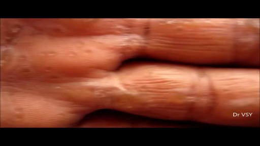
Keratoderma Blennorrhagicum is a manifestation on the skin that appears in patients diagnosed with reactive arthritis (this condition was previously known as Reiter syndrome). The condition manifests itself by lesions that appear on the skin, initially on the palm of the hands and soles of the feet. The lesions have the tendency to spread, affecting other parts of the body, such as the scrotum, scalp or trunk. Because of their appearance, the lesions might be easily confused with the ones from psoriasis. Keratoderma blennorrhagicum is one of the symptoms that can be used for the clinical diagnosis of reactive arthritis.
Transvenous cardiac pace maker, also called endocardial pacing, is a potentially life saving intervention used primarily to correct profound bradycardia. It can be used to treat symptomatic bradycardias that do not respond to transcutaneous pacing or to drug therapy.
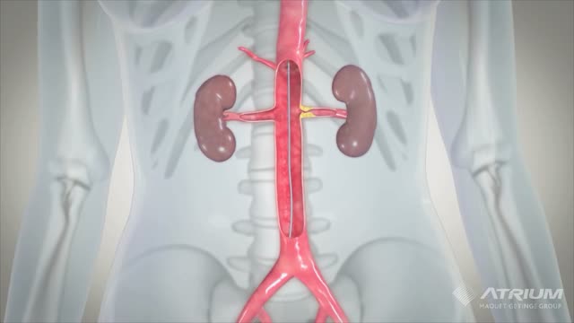
A ureteral stent, sometimes as well called ureteric stent, is a thin tube inserted into the ureter to prevent or treat obstruction of the urine flow from the kidney. The length of the stents used in adult patients varies between 24 to 30 cm.

Heart sounds are the noises generated by the beating heart and the resultant flow of blood through it. Specifically, the sounds reflect the turbulence created when the heart valves snap shut. In cardiac auscultation, an examiner may use a stethoscope to listen for these unique and distinct sounds that provide important auditory data regarding the condition of the heart. In healthy adults, there are two normal heart sounds often described as a lub and a dub (or dup), that occur in sequence with each heartbeat. These are the first heart sound (S1) and second heart sound (S2), produced by the closing of the atrioventricular valves and semilunar valves, respectively. In addition to these normal sounds, a variety of other sounds may be present including heart murmurs, adventitious sounds, and gallop rhythms S3 and S4.
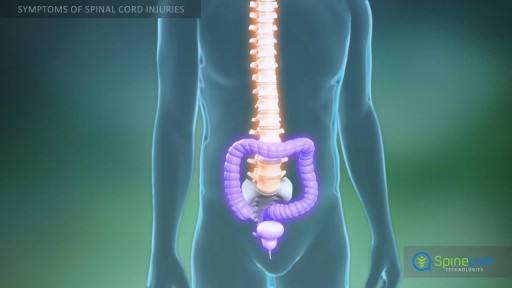
Though you might think of your spinal cord as one single piece, it's actually a column of nerves protected by a sheath of myelin and then further secured by 31 butterfly-shaped vertebrae (singular: vertebra). Medical providers divide the spinal cord into four distinct regions. Knowing the region in which the injury is located is often the key to understanding diagnosis and treatment. The four spinal cord regions are: The cervical spinal cord: This is the topmost portion of the spinal cord, where the brain connects to the spinal cord, and the neck connects to the back. This region consists of eight vertebrae, commonly referred to as C1-C8. All spinal cord numbers are descending, so C1 is the highest vertebra, while C8 is the lowest in this region. The thoracic spinal cord: This section forms the middle of the spinal cord, containing twelve vertebrae numbered T1-T12.
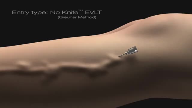
No - Knife Endovenous Laser

Bleeding usually occurs from only one nostril. If the bleeding is heavy enough, the blood can fill up the nostril on the affected side and overflow within the nasopharynx (the area inside the nose where the two nostrils merge), spilling into the other nostril to cause bleeding from both sides. Blood can also drip back into the throat or down into the stomach, causing a person to spit or even vomit blood. Signs of excessive blood loss include dizziness, light-headedness, confusion, and fainting. Excessive blood loss from nosebleeds is rare. Additional bleeding from other parts of the body, such as bleeding gums when brushing teeth, blood in urine or bowel movements, or easy bruising may indicate an inability of the blood to clot. Additional bleeding or easy bruising can be a sign of a more significant medical problem.
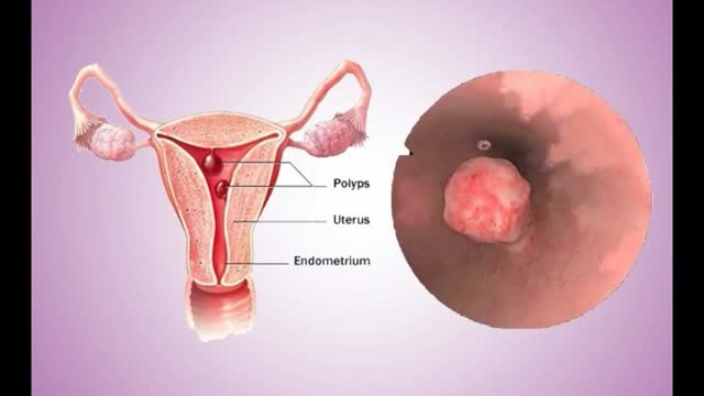
Uterine polyps are growths attached to the inner wall of the uterus that extend into the uterine cavity. Overgrowth of cells in the lining of the uterus (endometrium) leads to the formation of uterine polyps, also known as endometrial polyps. These polyps are usually noncancerous (benign), although some can be cancerous or can eventually turn into cancer (precancerous polyps). Uterine polyps range in size from a few millimeters — no larger than a sesame seed — to several centimeters — golf-ball-size or larger. They attach to the uterine wall by a large base or a thin stalk.
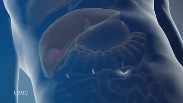
The most frequent incision utilized to open the abdomen for liver surgery is called a chevron incision. In this incision a cut is made on the abdomen below the rib cage. The cut starts under the armpit below the ribs on the right side of the abdomen and continues all the way across the abdomen to the opposite arm pit thereby the whole width of the abdomen is cut to provide access to the liver. The average length of the incision is approximately 24 to 30 inches. This is one of the longest incisions is utilized in abdominal surgery. The incision is frequently associated with significant discomfort after the surgery and in some patients the discomfort can continue for many months, particularly when some of the nerves in the abdominal wall have been cut during the surgery. Laparoscopic surgery provides advantages over open surgery for the liver since the chevron incision is completely avoided and the surgery is performed through tiny incisions. As a consequence the duration of stay in hospital, the amount and duration of post operative discomfort, and the length of recovery is much shorter after the laparoscopic procedure compared to open surgery
A lumbar puncture (also called a spinal tap) is a procedure to collect and look at the fluid (cerebrospinal fluid, or CSF) surrounding the brain and spinal cord. During a lumbar puncture, a needle is carefully inserted into the spinal canal low in the back (lumbar area). Samples of CSF are collected.
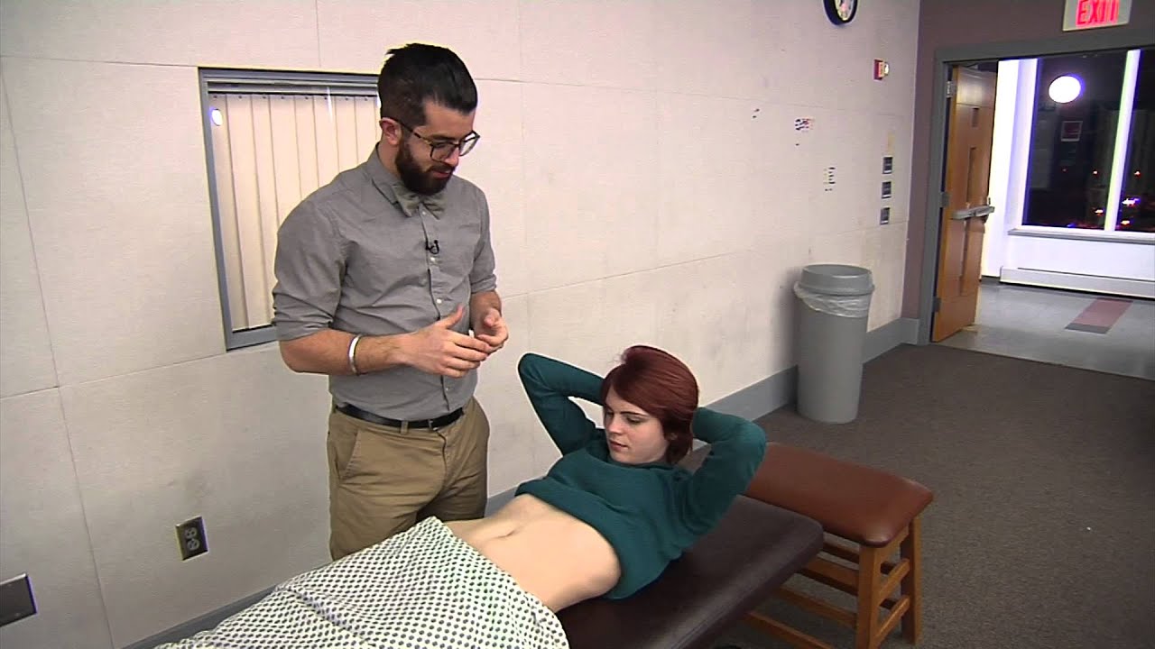
surgical examination of intra abdominal lump or mass
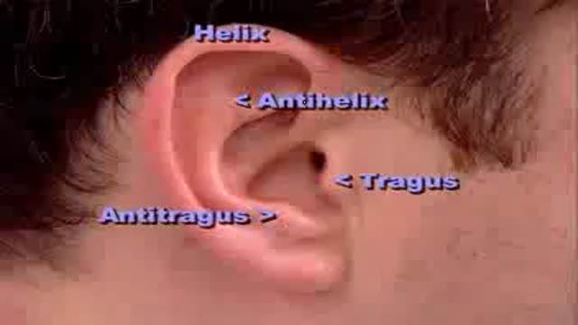
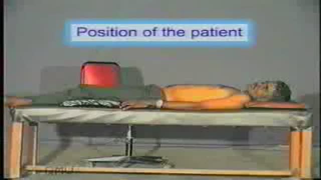
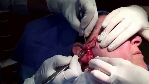
Complete clinical examination of the ears with all the associated tests
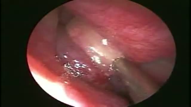
Drainage of a maxillary Sinus pyocoele

When United Airlines decides their employees flying to Kentucky is more important than a doctor or any passenger who paid for their ticket it is time to STOP FLYING UNITED!!! Here are United employees dragging the man off the plane like a criminal.
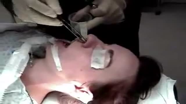
Open rhinoplasty without oseotomies peformed by Dr. Robert Dryden and Dr. Brett Kotlus. Basic steps for rasping of dorsal hump and cephalic trim with septoplasty and tip strut.
Thanks to a new, state-of-the-art procedure for total knee replacement developed by surgeons at the Detroit Medical Center's Sinai-Grace Hospital, the rehabilitation time for patients has been reduced from six months to six weeks. ~ Detroit Medical Center

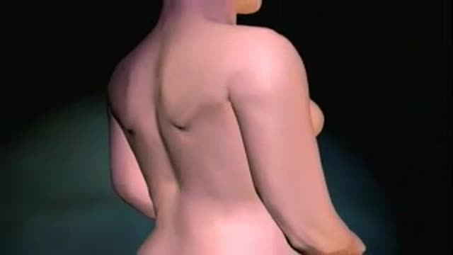
Orgasm is the sudden discharge of accumulated sexual excitement during the sexual response cycle, resulting in rhythmic muscular contractions in the pelvic region characterized by sexual pleasure

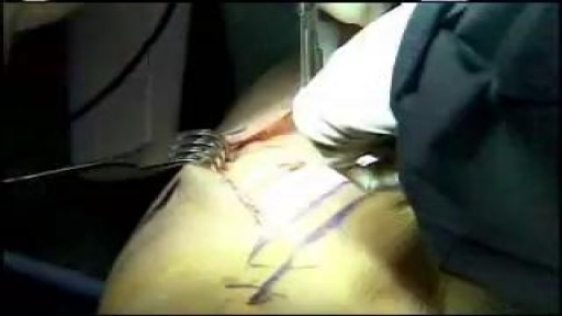
Examination of Varicose Veins
Mini Face Lift Surgery -- Short Scars -- No Anesthesia
