- Physical Examination
- Surgical Examination
- Ophthalmology
- Clinical Skills
- Orthopedics
- Surgery Videos
- Laparoscopy
- Pediatrics
- Funny Videos
- Cardiothoracic Surgery
- Nursing Videos
- Plastic Surgery
- Otorhinolaryngology
- Histology and Histopathology
- Neurosurgery
- Dermatology
- Pediatric Surgery
- Urology
- Dentistry
- Oncology and Cancers
- Anatomy Videos
- Health and Fitness
- Radiology
- Anaesthesia
- Physical Therapy
- Pharmacology
- Interventional Radiology
- Cardiology
- Endocrinology
- Gynecology
- Emergency Medicine
- Psychiatry and Psychology
- Childbirth Videos
- General Medical Videos
- Nephrology
- Physiology
- Diet and Food Health
- Diabetes Mellitus
- Neurology
- Women Health
- Osteoporosis
- Gastroenterology
- Pulmonology
- Hematology
- Rheumatology
- Toxicology
- Nuclear Medicine
- Infectious Diseases
- Vascular Disease
- Reproductive Health
- Burns and Wound Healing
- Other
Top videos
Care must be taken to prevent stenosis at the anastomotic site. If the diameter of the anastomosis is less than 2 cm, the anastomosis should be taken down and resected. A classic end-to-end anastomosis should be performed to ensure adequate diameter to the intestine. If the posterior wall of the colon has been preserved, care should be taken to close the colostomy prior to opening the peritoneal cavity. This will reduce intraperitoneal contamination from the stoma site. Copious irrigation of the wound should be made prior to primary closure. If gross contamination has occurred, delayed closure of the wound should be considered.
Incision and Drainage of a Huge Gluteal Abscess
The gold standard treatment for bladder outlet obstruction.This is an endoscopic procedure in which a resectoscope is placed transurethrally and the obstructing lobes of the prostate are removed as chips of tissue. TURP results in improvement of flow rate, and symptom scores are superior to that of other minimally invasive therapies
Watch that Medical Abortion Surgical Procedure

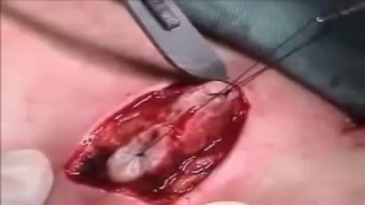
This video demonstrate Bilateral Salpingectomy for a patient suffering from hematosalpinx of one side and Hydrosalpinx other side in which one IVF has failed. Laparoscopic salpingectomy. In this less-invasive procedure, the surgeon makes 1-3 small incisions in the lower abdomen, and inserts a laparoscope into the pelvis through one of the incisions. The camera at the end of the laparoscope guides the surgeon through the procedure. The fallopian tube tissue is then removed. For more information https://www.laparoscopyhospital.com/
For more information please contact:
World Laparoscopy Hospital
Cyber City, Gurugram, NCR DELHI
INDIA 122002
Phone & WhatsApp: +919811416838, + 91 9999677788
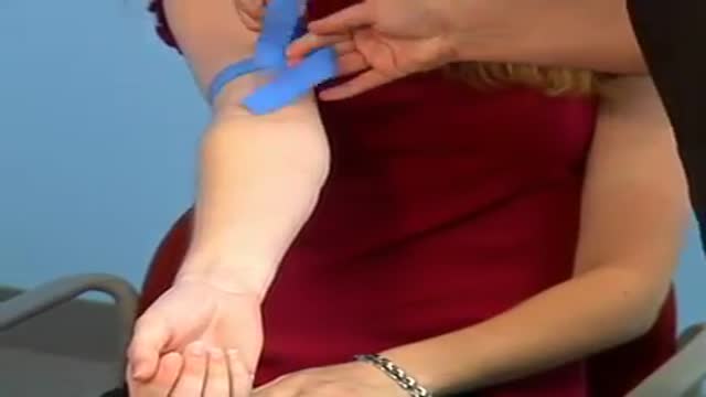
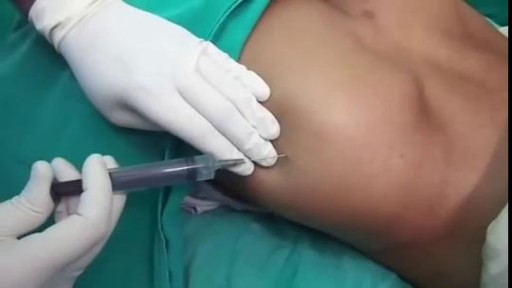
What is Venipuncture? While venipuncture can refer to a variety of procedures, including the insertion of IV tubes into a vein for the direct application of medicine to the blood stream, in phlebotomy venipuncture refers primarily to using a needle to create a blood evacuation point. As a phlebotomist, you must be prepared to perform venipuncture procedures on adults, children, and even infants while maintaining a supportive demeanor and procedural accuracy. Using a variety of blood extraction tools, you must be prepared to respond to numerous complications in order to minimize the risk to the patient while still drawing a clean sample. In its entirety, venipuncture includes every step in a blood draw procedure—from patient identification to puncturing the vein to labeling the sample. Patient information, needle placement, and emotional environment all play a part in the collection of a blood sample, and it's the fine details that can mean the difference between a definite result and a false positive. After placing the tourniquet and finding the vein, it's time for the phlebotomist to make the complex choice on what procedure will best suit the specific situation. Keeping this in mind, it should be noted that the following information is not an instructional guide on how to perform these phlebotomy procedures. Rather, the information below is intended to serve as an educational resource to inform you of the equipment and procedures you will use. Venipuncture Technqiues Venipuncture with an Evacuated or Vacuum Tube: This is the standard procedure for venipuncture testing. Using a needle and sheath system, this procedure allows multiple sample tubes to be filled through a single puncture. This procedure is ideal for reducing trauma to patients. After drawing the blood, the phlebotomist must make sure the test stopper is correctly coded and doesn't contact exposed blood between samples. Venipuncture with a Butterfly Needle : This is a specialized procedure that utilizes a flexible, butterfly needle adaptor. A butterfly needle has two plastic wings (one on either side of the needle) and is connected to a flexible tube, which is then attached to a reservoir for the blood. Due to the small gauge of the needle and the flexibility of the tube, this procedure is used most often in pediatric care, where the patients tend to have smaller veins and are more likely to move around during the procedure. After being inserted into a vein at a shallow angle, the butterfly needle is held in place by the wings, which allow the phlebotomist to grasp the needle very close to the skin. Phlebotomists should be careful to watch for blood clots in the flexible tubing. Venipuncture with a Syringe: This technique is typically only used when there is a supply shortage, or when a technician thinks it is the appropriate method. It uses the classic needle, tube, and plunger system, operating in a similar manner to the vacuum tube but requiring multiple punctures for multiple samples. Additionally, after the blood is drawn it must be transferred to the appropriate vacuum tube for testing purposes. If you choose to use this method, remember to check for a sterile seal, and use a safety device when transferring the sample. Fingerstick (or Fingerprick): This procedure uses a medical lance to make a small incision in the upper capillaries of a patient's finger in order to collect a tiny blood sample. It is typically used to test glucose and insulin levels. When performing a Fingerstick, the phlebotomist should remember to lance the third or fourth finger on the non-dominant arm. Never lance the tip or the center of the finger pad; instead, lance perpendicular to the fingerprint lines. Heelstick (or Heelprick): Similar to the Fingerstick procedure, this process is used on infants under six months of age. A medical lance is used to create a small incision on the side of an infant's heel in order to collect small amounts of blood for screening. As with a Fingerstick, the incision should be made perpendicular to the heel lines, and it should be made far enough to the left or right side of the heel to avoid patient agitation. Before performing a Heelstick, the infant's heel should be warmed to about 42 degrees Celsius in order to stimulate capillary blood and gas flow. Therapeutic Phlebotomy: This involves the actual letting of blood in order to relieve chemical and pressure imbalances within the blood stream. Making use of a butterfly needle, this therapy provides a slow removal of up to one pint of blood. Though the blood removed is not used for blood transfusions, the procedure and concerns are the same as with routine blood donation. As with any phlebotomy procedure, one should pay close attention to the patient in order to prevent a blood overdraw. Bleeding Time: A simple diagnostic test that is used to determine abnormalities in blood clotting and platelet production. A shallow laceration is made, followed by sterile swabbing of the wound every 30 seconds until the bleeding stops. Average bleed times range between one and nine minutes. As a phlebotomist, you should familiarize yourself with the application and cross-application of these procedures in order to recognize when a procedure is necessary, and what the risks are for each.
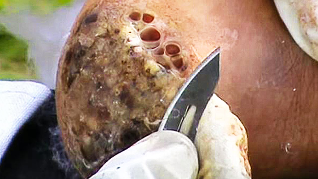
Watch that Skin Jiggers Removal Procedure

LASIK eye procedure for correcting vision
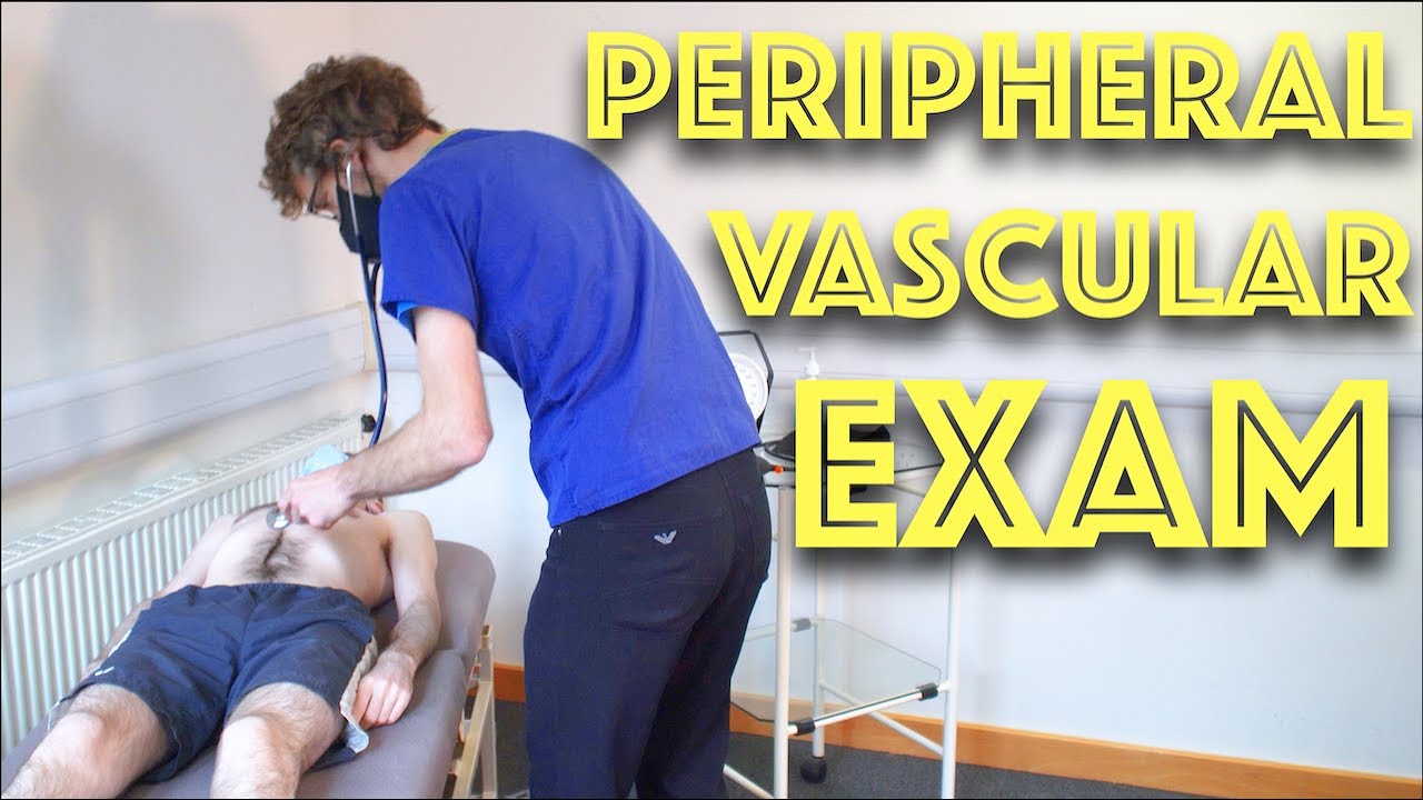
Peripheral Vascular Examination OSCE - Clinical Skills - Dr Gill
In the cardiovascular examination, particularly in the case of an OSCE station, we conclude the examination often by stating that the examiner would want to perform:
- An ECG
- Check full blood count
- and "do a peripheral vascular examination
In this video, we demonstrate that oft-talked about, but comparatively less common examination.
Starting off, with the examination of the hands, the radial, brachial and carotid pulses. before moving down to assess for a AAA, checking the femoral and popliteal pulses, before wrapping up around the ankle with the posterior tibial and dorsalis pedis pulses
For completeness, the cardiovascular examination is demonstrated here
https://www.youtube.com/watch?v=ECs9O5zl6XQ&t=2s
#PeripheralVascular #ClinicalSkills #DrGill

Traditional Liposuction VS Vaser Liposuction
A side-by-side comparison of traditional liposuction and a #Vaser liposuction. Both of these were performed by our skilled surgeons at Divine Cosmetic Surgery.
#vaserliposuction #liposuction #liposuctionDelhi #liposuctionresults #shorts #vaserliposuctionDelhi
Know more about liposuction
https://www.divinecosmeticsurg....ery.com/liposuction-
Traditional Liposuction vs 360 High Def Vaser Liposuction - https://www.youtube.com/watch?v=r_bBI2p9fVI&t=14s
-------------------------------------------------------------------------------
Why Vaser Is Best For Thigh Liposuction - https://youtu.be/dlzpdDEZcS4
-------------------------------------------------------------------------------
Abdomen Vaser Liposuction - Live - https://www.youtube.com/watch?v=_Cvl2Txn8LQ
-------------------------------------------------------------------------------
Back Vaser Liposuction In Female - https://youtu.be/OC60UdgtIWU
-------------------------------------------------------------------------------
For more details about Liposuction Visit - https://www.divinecosmeticsurgery.com/
-------------------------------------------------------------------------------
Dr. Amit Gupta
MBBS, M.S., DNB (Plastic & Cosmetic Surgery)
Divine Cosmetic Surgery | +91 9811994417
info@divinecosmeticsurgery.com | 01141828787
Delhi | Mumbai | Gurgaon
𝗦𝗼𝗰𝗶𝗮𝗹 𝗠𝗲𝗱𝗶𝗮 𝗮𝗻𝗱 𝗬𝗼𝘂𝘁𝘂𝗯𝗲 𝘃𝗶𝗱𝗲𝗼 𝗰𝗵𝗮𝗻𝗻𝗲𝗹 : -
🎦 https://www.youtube.com/c/DrAm....itGuptaBestPlasticCo
👍🏻 https://www.facebook.com/dramitguptaplasticsurgeon
📷 https://www.instagram.com/divineaesthetics_delhi/
🐥 https://twitter.com/dramitguptajee
🖇️ https://www.linkedin.com/compa....ny/divinecosmeticsur
📌 https://pinterest.com/divinesurgery
#Liposuction #vaserliposuction #liposuctioncostinindia #liposuctiondelhi #liposuction #liposuctioncost #liposuctioncostfactors #liposuctioncostindelhi #DrAmitGuptaPlasticSurgeon #DivineCosmeticSurgery #dramitgupta
Disclaimer: The information on our videos & social media is provided for informational purposes only and is not meant for the advice provided by your surgeon.
We are not responsible for any harm if anyone misguides you from our name. Our all-social media official handles are linked up on our website. All images & content used on our videos & social media are for illustrative concerns only, original results and processes may vary.

A patient at a British hospital played Mahler and Gershwin on the violin while surgeons removed a tumor from her brain, so doctors could preserve her ability to play music.
She left the hospital 3 days later and hopes to return to the symphony soon. https://abcn.ws/2SGY9mp
SUBSCRIBE to ABC NEWS: https://www.youtube.com/ABCNews/
Watch More on http://abcnews.go.com/
LIKE ABC News on FACEBOOK
https://www.facebook.com/abcnews
FOLLOW ABC News on TWITTER:
https://twitter.com/abc
GOOD MORNING AMERICA'S HOMEPAGE:
https://www.goodmorningamerica.com/

This animation demonstrates how a unilateral complete cleft lip repair is performed. This video is meant for educational purposes for patients and families. There are many ways to fix a complete cleft lip, but the technique shown here is the most common known as the Millard Rotation Advancement Repair.

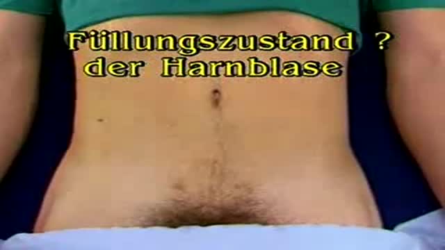
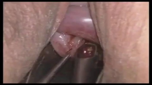
German Video showing examination of the urinary bladder

Watch that Human Baby Medical Abortion Surgery

At one time, women who had delivered by cesarean section in the past would usually have another cesarean section for any future pregnancies. The rationale was that if allowed to labor, many of these women with a scar in their uterus would rupture the uterus along the weakness of the old scar. Over time, a number of observations have become apparent: Most women with a previous cesarean section can labor and deliver vaginally without rupturing their uterus. Some women who try this will, in fact, rupture their uterus. When the uterus ruptures, the rupture may have consequences ranging from near trivial to disastrous. It can be very difficult to diagnose a uterine rupture prior to observing fetal effects (eg, bradycardia). Once fetal effects are demonstrated, even a very fast reaction and nearly immediate delivery may not lead to a good outcome. The more cesarean sections the patient has, the greater the risk of subsequent rupture during labor. The greatest risk occurs following a “classical” cesarean section (in which the uterine incision extends up into the fundus.) The least risk of rupture is among women who had a low cervical transverse incision. Low vertical incisions probably increase the risk of rupture some, but usually not as much as a classical incision. Many studies have found the use of oxytocin to be associated with an increased risk of rupture, either because of the oxytocin itself, or perhaps because of the clinical circumstances under which it would be contemplated. Pain medication, including epidural anesthetic, has not resulted greater adverse outcome because of the theoretical risk of decreasing the attendant’s ability to detect rupture early. The greatest risk of rupture occurs during labor, but some of the ruptures occur prior to the onset of labor. This is particularly true of the classical incisions. Overall successful vaginal delivery rates following previous cesarean section are in the neighborhood of 70 This means that about 30of women undergoing a vaginal trial of labor will end up requiring a cesarean section. Those who undergo cesarean section (failed VBAC) after a lengthy labor will frequently have a longer recovery and greater risk of infection than had they undergone a scheduled cesarean section without labor. Women whose first cesarean was for failure to progress in labor are only somewhat less likely to be succesful in their quest for a VBAC than those with presumably non-recurring reasons for cesarean section. For these reasons, women with a prior cesarean section are counseled about their options for delivery with a subsequent pregnancy: Repeat Cesarean Section, or Vaginal Trial of Labor. They are usually advised of the approximate 70successful VBAC rate (modified for individual risk factors). They are counseled about the risk of uterine rupture (approximately 1in most series), and that while the majority of those ruptures do not lead to bad outcome, some of them do, including fetal brain damage and death, and maternal loss of future childbearing. They are advised of the usual surgical risks of infection, bleeding, anesthesia complications and surgical injury to adjacent structures. After counseling, many obstetricians leave the decision for a repeat cesarean or VBAC to the patient. Both approaches have risks and benefits, but they are different risks and different benefits. Fortunately, most repeat cesarean sections and most vaginal trials of labor go well, without any serious complications. For those choosing a trial of labor, close monitoring of mother and baby, with early detection of labor abnormalities and preparation for

An inguinal hernia occurs when tissue, such as part of the intestine, protrudes through a weak spot in the abdominal muscles. The resulting bulge can be painful, especially when you cough, bend over or lift a heavy object. However, many hernias do not cause pain.
An inguinal hernia isn't necessarily dangerous. It doesn't improve on its own, however, and can lead to life-threatening complications. Your doctor is likely to recommend surgery to fix an inguinal hernia that's painful or enlarging. Inguinal hernia repair is a common surgical procedure.

Brain port surgery is a minimally invasive surgical technique performed through a specially designed tube about the size of a dime. Using neuronavigation GPS-like guidance, the brain port is inserted into the brain with millimeter accuracy and is used as a channel to guide the surgeon and his/her instruments to various regions of the brain. Colloid cysts, metastatic tumors, and a variety of tumors within the ventricles are often candidates for this approach.
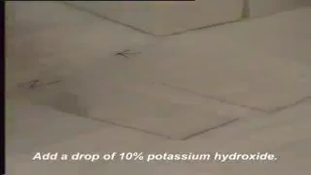
A wet mount is the suspension of a small amount of vaginal discharge in a liquid medium. Two liquids are commonly used, normal saline and potassium hydroxide. Each has it's own unique properties that make it useful in this setting
