- Physical Examination
- Surgical Examination
- Ophthalmology
- Clinical Skills
- Orthopedics
- Surgery Videos
- Laparoscopy
- Pediatrics
- Funny Videos
- Cardiothoracic Surgery
- Nursing Videos
- Plastic Surgery
- Otorhinolaryngology
- Histology and Histopathology
- Neurosurgery
- Dermatology
- Pediatric Surgery
- Urology
- Dentistry
- Oncology and Cancers
- Anatomy Videos
- Health and Fitness
- Radiology
- Anaesthesia
- Physical Therapy
- Pharmacology
- Interventional Radiology
- Cardiology
- Endocrinology
- Gynecology
- Emergency Medicine
- Psychiatry and Psychology
- Childbirth Videos
- General Medical Videos
- Nephrology
- Physiology
- Diet and Food Health
- Diabetes Mellitus
- Neurology
- Women Health
- Osteoporosis
- Gastroenterology
- Pulmonology
- Hematology
- Rheumatology
- Toxicology
- Nuclear Medicine
- Infectious Diseases
- Vascular Disease
- Reproductive Health
- Burns and Wound Healing
- Other
Top videos

How amblyopia develops in children. Basically, if one eye doesn't see well from an early age, the wiring never forms correctly back to the occipital cortex.
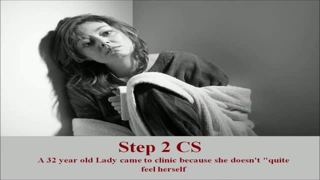
USMLE Step 2 CS - Fatigue This is just preview video. To get full access please visit our website : www.usmletutoring.com
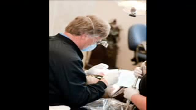
Dentist Kennewick WA Call (509) 783-8822
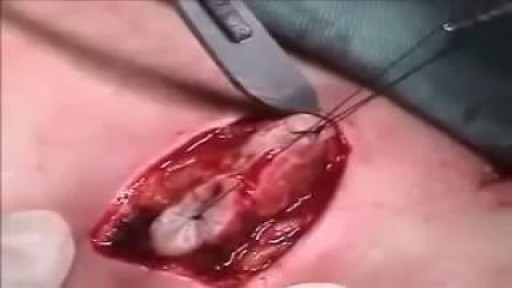
Care must be taken to prevent stenosis at the anastomotic site. If the diameter of the anastomosis is less than 2 cm, the anastomosis should be taken down and resected. A classic end-to-end anastomosis should be performed to ensure adequate diameter to the intestine. If the posterior wall of the colon has been preserved, care should be taken to close the colostomy prior to opening the peritoneal cavity. This will reduce intraperitoneal contamination from the stoma site. Copious irrigation of the wound should be made prior to primary closure. If gross contamination has occurred, delayed closure of the wound should be considered.
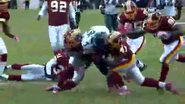
Brain Concussion in Sports
Cardiology Physical Examination Lecture
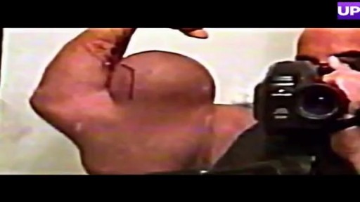
Bodybuilder Drains Synthol Hematoma From Bicep
Histology of Liver
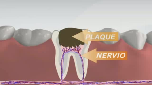
Root canals are common procedures and can help save your tooth from extraction. Dentists at Aspen Dental practices have been safely and expertly performing root canal procedures for over two decades.
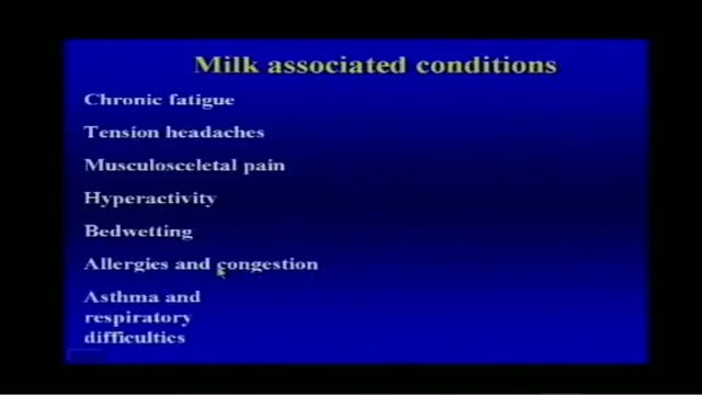
Milk Associated Diseases Information
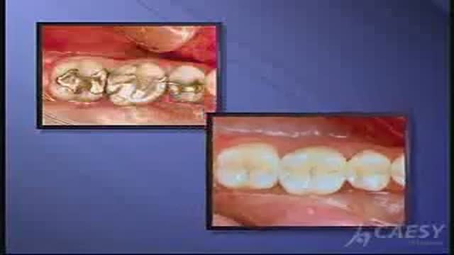
White Fillings
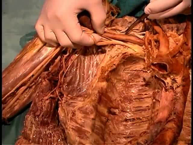
Anatomy of The Axillary Fossa

Blackheads are small bumps that appear on your skin due to clogged hair follicles. These bumps are called “blackheads” because the surface looks dark or black. Blackheads are a mild type of acne that usually form on the face, but they can also appear on the back, chest, neck, arms, and shoulders
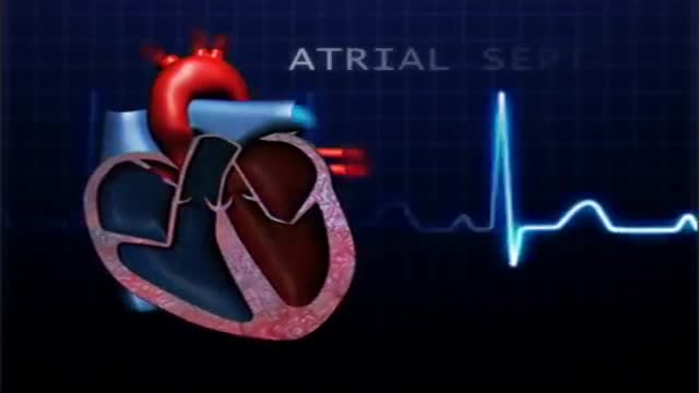
atrial septal defect (ASD) is a hole in the wall between the two upper chambers of your heart (atria). The condition is present from birth (congenital). Small atrial septal defects may close on their own during infancy or early childhood. Large and long-standing atrial septal defects can damage your heart and lungs. Small defects may never cause a problem and may be found incidentally. An adult who has had an undetected atrial septal defect for decades may have a shortened life span from heart failure or high blood pressure that affects the arteries in the lungs (pulmonary hypertension). Surgery may be necessary to repair atrial septal defects to prevent complications
Non-alcoholic fatty liver disease (NAFLD) is a very common disorder and refers to a group of conditions where there is accumulation of excess fat in the liver of people who drink little or no alcohol. The most common form of NAFLD is a non serious condition called fatty liver.
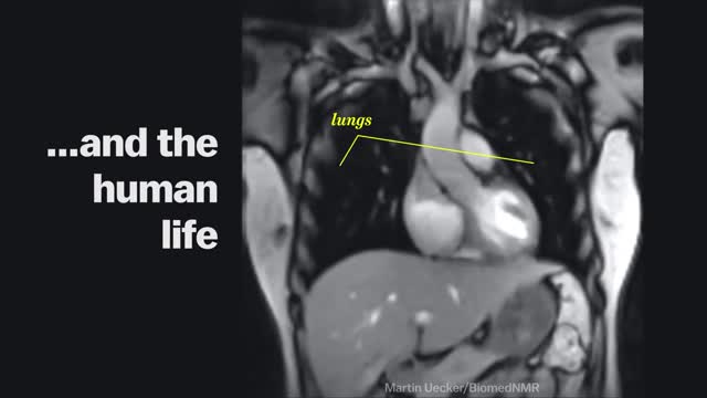
Magnetic Resonance Imaging (MRI) "sees" inside the body by mapping the position of water molecules, which exist at different densities in different types of tissue. Watch the video above for a sample of some impressive MRI images of the human body in action.
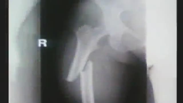
Treating osteoporosis with bisphosphonates, particularly for more than five years, has been linked to some side effects, including atypical femur fractures. Osteoporosis medications are supposed to prevent bone breaks. But if they are taken for too long, the opposite can happen. This video highlights what you need to know as a healthcare professional to educate patients
Several options are available to remove spider veins — thin red lines or weblike networks of blood vessels that appear on your legs and feet. Spider veins are usually harmless, though they can sometimes cause aching, burning or pain, especially when you've been standing for long periods. If you have symptoms or are concerned about the appearance of spider veins, treatment options include: Sclerotherapy. In this procedure, your doctor injects the veins with a solution that scars and closes those veins, causing the blood to reroute through healthier veins. In a few weeks, treated spider veins fade. Although the same vein may need to be injected more than once, sclerotherapy is usually effective if done correctly. Sclerotherapy doesn't require anesthesia and can be done in your doctor's office. Side effects include swelling, itching and skin color changes in the treated area. Laser surgery. Laser surgery works by sending strong bursts of light into the vein that make the vein slowly fade and disappear. No incisions or needles are used. The treatment is often less effective than sclerotherapy, particularly for larger veins. Side effects may include redness, bruising, itching, swelling and permanent skin tone changes. After treatment, blood vessels fade over several months, but they may not disappear completely. Also, new spider veins can develop in the same area.
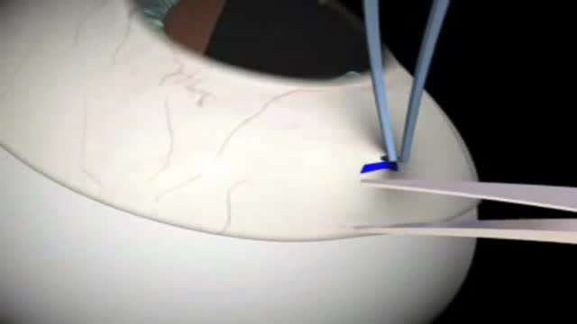
Glaucoma Surgery 3D Animation
Over 60 million Americans suffer from chronic heartburn. Get the basics on acid reflux
