- Physical Examination
- Surgical Examination
- Ophthalmology
- Clinical Skills
- Orthopedics
- Surgery Videos
- Laparoscopy
- Pediatrics
- Funny Videos
- Cardiothoracic Surgery
- Nursing Videos
- Plastic Surgery
- Otorhinolaryngology
- Histology and Histopathology
- Neurosurgery
- Dermatology
- Pediatric Surgery
- Urology
- Dentistry
- Oncology and Cancers
- Anatomy Videos
- Health and Fitness
- Radiology
- Anaesthesia
- Physical Therapy
- Pharmacology
- Interventional Radiology
- Cardiology
- Endocrinology
- Gynecology
- Emergency Medicine
- Psychiatry and Psychology
- Childbirth Videos
- General Medical Videos
- Nephrology
- Physiology
- Diet and Food Health
- Diabetes Mellitus
- Neurology
- Women Health
- Osteoporosis
- Gastroenterology
- Pulmonology
- Hematology
- Rheumatology
- Toxicology
- Nuclear Medicine
- Infectious Diseases
- Vascular Disease
- Reproductive Health
- Burns and Wound Healing
- Other
Top videos
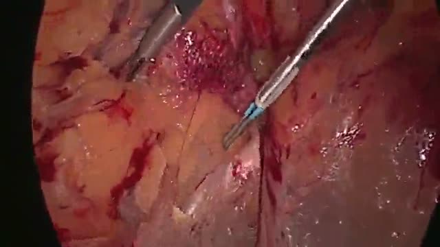
A high definition medical video showing the Laparoscopic inguinal hernia repair

When it comes to our health men over the age of 45 are in need of regular doctor visits and testing, as a large percentage of medical decisions are based on the lab test results.
The mainstay of treatment is usually medication, talk therapy, or a combination of the two. Increasingly, research suggests these treatments may normalize brain changes associated with depression.
Anatomy of The Ear
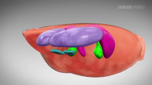
Anatomy of Anterior Abdominal Wall
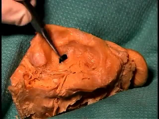
GIANT CELL TUMOR REMOVAL Plastic, Cosmetic and Reconstructive

Dialysis services at UC San Diego Health: https://health.ucsd.edu/care/kidney/dialysis
UC San Diego Health Licensed Clinical Social Worker, Norma Reggev, discusses hemodialysis as a treatment option for failing kidneys with patient testimonials. Discussion includes In Center Hemodialysis and Home Hemodialysis.
0:00 - Hemodialysis
1:34 - When Should Dialysis Begin?
2:00 - What is Dialysis?
2:25 - How Hemodialysis Works
3:15 - In-Center Hemodialysis Considerations
3:42 - Patient Shares Their Experience With In-Center Hemodialysis
7:30 - Home Hemodialysis Considerations
8:35 - Patient Shares Their Experience With Home Hemodialysis
12:23 - Types of Vascular Access
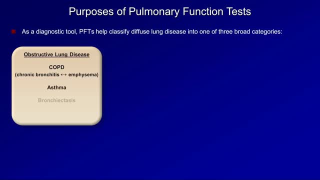
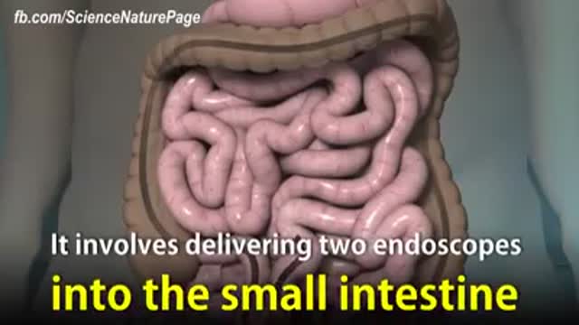
Pulmonary function tests are a broad range of tests that measure how well the lungs take in and exhale air and how efficiently they transfer oxygen into the blood. Spirometry measures how well the lungs exhale.

Prevent Stretch Marks During Pregnancy

Get rid of blackheads
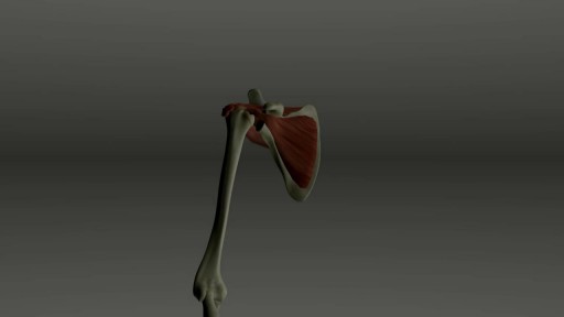
A rotator cuff tear is a common injury, especially in sports like baseball or tennis, or in jobs like painting or cleaning windows. It usually happens over time from normal wear and tear, or if you repeat the same arm motion over and over. But it also can happen suddenly if you fall on your arm or lift something heavy. Your rotator cuff is a group of four muscles and tendons that stabilize your shoulder joint and let you lift and rotate your arms. There are two kinds of rotator cuff tears. A partial tear is when the tendon that protects the top of your shoulder is frayed or damaged. The other is a complete tear. That’s one that goes all the way through the tendon or pulls the tendon off the bone.
Angioplasty Procedure Animation

Sclerotherapy is a procedure used to eliminate varicose veins and spider veins. Sclerotherapy involves an injection of a solution (generally a salt solution) directly into the vein. The solution irritates the lining of the blood vessel, causing it to collapse and stick together and the blood to clot.Sep 17, 2016

Initial symptoms may include: Pain or discomfort in the upper tummy (abdomen), especially after eating. Indigestion. (Note: most people who have indigestion do not have stomach cancer.) Feeling sick, and being off food. ... Weight loss and/or loss of appetite. You may pass blood out with your stools (faeces).
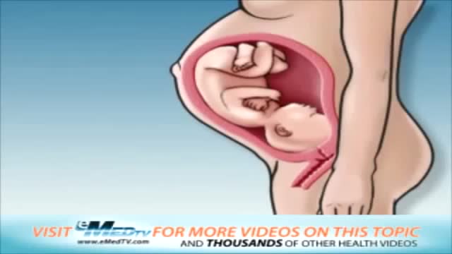
Your baby is still tiny, but already your body is changing. Your breasts start to swell and may feel tender. Tiredness, nausea, and a frequent need to pee are common pregnancy symptoms. In your second trimester, your growing uterus gradually rises up out of your pelvis.
Diabetes is a growing global health concern, as is obesity. Diabetes and obesity are intrinsically linked: obesity increases the risk of diabetes and also contributes to disease progression and cardiovascular disease. Although the benefits of weight loss in the prevention of diabetes and as a critical component of managing the condition are well established, weight reduction remains challenging for individuals with type 2 diabetes due to a host of metabolic and psychological factors. For many patients, lifestyle intervention is not enough to achieve weight loss, and alternative options, such as pharmacotherapy, need to be considered. However, many traditional glucose-lowering medications may lead to weight gain. This article focuses on the potential of currently available pharmacological strategies and on emerging approaches in development to support the glycemic and weight-loss goals of individuals with type 2 diabetes. Two pharmacotherapy types are considered: those developed primarily for blood glucose control that have a favorable effect on body weight and those developed primarily to induce weight loss that have a favorable effect on blood glucose control. Finally, the potential of combination therapies for the management of obese patients with type 2 diabetes is discussed.
Too much cholesterol in the blood can lead to cardiovascular disease. Heart disease is the No. 1 cause of death in the United States. Over 2,100 Americans die of cardiovascular disease each day, an average of one death every 40 seconds. The good news is, you can lower your cholesterol and reduce your risk of heart disease and stroke. Working with your doctor is key. It takes a team to develop and maintain a successful health program. You and your healthcare professionals each play an important role in maintaining and improving your heart health. Work with your doctor to determine your risk and the best approach to manage it. In all cases, lifestyle changes are important to reduce your risk for heart attack and stroke. In some cases, cholesterol-lowering statin medicines may also provide benefit. Learn how to make diet and lifestyle changes easy and lasting. Also make sure you understand instructions for taking medication because it won't work if you don't take it as directed. Lifestyle Changes Your diet, weight, physical activity and exposure to tobacco smoke all affect your cholesterol level. Know Your Fats Knowing which fats raise LDL cholesterol and which ones don't is the first step in lowering your risk of heart disease.
A heart attack occurs when the flow of blood to the heart is blocked, most often by a build-up of fat, cholesterol and other substances, which form a plaque in the arteries that feed the heart (coronary arteries). The interrupted blood flow can damage or destroy part of the heart muscle. A heart attack, also called a myocardial infarction, can be fatal, but treatment has improved dramatically over the years. It's crucial to call 911 or emergency medical help if you think you might be having a heart attack

TUMMY TUCK 🤩 Immediate OR Results
This patient wanted to get her abs back, but unfortunately NO diet or workout can tighten muscles that have been stretched apart from carrying a baby 👀 But we can fix that at Lemmon Avenue Plastic Surgery & Laser Center!
To learn more about the #tummytuck click here: https://drdeuber.com/procedures/tummy-tuck/
For #mommymakeover, click here: https://drdeuber.com/procedures/mommy-makeover/
👙
#MarkDeuberMD
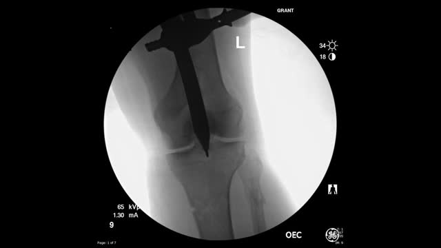
This video shows the technique of suprapatellar tibial nailing as used for a segmental tibia fracture. The broken leg was treated with the nail to allow immediate mobility and range of motion; no cast was needed for this injury.
