- Physical Examination
- Surgical Examination
- Ophthalmology
- Clinical Skills
- Orthopedics
- Surgery Videos
- Laparoscopy
- Pediatrics
- Funny Videos
- Cardiothoracic Surgery
- Nursing Videos
- Plastic Surgery
- Otorhinolaryngology
- Histology and Histopathology
- Neurosurgery
- Dermatology
- Pediatric Surgery
- Urology
- Dentistry
- Oncology and Cancers
- Anatomy Videos
- Health and Fitness
- Radiology
- Anaesthesia
- Physical Therapy
- Pharmacology
- Interventional Radiology
- Cardiology
- Endocrinology
- Gynecology
- Emergency Medicine
- Psychiatry and Psychology
- Childbirth Videos
- General Medical Videos
- Nephrology
- Physiology
- Diet and Food Health
- Diabetes Mellitus
- Neurology
- Women Health
- Osteoporosis
- Gastroenterology
- Pulmonology
- Hematology
- Rheumatology
- Toxicology
- Nuclear Medicine
- Infectious Diseases
- Vascular Disease
- Reproductive Health
- Burns and Wound Healing
- Other
Top videos

Dealing with Anxiety and Panic Attacks
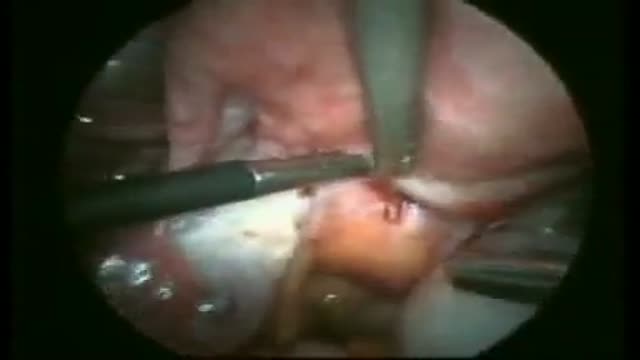
Excision of Rectovaginal Nodule

Fast Lower Back Pain & Sciatica Pain Relief – Beginners Yoga Stretches and Poses
Laparoscopic Gastric Bypass Surgery
A new well designed randomized study has suggested that long term baby aspirin usage may aid in fight against cancer. The suggested mechanism is that cancers induce inflammatory responses so the anti-inflammatory mechanism of prostaglandins inhibitors may cease the progress of many cancers. There are some concerns about the study because despite the well-designed randomized study; the study didn't include a satisfying number of female participants. The study was also conducted on esophageal, colorectal and lung cancers.
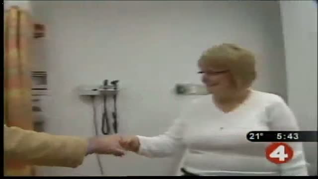
Pancreatic cysts are saclike pockets of fluid on or in your pancreas, a large organ behind the stomach that produces hormones and enzymes that help digest food. Most pancreatic cysts aren't cancerous, and many don't cause symptoms. They're typically found during imaging testing for another problem. Some are actually noncancerous (benign) pockets of fluids lined with scar or inflammatory tissue, not the type of cells found in true cysts (pseudocysts). But some pancreatic cysts can be or can become cancerous. Your doctor might take a sample of the pancreatic cyst fluid to determine if cancer cells are present. Or your doctor might recommend monitoring a cyst over time for changes that indicate cancer.

Head Cyst watch to see more

Getting Out of Bed after Surgery
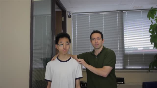
Romberg Test
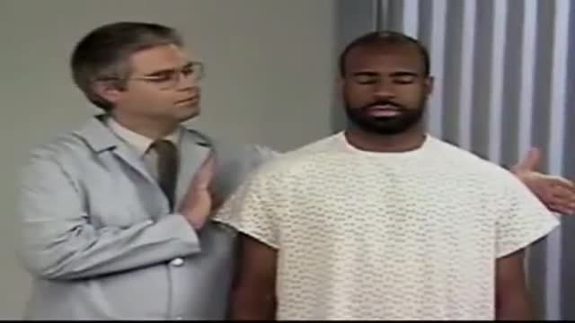
Therapeutic Interaction

bnbmedispa.com offers Titan Laser, Zerona Laser, Zerona Laser Treatment, Titan skin tightening in NJ, Monmouth, and Shrewsbury. call us at (732) 460-0600 to get discout on any laser treatment.

Ventricular Assist Devices

This video illustrates several forms of catatonia including waxy flexibility, forced grasping, opposition, negativism and aversion.
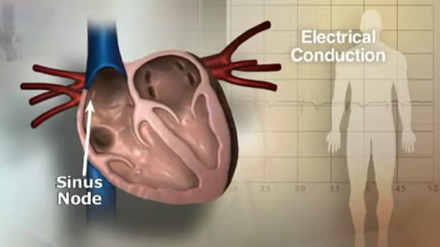
If you or someone you love has atrial fibrillation, learn more about what AFib is, why treatment can save lives, and what you can do to reach your goals, lower your risks and live a healthy life.
Scoliosis is a sideways curvature of the spine that occurs most often during the growth spurt just before puberty. While scoliosis can be caused by conditions such as cerebral palsy and muscular dystrophy, the cause of most scoliosis is unknown.

Austin Vampire Facelift provider Dr. David Sneed gives us a comprehensive overview of this new facial rejuvenation technology also known as PRP. This nonsurgical anti-aging treatment utilizes the patient's own blood to stimulate the growth of new collagen, tighten skin and smooth out wrinkles.
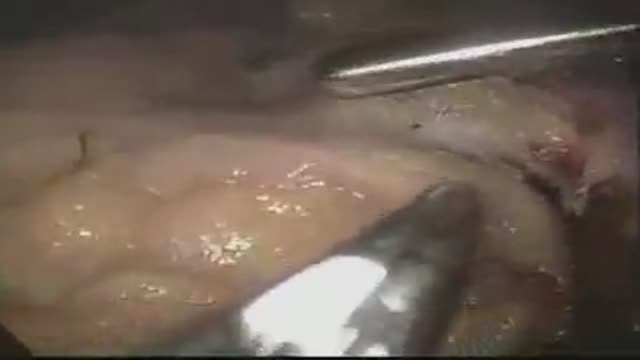
Spirotome macrobiopsy of a lung as a minimal invasive way to complete the diagnosis of lung lesions.

The type of operation performed for removal of pancreatic cancer is based on the location of the tumor. For tumors of the head and neck of the pancreas a Whipple procedure, (also called a pancreaticoduodenectomy) is performed. This is a complex operation perfected at Johns Hopkins. This video will explain the surgery and what patients can expect.
Indications for intervention in patients with a renal artery aneurysm (RAA) include the following [20, 8, 13, 14] : Rupture Symptomatic RAA - Hypertension (from associated renal artery stenosis, refractory to medical management), pain, renal ischemia or infarction secondary to embolization from the aneurysm sac RAAs in females who are pregnant or are contemplating pregnancy Diameter greater than 2 cm Enlarging RAA RAA associated with acute dissection Currently, there is no consensus regarding the size at which an RAA should be repaired in an asymptomatic patient. Experts have recommended RAA repair at diameters ranging from 1.5 to 3 cm, [8] though most suggest 2 cm. Some reports have even suggest that larger asymptomatic saccular aneurysms may be managed expectantly. Note that aneurysm rupture at a diameter of 1.5 cm has been reported. Complete calcification of the wall of the aneurysm sac manifests in about 40% of patients. This was once believed to confer protection against rupture [21] ; however, this belief has since been questioned. [30] Asymptomatic, small (<2 cm in diameter) RAAs do not usually require treatment. One notable exception is an RAA in a woman who is pregnant or contemplating pregnancy. In view of the increased risk of rupture in such cases, even small asymptomatic aneurysms should be repaired in this population. For diagnosis and preinterventional planning, gadolinium-enhanced magnetic resonance angiography (MRA) and computed tomography (CT) angiography (CTA) with three-dimensional (3D) reconstruction have essentially replaced conventional arteriography. Regular follow-up examination with ultrasonography (US) or CT) is recommended in patients who are treated expectantly. Spontaneous cure by thrombosis of small aneurysms has been described. Further refinements in endovascular techniques may allow more RAAs to be treated in this manner. So far, excellent short- and intermediate-term results have been described in the literature [40] ; however, there remains a need for further long-term outcome data.

http://www.landging.com/accident-animation-workers-compensation-desk-job.html
This desk job accident animation demonstrates the injury covered by workers compensation program.
