- Physical Examination
- Surgical Examination
- Ophthalmology
- Clinical Skills
- Orthopedics
- Surgery Videos
- Laparoscopy
- Pediatrics
- Funny Videos
- Cardiothoracic Surgery
- Nursing Videos
- Plastic Surgery
- Otorhinolaryngology
- Histology and Histopathology
- Neurosurgery
- Dermatology
- Pediatric Surgery
- Urology
- Dentistry
- Oncology and Cancers
- Anatomy Videos
- Health and Fitness
- Radiology
- Anaesthesia
- Physical Therapy
- Pharmacology
- Interventional Radiology
- Cardiology
- Endocrinology
- Gynecology
- Emergency Medicine
- Psychiatry and Psychology
- Childbirth Videos
- General Medical Videos
- Nephrology
- Physiology
- Diet and Food Health
- Diabetes Mellitus
- Neurology
- Women Health
- Osteoporosis
- Gastroenterology
- Pulmonology
- Hematology
- Rheumatology
- Toxicology
- Nuclear Medicine
- Infectious Diseases
- Vascular Disease
- Reproductive Health
- Burns and Wound Healing
- Other
Top videos
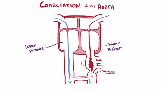
Coarctation of the aorta (CoA[1][2] or CoAo), also called aortic narrowing, is a congenital condition whereby the aorta is narrow, usually in the area where the ductus arteriosus (ligamentum arteriosum after regression) inserts. The word “coarctation” means narrowing. Coarctations are most common in the aortic arch. The arch may be small in babies with coarctations. Other heart defects may also occur when coarctation is present, typically occurring on the left side of the heart. When a patient has a coarctation, the left ventricle has to work harder. Since the aorta is narrowed, the left ventricle must generate a much higher pressure than normal in order to force enough blood through the aorta to deliver blood to the lower part of the body. If the narrowing is severe enough, the left ventricle may not be strong enough to push blood through the coarctation, thus resulting in lack of blood to the lower half of the body. Physiologically its complete form is manifested as interrupted aortic arch
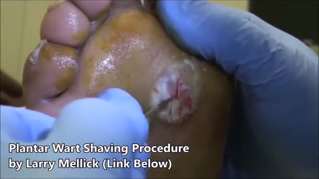
Plantar warts are hard, grainy growths that usually appear on the heels or balls of your feet, areas that feel the most pressure. This pressure also may cause plantar warts to grow inward beneath a hard, thick layer of skin (callus). Plantar warts are caused by the human papillomavirus (HPV). The virus enters your body through tiny cuts, breaks or other weak spots on the bottom of your feet. Most plantar warts aren't a serious health concern and may not require treatment. But plantar warts can cause discomfort or pain. If self-care treatments for plantar warts don't work, you may want to see your doctor to have them removed.
The mainstay of treatment is usually medication, talk therapy, or a combination of the two. Increasingly, research suggests these treatments may normalize brain changes associated with depression.
Patent ductus arteriosus (PDA), in which there is a persistent communication between the descending thoracic aorta and the pulmonary artery that results from failure of normal physiologic closure of the fetal ductus (see image below), is one of the more common congenital heart defects.
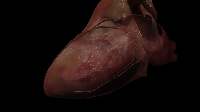
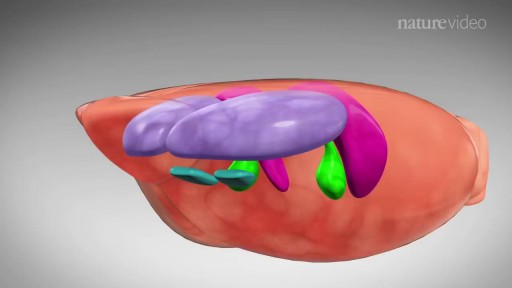
The heart is the body's engine room, responsible for pumping life-sustaining blood via a 60,000-mile-long (97,000-kilometer-long) network of vessels. The organ works ceaselessly, beating 100,000 times a day, 40 million times a year—in total clocking up three billion heartbeats over an average lifetime. It keeps the body freshly supplied with oxygen and nutrients, while clearing away harmful waste matter.
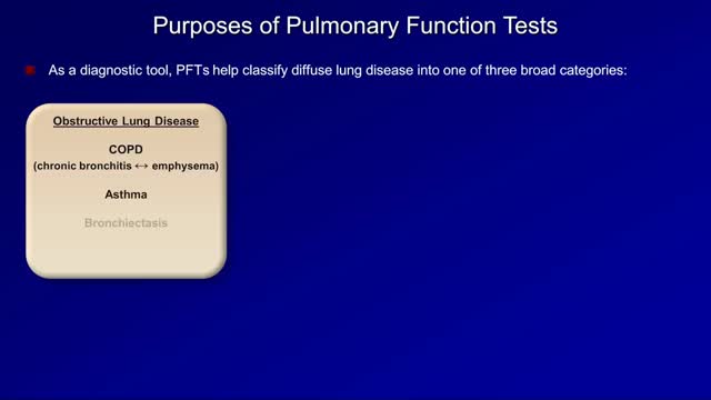
Pulmonary function tests are a broad range of tests that measure how well the lungs take in and exhale air and how efficiently they transfer oxygen into the blood. Spirometry measures how well the lungs exhale.

Prevent Stretch Marks During Pregnancy
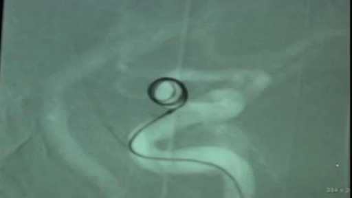
Endovascular Coiling of Unruptured Ophthalmic Artery Aneurysm

Get rid of blackheads

Things nurses should know about their patients. As a new nurse, it can be hard trying to determine what information you need to know during your shift. In addition, nurses can get extremely busy and strapped for time, so how do you keep up with all of the things you need to know?
🟣Nursing Resume Templates and Job Guide🟣
eBook: https://registerednursern.creator-spring.com/
Paperback: https://amzn.to/3QvzH3W (affiliate ad)
Free Report Sheet Templates: https://www.registerednursern.....com/nursing-report-s
In this video, Nurse Sarah explains some of the most important things nurses need to know about their patients. However, these things can vary depending on your specialty and patient population. These tips are designed to help new nurses begin to think like a nurse.
Some examples of thing nurses should know about their patients include their allergies, code status, diagnosis, medications, vital signs, and much more.
Website: https://www.registerednursern.com/
More Videos: https://www.youtube.com/watch?v=R2XMro13dD0&list=UUPyMN8DzkFl2__xnTEiGZ1w
Nursing Gear: https://teespring.com/stores/registerednursern
Instagram: https://www.instagram.com/registerednursern_com/
Facebook: https://www.facebook.com/RegisteredNurseRNs
Twitter: https://twitter.com/NursesRN
Popular Playlists:
NCLEX Reviews: https://www.youtube.com/playli....st?list=PLQrdx7rRsKf
Fluid & Electrolytes: https://www.youtube.com/playli....st?list=PLQrdx7rRsKf
Nursing Skills: https://www.youtube.com/playli....st?list=PLQrdx7rRsKf

Sclerotherapy is a procedure used to eliminate varicose veins and spider veins. Sclerotherapy involves an injection of a solution (generally a salt solution) directly into the vein. The solution irritates the lining of the blood vessel, causing it to collapse and stick together and the blood to clot.Sep 17, 2016
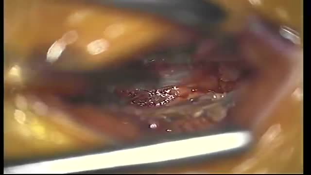
An epidural abscess is a collection of pus (infected material) between the outer covering of the brain and spinal cord and the bones of the skull or spine. The abscess causes swelling in the area. Spinal cord abscess (SCA) is a rare condition capable of causing permanent damage to the spinal cord. Abscesses are caused when injured tissue becomes infected. The body's immune system sends white blood cells to help fight off the infection. They begin to fill the damaged tissue, causing pus to build up.
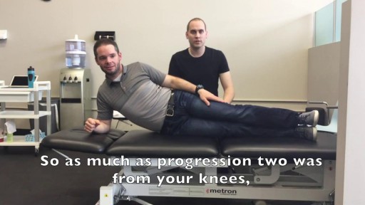
We get excited when people graduate! May it be graduating from physiotherapy or even graduating onto a new progression of an exercise! Today you move onto new challenges as Mike & Tyler demonstrate the final side plank progression. Kitchener Massage Therapy - http://www.strivept.ca/massage-therapy-kitchener.html

Initial symptoms may include: Pain or discomfort in the upper tummy (abdomen), especially after eating. Indigestion. (Note: most people who have indigestion do not have stomach cancer.) Feeling sick, and being off food. ... Weight loss and/or loss of appetite. You may pass blood out with your stools (faeces).

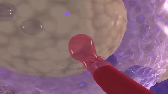
Too much cholesterol in the blood can lead to cardiovascular disease. Heart disease is the No. 1 cause of death in the United States. Over 2,100 Americans die of cardiovascular disease each day, an average of one death every 40 seconds. The good news is, you can lower your cholesterol and reduce your risk of heart disease and stroke. Working with your doctor is key. It takes a team to develop and maintain a successful health program. You and your healthcare professionals each play an important role in maintaining and improving your heart health. Work with your doctor to determine your risk and the best approach to manage it. In all cases, lifestyle changes are important to reduce your risk for heart attack and stroke. In some cases, cholesterol-lowering statin medicines may also provide benefit. Learn how to make diet and lifestyle changes easy and lasting. Also make sure you understand instructions for taking medication because it won't work if you don't take it as directed. Lifestyle Changes Your diet, weight, physical activity and exposure to tobacco smoke all affect your cholesterol level. Know Your Fats Knowing which fats raise LDL cholesterol and which ones don't is the first step in lowering your risk of heart disease.
A heart attack occurs when the flow of blood to the heart is blocked, most often by a build-up of fat, cholesterol and other substances, which form a plaque in the arteries that feed the heart (coronary arteries). The interrupted blood flow can damage or destroy part of the heart muscle. A heart attack, also called a myocardial infarction, can be fatal, but treatment has improved dramatically over the years. It's crucial to call 911 or emergency medical help if you think you might be having a heart attack

Knee replacement involves replacing a knee joint that has been damaged or worn away, usually by arthritis or injury. Find out more here: https://www.bupa.co.uk/health-....information/knee-cli
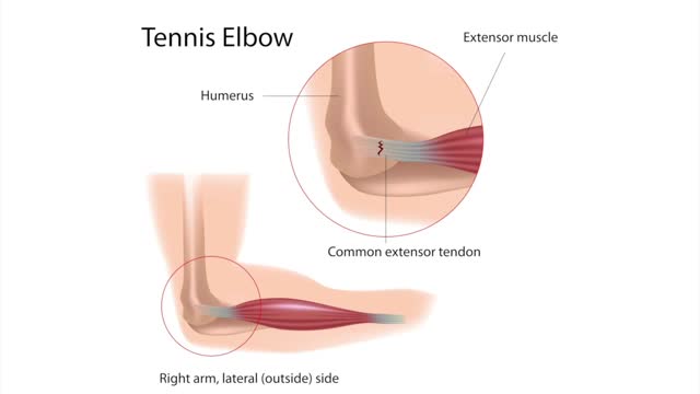
Tennis elbow or lateral epicondylitis is a condition in which the outer part of the elbow becomes sore and tender. The forearm muscles and tendons become damaged from overuse — repeating the same strenuous motions again and again.
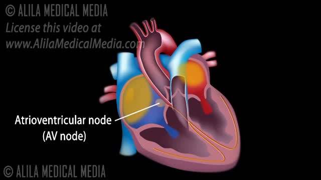
The normal electrical conduction in the heart allows the impulse that is generated by the sinoatrial node (SA node) of the heart to be propagated to (and stimulate) the cardiac muscle (myocardium). The myocardium contracts after stimulation.
What does the placenta do? The placenta is an organ that develops in your uterus during pregnancy. This structure provides oxygen and nutrients to your growing baby and removes waste products from your baby's blood. The placenta attaches to the wall of your uterus, and your baby's umbilical cord arises from it. In most pregnancies, the placenta attaches at the top or side of the uterus.
