- Physical Examination
- Surgical Examination
- Ophthalmology
- Clinical Skills
- Orthopedics
- Surgery Videos
- Laparoscopy
- Pediatrics
- Funny Videos
- Cardiothoracic Surgery
- Nursing Videos
- Plastic Surgery
- Otorhinolaryngology
- Histology and Histopathology
- Neurosurgery
- Dermatology
- Pediatric Surgery
- Urology
- Dentistry
- Oncology and Cancers
- Anatomy Videos
- Health and Fitness
- Radiology
- Anaesthesia
- Physical Therapy
- Pharmacology
- Interventional Radiology
- Cardiology
- Endocrinology
- Gynecology
- Emergency Medicine
- Psychiatry and Psychology
- Childbirth Videos
- General Medical Videos
- Nephrology
- Physiology
- Diet and Food Health
- Diabetes Mellitus
- Neurology
- Women Health
- Osteoporosis
- Gastroenterology
- Pulmonology
- Hematology
- Rheumatology
- Toxicology
- Nuclear Medicine
- Infectious Diseases
- Vascular Disease
- Reproductive Health
- Burns and Wound Healing
- Other
Top videos
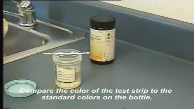
This video demonstrates how use a commercially-prepared "dip-stick" to test a random urine specimen for the presence of protein or glucose.
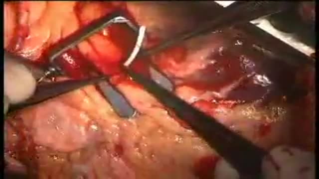
Beating heart or "off pump" coronary artery surgery is the latest revolution in the management coronary disease. It is being embraced world-wide by increasing numbers of surgeons. Many of the advantages are subtle but reduced mortality, stroke, and bleeding as well as earlier discharge are well-established benefits. A cardiac stabiliser is mandatory for this surgery, most are single use only and very expensive, this one is multiple use and is saving many healthcare dollars
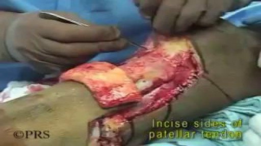
Pathology: Previous spinal cord injury, diabetes, renal failure, dynamic knee contracture, open left ankle disarticulation for sepsis and severe foot infection
Laparoscopic anterior resection for cancer colon in Qatar by Dr. Al-Emadi

This membrane could prevent heart attacks and keep the heart alive.
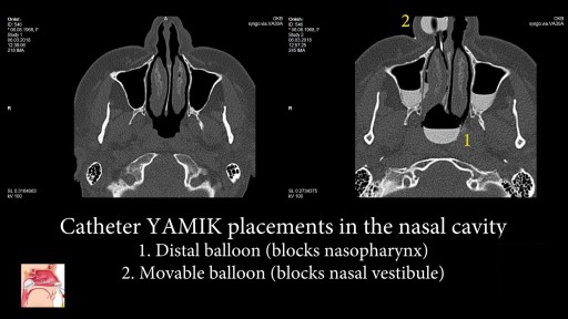
Nasal catheter YAMIK - is a new drug delivery sistem for topical treatment for sinusitis. The introduction of a large volume of the #drugsolution directly into all the paranasal #sinuses - provides new opportunities in the #treatment of #sinusitis! #YamikprocedureNasal catheter YAMIK is a new device for topical sinonasal delivery of medication during rhinosinusitis treatment. Administration of therapeutic solution with YAMIK catheter is called YAMIK procedure. The following features differs YAMIK procedure from all other topical sinonasal delivery techniques: - Medication is delivered into the all paranasal sinuses at one side of nose regardless of their involvement in the inflammatory disease. -Specific position of patient’s head. Patient should lay on the side of of the sinuses, into which solution will be administered. This position is physiological and comfortable for patients, including children and elders. The LHL position was suggested to be the most favorable position for patients to adopt - Therapeutic solutions reliably penetrates into without previous sinus surgery sinuses with natural ostia size. - Paranasal sinuses are filling with medicinal solution by gravity. To accelerate process, it is used small pressure gradient, which created by motion of syringe plunger with amplitude 1 - 2 ml during administration of solution. - It is provided contact of the whole sino-nasal mucosa with medication. - Prolonged time of the contact of sino-nasal mucosa with medicine provides administration of the therapeutically significant dose. Therapeutic solution administered into paranasal sinuses is considered as a STORE. Thanks to affect mucociliary clearance, therapeutic solution is gradually evacuated from sinuses through ostia. Thereby, prolonged nasal irrigation is performed. - Due to extended contact with saline (NaCl 0,9%), viscous colloidal pathological substance filling paranasal sinuses is dissolved. As a result, its viscosity decreases, and substance is removing by mucociliary clearance. Thereby, drainage function of the ostia are returned some time after finish YAMIK method procedure. -The procedure is performing under local anesthesia. - There is no need in active involvement of the patient. Blowing, pronouncing any sounds like “cuckoo”, holding any things and so on is unnecessary. If is performed by a qualified medical professional the procedure is more effective. - Medication contacts only with nasal passages and paranasal sinuses. Thus, it is provided topical drug therapy. - YAMIK procedure is call sinonasal delivery techniques of a therapeutic solution. It differs from nasal techniques, because medicinal solution contacts not only with nasal mucosa, but with mucous membrane of paranasal sinuses. - The only used drug formulation is a solution. - It is possible non-invasive sample extraction from mucosa of paranasal sinuses (for bacteriological, immunological, cytological and a number of others investigation methods).
METHODS:
Previously existing methods are characterized by unpleasant scars that, despite surgeons promises, remain for life.
Incisions are:
- around the areola (Round block) leading to a flat areola, often unpleasant hypertophic skars, skin rippling.
- inverted T (around the areola, vertically down and in the fold under the breast).
- Vertical (around the areola and vertically down). Due to the extess skin, incisions often turn into inverted L or T. Rearrangement of glandular tissue and skin changes the shape of the breasts and may be different from expectations. Scars worry patients and sometimes cause disturbances in the relationship with their partner.
- No scars. The "Serdev Suture" lifting technique for breast lifting without scars (only points - needle perforations in the skin) is created by the Bulgarian cosmetic surgeon Prof. Dr. Nikolay Serdev. It is a novelty that had changed the cosmetic surgery world in the last 10-14 years for young patients. The technique is especially important in Asia and Latin America, for Asians, African-Americans, Indians, and others who form keloids and lumpy scars after operations.
The Serdev suture method can achieve lift upto and over 14 centimeters and is most suitable for the following types of breasts:
- not very heavy full breasts.
- in the presence of subpectoral implants with subsequent drooping of the breasts after childbirth and lactation.
- empty and loose breasts after childbirth and breastfeeding. In such cases this technique is combined with subpectoral implants. In sagging breasts implants should not be placed in the skin over the pectoral muscles, because thus will lead to even more drooping. Therefore, breast lift requires breast fixation to the level of the pectoral muscle (the normal position in young women), and then placement of appropriate implants under the muscle, to hold them in the appropriated position.
- in drooping breasts after subglandular augmentation (over the muscle). In such cases, patients should not wait until the skin elongation becomes visible. The implants should be removed, the capsule removed - a difficult but a necessary operation, preventing postop seromas and infection. Implants should be placed under the pectoralis muscle to wear them. Patients should orient the cosmetic surgeon at what level they want the nipples - in the middle of the implant, higher or lower.
Implants should be generally replaced - below the muscle implants should be smooth, move naturally without hurting the muscle.
Because of modern anesthetics and new methods without trauma, pain and swelling after surgery are not significant. In 3-4 days, patients can return to social life, even the next day, but it is preferable to rest for 2-3 days.
Exercises with the arms and weight lifting is prohibited for a month and a half.
Due to lack of scars, the breast lift using the Serdev sutures can be repeated to maintain the aesthetic appearence of the breasts even in advanced age.
Gigantomastia i.e. very large, very heavy and drooping breasts can not be operated in this manner, because of gravity and overskin.
Early mastopexy using Serdev sutures is recommended before too much changes in the tissues. If late, more and more complex interventions are required.
"A lot of people are opting for various breast procedures and one of the most common among them is “mastopexy”. This is the surgery that involved uplifting of sagging breasts and, in certain cases, repositioning of the nipple and areola in order to restore normality and beauty. The excess skin is removed and firmness is provided to the breasts. Though mastopexy can be done as a stand alone surgery, many people combine it with breast augmentation which involves inserting implants inside the b
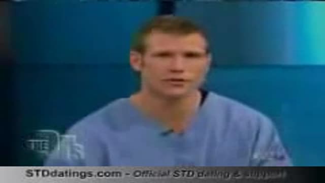
TV interview with Adina Nack, Ph.D. about her own cervical HPV experiences, STD research, her new book (Damaged Goods? Women Living with Incurable Sexually Transmitted Diseases), and women's lives after genital warts, HPV and herpes infections. More info is available on STDdatings.com, which is the official STD dating & support site.
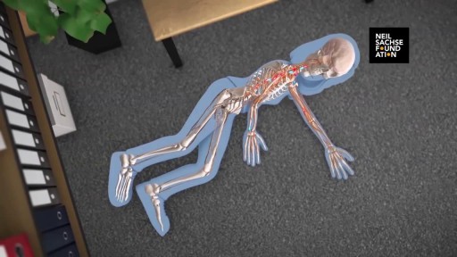
A detailed animation video explaining a spinal cord injury.
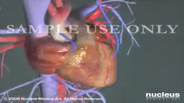
Coronary Artery Bypass Surgery CABG Heart
Examination of Neck Swelling

An antecedent upper respiratory infection is present in 50% of patients. Abdominal pain is a presenting symptom in 1 0-15% of patients. The skin lesions are symmetric, involve dependent parts of the body, and classically progress from an erythematous, macular rash to papular purpura. The joints and kidneys are also commonly involved
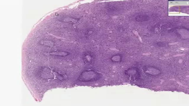
Histology of Spleen
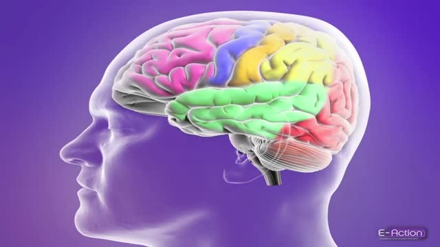
A brief demonstration of the different types of epileptic seizures based on the International Classification of Epileptic Seizures.
Methotrexate anti-tumor activity is a result of the inhibition of folic acid reductase, leading to inhibition of DNA synthesis and inhibition of cellular replication. The mechanism involved in its activity against rheumatoid arthritis is not known.
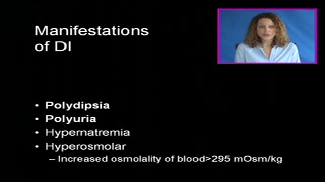
Diabetes insipidus (die-uh-BEE-teze in-SIP-uh-dus) is an uncommon disorder that causes an imbalance of water in the body. This imbalance leads to intense thirst even after drinking fluids (polydipsia), and excretion of large amounts of urine (polyuria). While the names diabetes insipidus and diabetes mellitus sound similar, they're not related. Diabetes mellitus — which can occur as type 1 or type 2 — is the more common form of diabetes. There's no cure for diabetes insipidus, but treatments are available to relieve your thirst and normalize your urine output.
A knee revision, from Pakistan!!
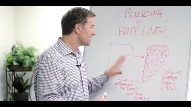
If severe, it can eventually lead to cirrhosis and liver failure. How would you know if you have a fatty liver? ... Luckily fatty liver is reversible. ... Eat less carbohydrate. ... Drink less alcohol. ... Eat more vegetables, protein and the right fats. ... Drink raw vegetable juices. ... Take a good liver tonic.
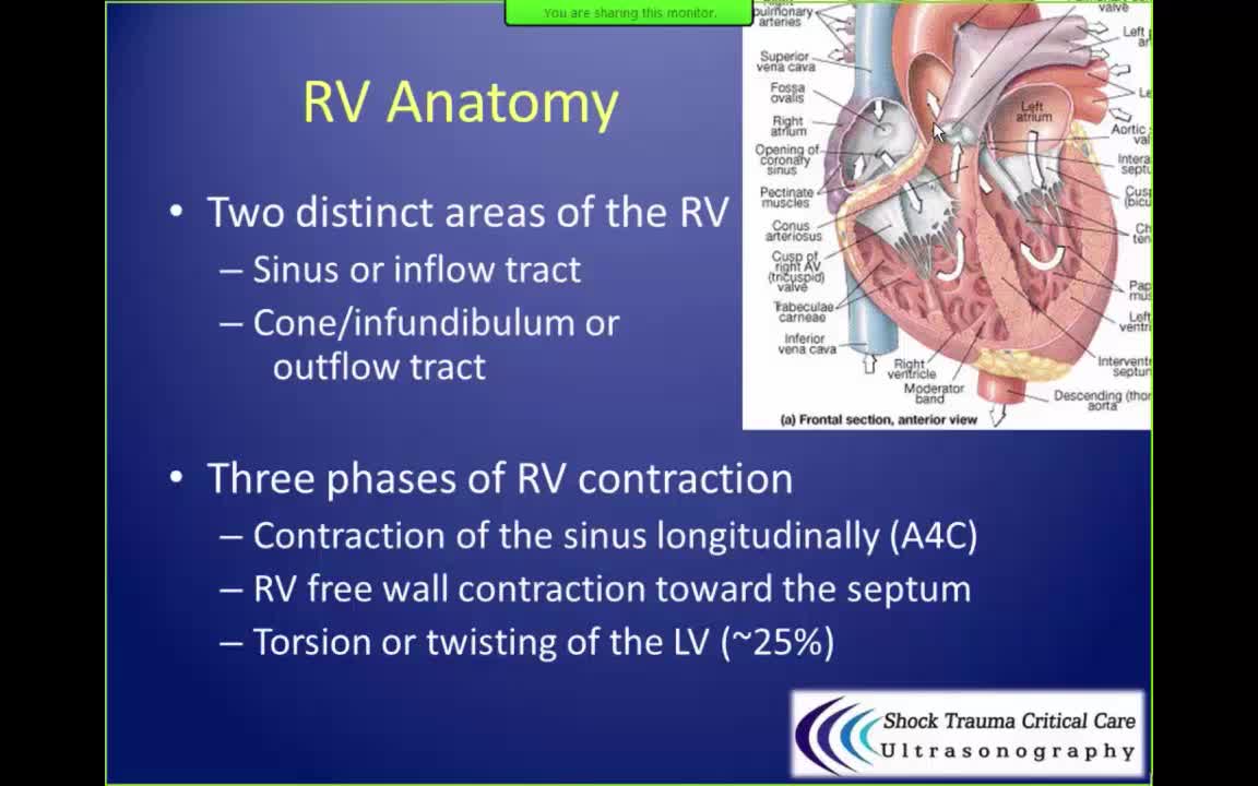
In patients with advanced congestive heart failure due to cardiomyopathy or ischemia, right ventricle shortening is the only significant independent associate of survival by multivariate analysis (as opposed to other parameters including left ventricular ejection fraction, cardiac index, and pulmonary resistance).

