- Physical Examination
- Surgical Examination
- Ophthalmology
- Clinical Skills
- Orthopedics
- Surgery Videos
- Laparoscopy
- Pediatrics
- Funny Videos
- Cardiothoracic Surgery
- Nursing Videos
- Plastic Surgery
- Otorhinolaryngology
- Histology and Histopathology
- Neurosurgery
- Dermatology
- Pediatric Surgery
- Urology
- Dentistry
- Oncology and Cancers
- Anatomy Videos
- Health and Fitness
- Radiology
- Anaesthesia
- Physical Therapy
- Pharmacology
- Interventional Radiology
- Cardiology
- Endocrinology
- Gynecology
- Emergency Medicine
- Psychiatry and Psychology
- Childbirth Videos
- General Medical Videos
- Nephrology
- Physiology
- Diet and Food Health
- Diabetes Mellitus
- Neurology
- Women Health
- Osteoporosis
- Gastroenterology
- Pulmonology
- Hematology
- Rheumatology
- Toxicology
- Nuclear Medicine
- Infectious Diseases
- Vascular Disease
- Reproductive Health
- Burns and Wound Healing
- Other
Top videos
40 years old patient, Parity 3, wanted to have a sterilization. The surgery was perfomed laparoscopically with coagulation technique. This video is not edited and presented in full length.
This was a Nasogastric Intubation that went very wrong. The tube went up into the brain, causing severe damage, instead of going down through the throat.

How to use a Chlamydia rapid test kit for self-diagnosis of Chlamydia (swab test). Convenient, Easy to Use, and over 95% Accurate. Certified GMP and ISO13485. Test yourself at home with Complete Privacy. Buy online today at: http://www.stdrapidtest.com
Exercises. Light exercises in which you move your affected limb may encourage lymph fluid drainage and help prepare you for everyday tasks, such as carrying groceries. ...
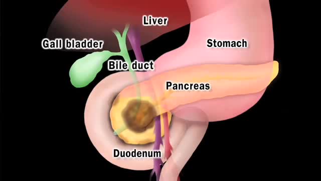
Pancreatic cancer begins in the tissues of your pancreas — an organ in your abdomen that lies horizontally behind the lower part of your stomach. Your pancreas secretes enzymes that aid digestion and hormones that help regulate the metabolism of sugars. Pancreatic cancer often has a poor prognosis, even when diagnosed early. Pancreatic cancer typically spreads rapidly and is seldom detected in its early stages, which is a major reason why it's a leading cause of cancer death. Signs and symptoms may not appear until pancreatic cancer is quite advanced and complete surgical removal isn't possible.
The temporomandibular joint (TMJ), located just in front of the lower part of the ear, allows the lower jaw to move. The TMJ is a ball-and-socket joint, just like the hip or shoulder. When the mouth opens wide, the ball (called the condyle) comes out of the socket and moves forward, going back into place when the mouth closes. TMJ becomes dislocated when the condyle moves too far and gets stuck in front of a bony prominence called the articular eminence. The condyle can't move back into place. This happens most often when the ligaments that normally keep the condyle in place are somewhat loose, allowing the condyle to move beyond the articular eminence. The surrounding muscles often go into spasm and hold the condyle in the dislocated position.

Reactive arthritis can affect the heels, toes, fingers, low back, and joints, especially of the knees or ankles. Though it often goes away on its own, reactive arthritis can be prolonged and severe enough to require seeing a specialist. Effective treatment is available for reactive arthritis. Reactive arthritis tends to occur most often in men between ages 20 and 50. Most cases of reactive arthritis appear as a short episode. Occasionally, it becomes chronic. Reactive arthritis is a painful form of inflammatory arthritis (joint disease due to inflammation). It occurs in reaction to an infection by certain bacteria. Most often, these bacteria are in the genitals (Chlamydia trachomatis) or the bowel (Campylobacter, Salmonella, Shigella and Yersinia). Chlamydia most often transmits by sex. It often has no symptoms, but can cause a pus-like or watery discharge from the genitals. The bowel bacteria can cause diarrhea. If you develop arthritis within one month of diarrhea or a genital infection – especially with a discharge – see a health care provider. You may have reactive arthritis. - See more at: http://www.rheumatology.org/I-Am-A/Patient-Caregiver/Diseases-Conditions/Reactive-Arthritis#sthash.VNgDSOOY.dpuf
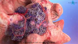
This cancer development medical video is devoted to elaborating the basics of cancer growth. We used advanced medical animation techniques to display such a complicated process.
What is happening in cancer development medical video
The fundamental abnormality described in the cancer development medical video is the nonstop unregulated multiplication of cancer cells. Being uncontrollable by body’s signals that regulate normal cell behavior; cancerous cells divide and grow populating neighboring normal tissues or even spread throughout the body. The overall lack of growth control acquired by cancer cells is due to the accumulated abnormalities in numerous cell regulatory mechanisms and is considered in some aspects of cell behavior that differs them from their healthy counterparts. The interaction of these cells is shown in our previous medical animation video.
Read full article on our webpage http://bit.ly/2LQj9ln
Follow us on Facebook https://www.facebook.com/Nanob....ot.Medical.Animation
Follow us on LinkedIn https://www.linkedin.com/compa....ny/nanobotmodels-med
Follow us on Twitter https://twitter.com/Nanobot_Studio
Follow us on Instagram https://www.instagram.com/nano....bot_medical_animatio
Follow us on Clutch https://clutch.co/profile/nano....bot-medical-animatio
Follow us on Behance https://www.behance.net/NanobotStudio
#cancer #tumor #oncology #metatastic #nanobot #visualscience #scientificcommunication #medicalanimation #animationvideo #animationdesign #animationstudio #animationmovie #nanotechnology #medicine #health #science #education #medschool #medicaleducation #animation_studio #animationstudio
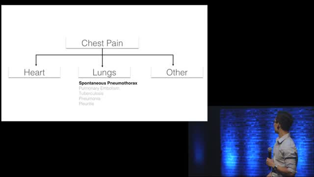
Chest pain is a frequent complaint of patients seeking urgent medical assistance, and accounts for an estimated 2-4 per cent of all A&E visits in the UK (Becker, 2000). Generally, acute chest pain should be considered cardiovascular in origin until proven otherwise and it is common in clinical practice to err on the conservative or ‘safe’ side when evaluating people with chest pain. Individuals with suspected ischaemic chest pain must be evaluated rapidly for several reasons: - Myocardial ischaemia, if prolonged and severe, can cause myocardial infarction (necrosis); - Treatment strategies that achieve myocardial salvage (thrombolytic therapy or primary coronary angioplasty) are available for patients with acute coronary syndromes and these treatments reduce morbidity and mortality;
Rotator cuff repair is surgery to repair a torn tendon in the shoulder. The procedure can be done with a large (open) incision or with shoulder arthroscopy, which uses small buttonhole-sized incisions.
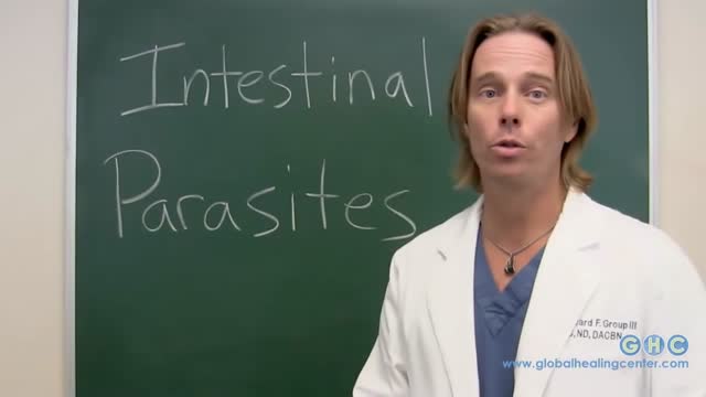
One of the most common parasites to infect human beings is the yeast-like Blastocystis hominis, a single-celled parasitic organism that causes abdominal cramping, bloating, gas, and sometimes anal itching. Other common parasites are: Tapeworms, which can grow as long as 60 feet while living in the human intestines.

Inner Workings tells the story of the ceaseless pull of the human heart — even as it works against the very stoic realism of the brain.
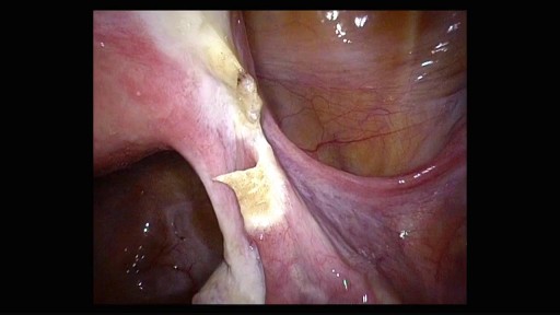
Surgical drainage of dental abscess extending Into the Sub mandibular Space
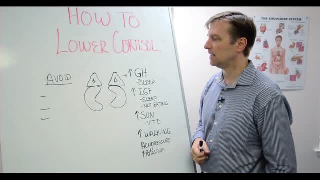
Assuming you haven't been diagnosed with Cushing's disease by your doctor, here are steps you can take to help lower high cortisol levels naturally: Switch to a Whole Foods, Anti-inflammatory Diet. Reduce and Manage Stress. Exercise Regularly. Use Adaptogen Herbs and Superfoods. Try Essential Oils to Promote Relaxation.
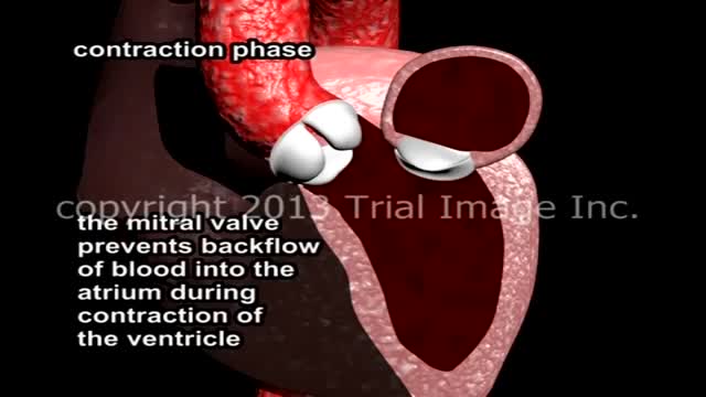
Mitral Valve Prolapse and Mitral Regurgitation. Review of mitral valve anatomy and function, including papillary muscle structure and function, with severe mitral valve prolapse and mitral regurgitation due to a flail segment caused by ruptured papillary muscle and chorda tendinae attachment.

Gastroschisis is a birth defect that develops in a baby while a woman is pregnant. This condition occurs when an opening forms in the baby's abdominal wall. The baby's bowel pushes through this hole. It then develops outside of the baby's body in the amniotic fluid.

Watch that video to know if it is safe to have intercourse during her period

Watch that video of Super Model's Butt and Leg Implants Exploded
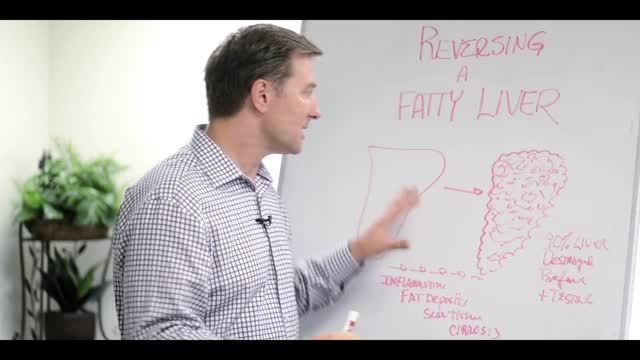
If severe, it can eventually lead to cirrhosis and liver failure. How would you know if you have a fatty liver? ... Luckily fatty liver is reversible. ... Eat less carbohydrate. ... Drink less alcohol. ... Eat more vegetables, protein and the right fats. ... Drink raw vegetable juices. ... Take a good liver tonic.

It is a specialized bodily fluid that supplies essential substances and nutrients, such as sugar, oxygen, and hormones to our cells, and carries waste away from those cells, this waste is eventually flushed out of the body in urine, feces, sweat, and lungs (carbon dioxide). Blood also contains clotting agents.
