- Physical Examination
- Surgical Examination
- Ophthalmology
- Clinical Skills
- Orthopedics
- Surgery Videos
- Laparoscopy
- Pediatrics
- Funny Videos
- Cardiothoracic Surgery
- Nursing Videos
- Plastic Surgery
- Otorhinolaryngology
- Histology and Histopathology
- Neurosurgery
- Dermatology
- Pediatric Surgery
- Urology
- Dentistry
- Oncology and Cancers
- Anatomy Videos
- Health and Fitness
- Radiology
- Anaesthesia
- Physical Therapy
- Pharmacology
- Interventional Radiology
- Cardiology
- Endocrinology
- Gynecology
- Emergency Medicine
- Psychiatry and Psychology
- Childbirth Videos
- General Medical Videos
- Nephrology
- Physiology
- Diet and Food Health
- Diabetes Mellitus
- Neurology
- Women Health
- Osteoporosis
- Gastroenterology
- Pulmonology
- Hematology
- Rheumatology
- Toxicology
- Nuclear Medicine
- Infectious Diseases
- Vascular Disease
- Reproductive Health
- Burns and Wound Healing
- Other
Top videos
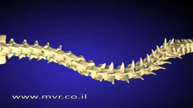
There are several approaches to scoliosis surgery, but all use modern instrumentation systems in which hooks and screws are applied to the spine to anchor long rods. The rods are then used to reduce and hold the spine while bone that is added fuses together with existing bone.

The oral contraceptive pill, commonly known as "the pill," is a hormone-based method of preventing pregnancy. It can also help resolve irregular menstruation, painful or heavy periods, endometriosis, acne, and premenstrual syndrome (PMS). Birth control pills work by preventing ovulation. No egg is produced, so there is nothing for the sperm to fertilize. Pregnancy cannot occur. "The pill" is used by nearly 16 percent of women aged 15 to 44 years in the United States, and it has both advantages and disadvantages. People with different risk factors may be advised to use a particular kind of pill. There are different types of contraceptive pills. They all contain synthetic forms of the hormones estrogen, progesterone, or both. Synthetic progesterone is called progestin. Combination pills contain progestin and estrogen. The "mini pill," contains only progestin. Monophasic pills all contain the same balance of hormones. With phasic pills, two or three different types of pill are taken each month, each with a different balance of hormones.
This test stimulates your acoustic nerve by delivering cold or warm water or air into your ear canal. When cold water or air enters your ear and the inner ear changes temperature, it should cause fast, side-to-side eye movements called nystagmus. The test is done in the following way: Before the test, your ear, especially the eardrum, will be checked. This is to make sure it is normal. One ear is tested at a time. A small amount of cold water or air is gently delivered into one of your ears. Your eyes should show an involuntary movement called nystagmus. Then they should turn away from that ear and slowly back. If water is used, it is allowed to drain out of the ear canal. Next, a small amount of warm water or air is gently delivered into the same ear. Again, your eyes should show nystagmus. Then they should turn toward that ear and slowly back. Your other ear is tested in the same way.

There are a number of different causes of vertigo. Vertigo can be defined based upon whether the cause is peripheral or central. Central causes of vertigo arise in the brain or spinal cord while peripheral vertigo is due to a problem within the inner ear. The inner ear can become inflamed because of illness, or small crystals or stones found normally within the inner ear can become displaced and cause irritation to the small hair cells within the semicircular canals, leading to vertigo. This is known as benign paroxysmal positional vertigo (BPPV).
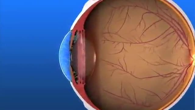
Glaucoma Pathogenesis Simplified
Ellie was born with a rare condition which stopped her jawbones from growing properly. At first, her parents didn't realize there was a problem, apart from the fact that her teeth were not aligned. But when she went to have braces fitted to straighten her teeth when she was 14, orthodontist Joy Hickman realized her jaw had not grown since she was eight. Over the next six years Hickman worked with a maxillofacial surgeon to transform Ellie's looks. Ellie, who is now 20, said the surgery was painful but paid almost immediate dividends. "About six months after it was my year 11 prom and it looked good." Ellie told the Daily Post the change in her appearance has been matched by an increase in confidence.
This video is really sad. You can literally watch this man dying. He was shot in the chest and rushed to the emergency room. His heart has stopped beating or has arrested. As a last resort, surgeons did an extreme procedure called an open thoracotomy which is that crazy tool you see there that basically splits the ribs open and allows easy open access to the heart. They did this so they could give him a cardiac massage. A cardiac massage is when surgeons are manually trying to pump the heart after it has stopped working on its own (cardiac arrest). Unfortunately he lost so much blood from his gun shot wound and he was pronounced dead. There are cases of patients surviving after having this kind of invasive resuscitation but it is rare.
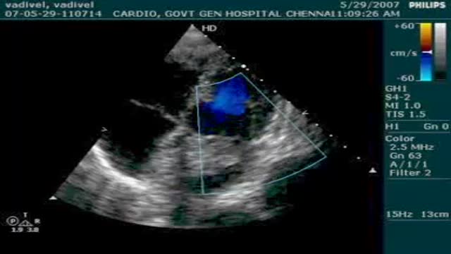
Symptoms range from nonspecific and constitutional to sudden cardiac death. [18] In about 20% of cases, myxomas may be asymptomatic and discovered as an incidental finding. Signs and symptoms of mitral stenosis, endocarditis, mitral regurgitation, and collagen vascular disease can simulate those of atrial myxoma. A high index of suspicion aids in diagnosis. Symptoms of left-sided heart failure include the following: Dyspnea on exertion (75%) that may progress to orthopnea, paroxysmal nocturnal dyspnea, and pulmonary edema is observed. [19, 20] Symptoms are caused by obstruction at the mitral valve orifice. Valve damage may result in mitral regurgitation.

Most folks remember puberty – and not always in a good way. It can be an awkward stage of budding breasts, unwanted hair, acne and unexpected body odor. Puberty, when a child undergoes physical changes and becomes sexually mature, typically begins around age 8 in girls and age 9 in boys. But imagine, say, a 6- or 7-year-old undergoing such changes? Studies are showing that the onset of puberty for both boys and girls is occurring earlier and earlier, a phenomenon defined as precocious puberty. A study published in Pediatrics in 2010 found that among a population of 1,200 American girls, about 23 percent of African-Americans,15 percent of Latinas and 10 percent of Caucasian girls had begun puberty (marked by breast development) at age 7. In 2012, another study published in Pediatrics found that puberty in American boys – measured by testicular enlargement and pubic hair growth – was beginning six months to two years earlier than what research in previous decades had documented, particularly among African-American children.
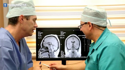
A subdural hematoma (SDH) is a collection of blood below the inner layer of the dura but external to the brain and arachnoid membrane (see the images below). Subdural hematoma is the most common type of traumatic intracranial mass lesion. Subdural hematoma occurs not only in patients with severe head injury but also in patients with less severe head injuries, particularly those who are elderly or who are receiving anticoagulants. Subdural hematoma may also be spontaneous or caused by a procedure, such as a lumbar puncture (see Etiology). Rates of mortality and morbidity can be high, even with the best medical and neurosurgical care (see Prognosis). Subdural hematomas are usually characterized on the basis of their size and location and the amount of time elapsed since the inciting event age (ie, whether they are acute, subacute, or chronic). When the inciting event is unknown, the appearance of the hematoma on neuroimaging studies can help determine when the hematoma occurred. These factors, as well as the neurologic and medical condition of the patient, determine the course of treatment and may also influence the outcome. Generally, acute subdural hematomas are less than 72 hours old and are hyperdense compared with the brain on computed tomography scans. The subacute phase begins 3-7 days after acute injury. Chronic subdural hematomas develop over the course of weeks and are hypodense compared with the brain. However, subdural hematomas may be mixed in nature, such as when acute bleeding has occurred into a chronic subdural hematoma. Presentation varies widely in acute subdural hematoma (see Clinical). Many of these patients are comatose on admission. However, approximately 50% of patients with head injuries who require emergency neurosurgery present with head injuries that are classified as moderate or mild (Glasgow Coma Scale scores 9-13 and 14-15, respectively). Many of these patients harbor intracranial mass lesions. In a large series of patients who developed intracranial hematomas requiring emergent decompression, more than half had lucid intervals and were able to make conversation between the time of their injury and subsequent deterioration. In a more comprehensive review of the literature on the surgical treatment of acute subdural hematomas, lucid intervals were noted in up to 38% of cases. These patients may be more likely to benefit from medical and surgical intervention when instituted in a timely fashion (ie, before further neurological deterioration).
One of the criteria to determine brain death is the irreversible absence of cerebral and brainstem reflexes including pupillary, oculocephalic, oculovestibular (caloric), corneal, gag, sucking, swallowing, and extensor posturing. Some of the other criteria for determination of brain death include: 1. Absence of respiratory drive (apnea) off the ventilator for a duration that is sufficient to produce hypercarbic drive (usually 10 to 20 minutes to achieve pC02 of 50 to 60 mmHg) ( 2. Body temperature below 34 C (93.2 F) 3. EEG isoelectric for 30 minutes at maximal gain 4. Absence of cerebral circulation by Doppler or magnetic resonance angiography 5. At least 24 hours of observation in adults with anoxic-ischemic brain damage with a negative drug screen
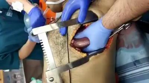
WHAT IS BURN DEBRIDEMENT? A burn is damage to body tissues caused by sunlight, heat, fire, electricity, friction, radiation, chemicals, hot water or steam. Burns may become infected. Infected burns and the swelling that happens as a result can cause severe damage to the organs and tissues underneath the burned area by putting pressure on the tissues, nerves, and blood vessels. To allow healthy tissue to heal and to prevent more damage or infection, burned tissue is removed in a procedure called burn debridement. Burn debridement can be done by several different methods. They include surgical, chemical, mechanical, or autolytic tissue removal. Debridement may need to be done multiple times as the burned area heals.

Meningococcal meningitis - causes, features, symptoms and treatment
Pelvic ureter. The ureter enters the pelvis, where it crosses anteriorly to the iliac vessels, which usually occurs at the bifurcation of the common iliac artery into the internal and external iliac arteries. Here, the ureters are within 5 cm of one another before they diverge laterally.
Anytime you're having unprotected sex, there's always a chance that a woman can get pregnant. Pregnancy requires sperm and egg to meet up together so a woman needs to be during her most fertile time of the month, which is usually 6 days out of the month; 5 days leading up to ovulation and on the day of ovulation. For most women, ovulation happens 12-16 days before her period's going to start. So a woman is usually most fertile for a week to a week and a half after her period has ended generally speaking, if you don't want to count each and every single day. So if you have unprotected intercourse during this time, then there's a high probability that a woman can get pregnant. Now, you mentioned that your girlfriend is supposed to start her period in about five days or so. If you've had intercourse any time leading up to this, there's always a chance that she could get pregnant. But as for the mechanics of it all, in order to get pregnant, semen needs to be inserted inside the vaginal canal where the egg and sperm can then meet. So if that did not happen, then the chances of her getting pregnant are slim. But if that has happened, the chances of her getting pregnant are great. So it would be best for you and her to just wait until her period is supposed to start and if she's late, then take an over-the-counter pregnancy test and if it's positive, congratulations to both of you! If it's negative and she still doesn't start her period, then tell her to wait about 5-7 days. Take another test and then maybe at that point, it will be positive if she is indeed pregnant. If she continues to not have a period or she's concerned about anything, it would be best for her to follow up with her doctor and they can decide if further investigation or treatment is warranted. If you have any other questions for me, feel free to ask them on our Facebook page at facebook.com/intermountainmoms and recommend us to your friends and family, too.
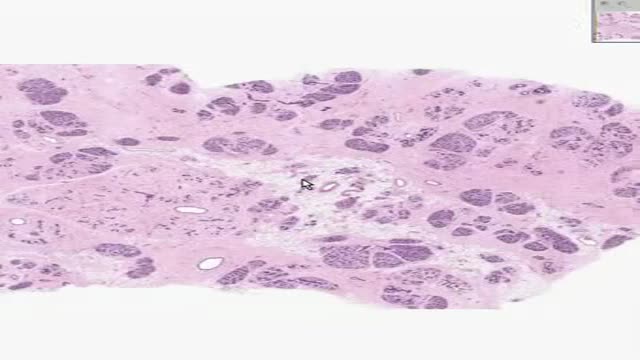
Histology of Inactive Breast

The deep veins play a significant role in propelling blood toward the heart. The one-way valves in deep veins prevent blood from flowing backward, and the muscles surrounding the deep veins compress them, helping force the blood toward the heart, just as squeezing a toothpaste tube ejects toothpaste.

Anatomy of The Abdominal Autonomic Nerve Supply
Head Eye and ENT Physical Examination
