- Physical Examination
- Surgical Examination
- Ophthalmology
- Clinical Skills
- Orthopedics
- Surgery Videos
- Laparoscopy
- Pediatrics
- Funny Videos
- Cardiothoracic Surgery
- Nursing Videos
- Plastic Surgery
- Otorhinolaryngology
- Histology and Histopathology
- Neurosurgery
- Dermatology
- Pediatric Surgery
- Urology
- Dentistry
- Oncology and Cancers
- Anatomy Videos
- Health and Fitness
- Radiology
- Anaesthesia
- Physical Therapy
- Pharmacology
- Interventional Radiology
- Cardiology
- Endocrinology
- Gynecology
- Emergency Medicine
- Psychiatry and Psychology
- Childbirth Videos
- General Medical Videos
- Nephrology
- Physiology
- Diet and Food Health
- Diabetes Mellitus
- Neurology
- Women Health
- Osteoporosis
- Gastroenterology
- Pulmonology
- Hematology
- Rheumatology
- Toxicology
- Nuclear Medicine
- Infectious Diseases
- Vascular Disease
- Reproductive Health
- Burns and Wound Healing
- Other
Top videos
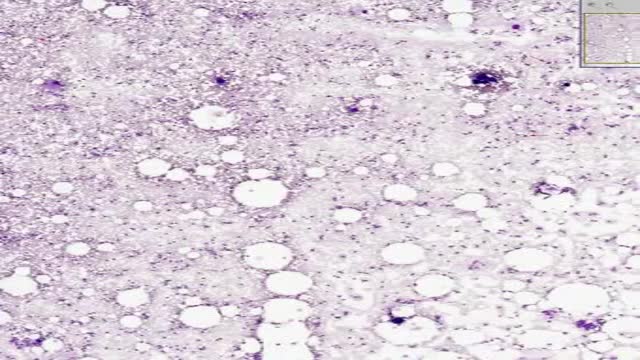
Histology of Bone Marrow Smear
Rehydration Tips: Kids & Teens (Ages 1+) Give clear liquids (avoid milk and milk products) in small amounts every 15 minutes. ... If your child vomits, start over with a smaller amount of fluid (2 teaspoons, or about 10 milliliters) and continue as above. ... After no vomiting for about 8 hours, introduce solid foods slowly.

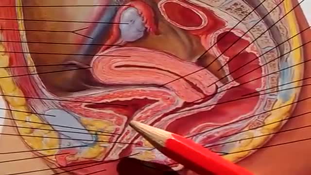
Absence of a woman's monthly menstrual period is called amenorrhea. Secondary amenorrhea is when a woman who has been having normal menstrual cycles stops getting her periods for 6 months or longer. Causes Secondary amenorrhea can occur due to natural changes in the body. For example, the most common cause of secondary amenorrhea is pregnancy. Breastfeeding and menopause are also common, but natural, causes. Women who take birth control pills or who receive hormone shots such as Depo-Provera may not have any monthly bleeding. When they stop taking these hormones, their periods may not return for more than 6 months. You are more likely to have absent periods if you: Are obese Exercise too much and for long periods of time Have very low body fat (less than 15 to 17%) Have severe anxiety or emotional distress Lose a lot of weight suddenly (for example, from strict or extreme diets or after gastric bypass surgery) Other causes include: Brain (pituitary) tumors Drugs for cancer treatment Drugs to treat schizophrenia or psychosis Overactive thyroid gland Polycystic ovarian syndrome Reduced function of the ovaries

DIRTIEST PARTS OF YOUR BODY
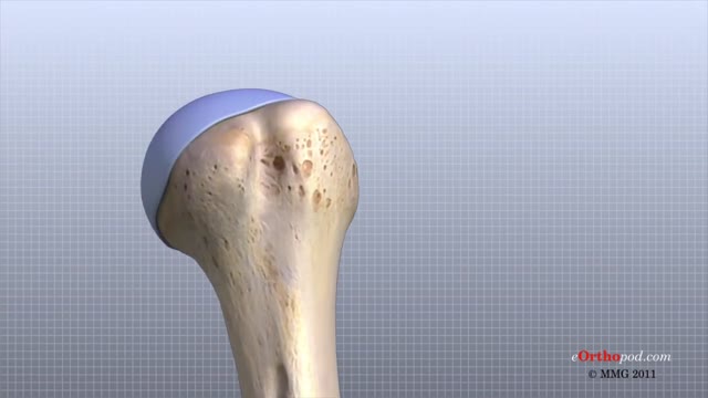
The shoulder joint is formed where the humerus (upper arm bone) fits into the scapula (shoulder blade), like a ball and socket. Other important bones in the shoulder include: The acromion is a bony projection off the scapula. The clavicle (collarbone) meets the acromion in the acromioclavicular joint.
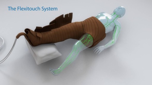
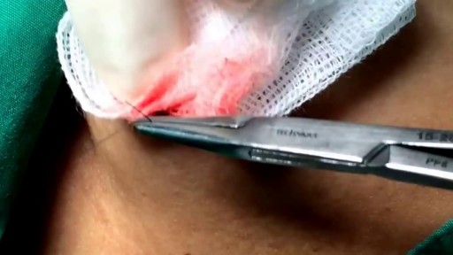
Treating Lymphedema -

A man's age matters. As men get older, the chances of conceiving and having a healthy child decline. Male fertility starts to decline after 40 when sperm quality decreases. This means it takes longer for their partners to conceive and when they do, there's an increased risk of miscarriage.
Spina bifida is a condition that affects the spine and is usually apparent at birth. It is a type of neural tube defect (NTD). Spina bifida can happen anywhere along the spine if the neural tube does not close all the way. When the neural tube doesn’t close all the way, the backbone that protects the spinal cord doesn’t form and close as it should. This often results in damage to the spinal cord and nerves. Spina bifida might cause physical and intellectual disabilities that range from mild to severe. The severity depends on: The size and location of the opening in the spine. Whether part of the spinal cord and nerves are affected.
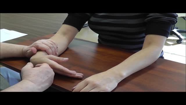
Hold your elbows at shoulder level and place the backs of your hands together with your wrists bent at 90 degrees. This position increases the pressure on the median nerve. If the test reproduces or worsens your symptoms (pain and tingling in your hands), you may have carpal tunnel syndrome.

Progeria (pro-JEER-e-uh), also known as Hutchinson-Gilford syndrome, is an extremely rare, progressive genetic disorder that causes children to age rapidly, beginning in their first two years of life. Children with progeria generally appear normal at birth. During the first year, signs and symptoms, such as slow growth and hair loss, begin to appear. Heart problems or strokes are the eventual cause of death in most children with progeria. The average life expectancy for a child with progeria is about 13 years, but some with the disease die younger and some live 20 years or longer. There's no cure for progeria, but ongoing research shows some promise for treatment.
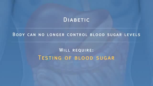
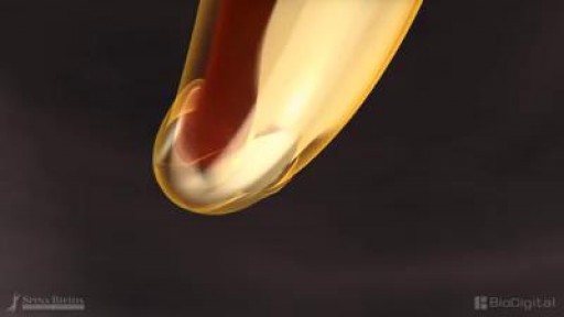
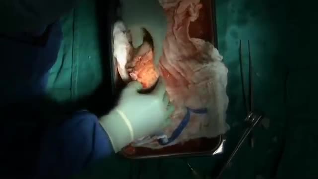
Pancreatic Auto Islet Transplantation is a procedure used to maintain insulin production and secretion in patients with chronic pancreatitis that are undergoing a total pancreatectomy, or removal of the entire pancreas. When all other medical therapies fail to control the pain, removal of the pancreas may be an option; however it can leave a person diabetic, which means that the body can no longer control blood sugar levels, and will require intensive testing of blood sugar and injections of insulin. The pancreas is an organ, located in the upper abdominal cavity, behind the stomach, liver and colon. Within the pancreas, specialized clusters of cells known as islets produce insulin, which maintain healthy blood sugar levels. The pancreas also produces enzymes to help digest food. In order to alleviate pain and maintain insulin production, the pancreas is removed from the body, processed and the islets are harvested. Once the pancreas is removed, it is placed in a solution and put into a machine where the pancreas is digested. The islets are then infused into the patient’s liver. Within a short time, the islets are expected to start producing insulin. In 80% of patients, the pain from pancreatitis is relieved by a total pancreatectomy. Over time, some patients may be diabetic and will need to take insulin to maintain healthy blood sugar levels. All patients will take pancreatic enzymes to help digest food after surgery.
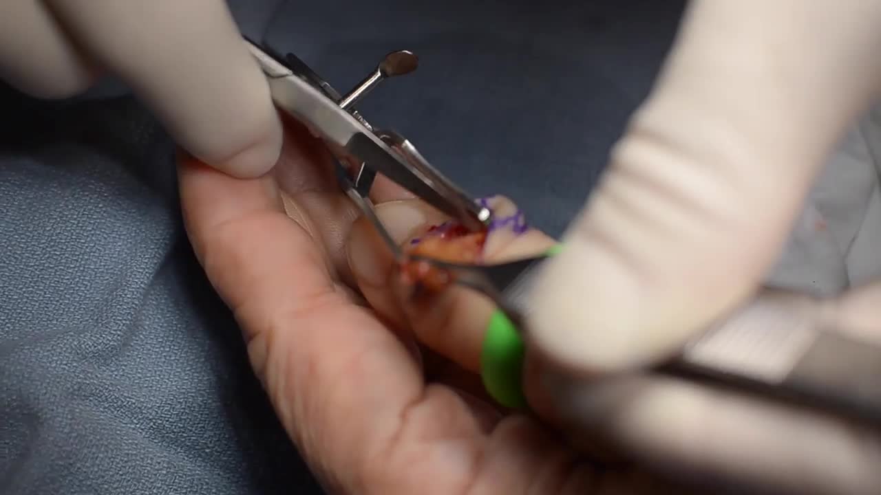
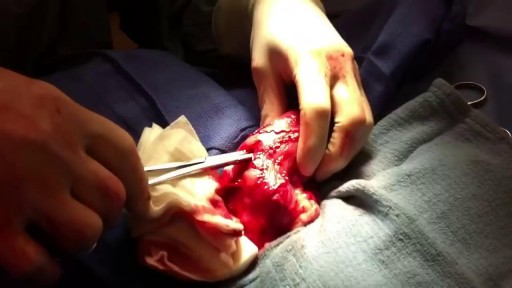
GIANT CELL TUMOR REMOVAL Plastic, Cosmetic and Reconstructive
The urinary bladder is a hollow muscular organ that collects urine from the kidneys before disposal by urination. A hollow muscular, and distensible (or elastic) organ, the bladder sits on the pelvic floor. Urine enters the bladder via the ureters and exits via the urethra.

Your egg usually lives for just 12 to 24 hours, but sperm will live inside you for anything from a few hours to seven days, with one to three days the optimum time. ... But because a small number of sperm are long-living, having sex up to six days before ovulation can also result in pregnancy.

This could be caused by an infection, food poisoning, parasites, Crohn's disease, or reduced blood flow in the colon. Hemorrhoids are another common cause of GI or rectal bleeding. A hemorrhoid is an enlarged vein in your rectum or anus. These enlarged veins can rupture and bleed, causing rectal bleeding.
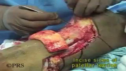
Pathology: Previous spinal cord injury, diabetes, renal failure, dynamic knee contracture, open left ankle disarticulation for sepsis and severe foot infection
Unbelievable Bladder Stone Removal

Forzest is the erectile dysfunction tablet meant for treating mens ED disorder, it is approved by FDA and comes in strenght dose 20mg. For more information kindly visit to http://www.medstorerx.com/forzest.aspx
When you get a kidney transplant, a healthy kidney is placed inside your body to do the work your own kidneys can no longer do. On the plus side, there are fewer limits on what you can eat and drink, but you should follow a heart-healthy diet. Your health and energy should improve. In fact, a successful kidney transplant may allow you to live the kind of life you were living before you got kidney disease. Studies show that people with kidney transplants live longer than those who remain on dialysis. On the minus side, there are the risks of surgery. You will also need to take anti-rejection medicines for as long as your new kidney is working, which can have side effects. You will have a higher risk for infections and certain types of cancer.
