- Physical Examination
- Surgical Examination
- Ophthalmology
- Clinical Skills
- Orthopedics
- Surgery Videos
- Laparoscopy
- Pediatrics
- Funny Videos
- Cardiothoracic Surgery
- Nursing Videos
- Plastic Surgery
- Otorhinolaryngology
- Histology and Histopathology
- Neurosurgery
- Dermatology
- Pediatric Surgery
- Urology
- Dentistry
- Oncology and Cancers
- Anatomy Videos
- Health and Fitness
- Radiology
- Anaesthesia
- Physical Therapy
- Pharmacology
- Interventional Radiology
- Cardiology
- Endocrinology
- Gynecology
- Emergency Medicine
- Psychiatry and Psychology
- Childbirth Videos
- General Medical Videos
- Nephrology
- Physiology
- Diet and Food Health
- Diabetes Mellitus
- Neurology
- Women Health
- Osteoporosis
- Gastroenterology
- Pulmonology
- Hematology
- Rheumatology
- Toxicology
- Nuclear Medicine
- Infectious Diseases
- Vascular Disease
- Reproductive Health
- Burns and Wound Healing
- Other
Top videos
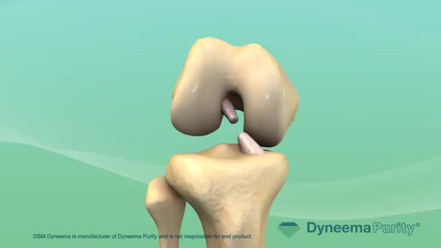
Most people have general anesthesia right before surgery. This means you will be asleep and pain-free. Other kinds of anesthesia, like regional anesthesia or a block, may also be used for this surgery. The tissue to replace your damaged ACL will come from your own body or from a donor. A donor is a person who has died and chose to give all or part of his or her body to help others. Tissue taken from your own body is called an autograft. The two most common places to take tissue from are the knee cap tendon or the hamstring tendon. Your hamstring is the muscle behind your knee. Tissue taken from a donor is called an allograft. The procedure is usually performed with the help of knee arthroscopy. With arthroscopy, a tiny camera is inserted into the knee through a small surgical cut. The camera is connected to a video monitor in the operating room. Your surgeon will use the camera to check the ligaments and other tissues of your knee. Your surgeon will make other small cuts around your knee and insert other medical instruments. Your surgeon will fix any other damage found, and then will replace your ACL by following these steps: The torn ligament will be removed with a shaver or other instruments. If your own tissue is being used to make your new ACL, your surgeon will make a larger cut. Then, the autograft will be removed through this cut. Your surgeon will make tunnels in your bone to bring the new tissue through. This new tissue will be in the same place as your old ACL. Your surgeon will attach the new ligament to the bone with screws or other devices to hold it in place. As it heals, the bone tunnels fill in. This holds the new ligament in place. At the end of the surgery, your surgeon will close your cuts with sutures (stitches) and cover the area with a dressing. You may be able to view pictures after the procedure of what the doctor saw and what was done during the surgery.
Three cholinesterase inhibitors are commonly prescribed: Donepezil (Aricept) is approved to treat all stages of Alzheimer's. Rivastigmine (Exelon) is approved to treat mild to moderate Alzheimer's. Galantamine (Razadyne) is approved to treat mild to moderate Alzheimer's. Currently, there is no cure for Alzheimer's. But drug and non-drug treatments may help with both cognitive and behavioral symptoms. Researchers are looking for new treatments to alter the course of the disease and improve the quality of life for people with dementia. ... Medications for Memory Loss.

Flexor compartment synovectomy in a patient with rheumatoid arthritis presenting with loss of finger movement and local pain due to synovitis. Performed at the Queen Victoria Hospital, East Grinstead.

I call this technique deep rendering. I basically stacked graphical cross-sections (in this case, MRI rendering data), using proper increments and clip through them with the camera. This way I am able to explore all internal components in full 3D real-time.
I actually was able to figure out how to colorize different organs to help distinguish them apart from each other but couldn't get the shader to render real-time in Maya.
Credit: MRI scans courtesy of University of Washington Digital Anatomist Program
Nerve damage can start as numbness or tingling and progress to an intense feeling of burning or stabbing. What to know about treatment options:
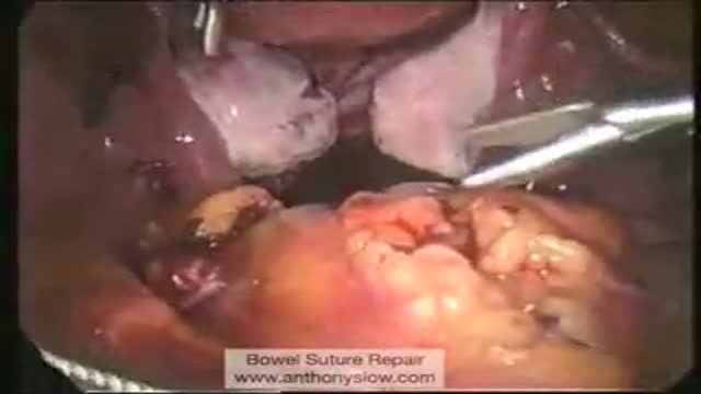
Laparoscopic Suture Repair of Bowel
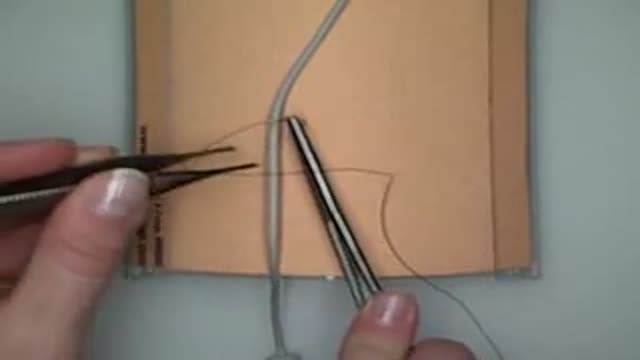
Demonstration of the technique used to insert a drain using an air knot in the operating room.

Harper University Hospital has been accredited as a Bariatric Center of Excellence by the American Society of Bariatric Surgeons. By employing laparoscopy, this bariatric procedure is minimally invasive and results in quicker recovery time, as well as less scarring. ~ Detroit Medical Center
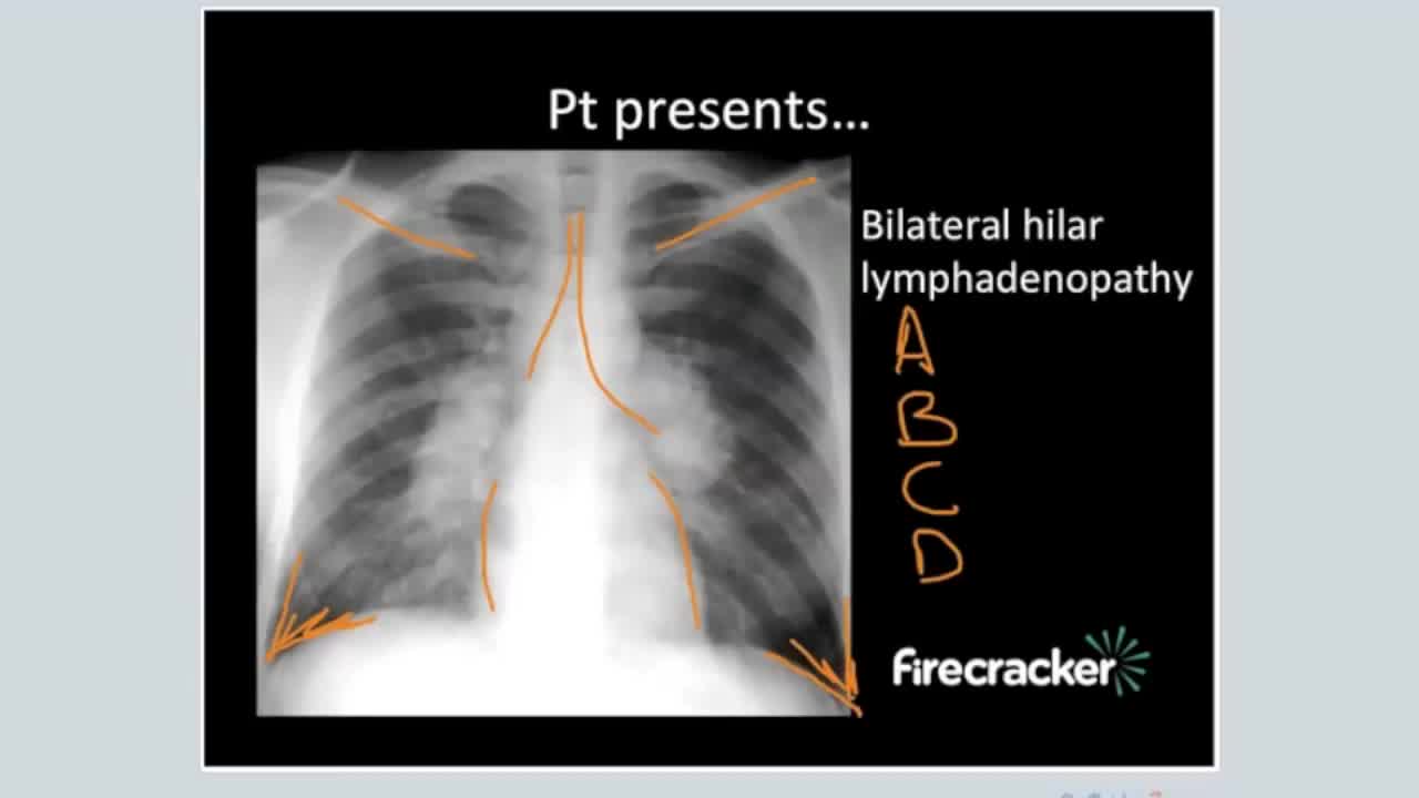
Sarcoidosis is an inflammatory disease that affects multiple organs in the body, but mostly the lungs and lymph glands. In people with sarcoidosis, abnormal masses or nodules (called granulomas) consisting of inflamed tissues form in certain organs of the body. These granulomas may alter the normal structure and possibly the function of the affected organ(s).
A Medical Video showing an overview of the endocrine and gland system of the human body
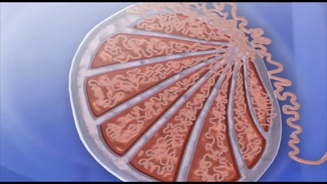
Sound waves enter the ear canal and make the ear drum vibrate. This action moves the tiny chain of bones (ossicles – malleus, incus, stapes) in the middle ear. The last bone in this chain 'knocks' on the membrane window of the cochlea and makes the fluid in the cochlea move.
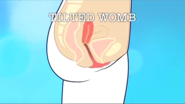
A retroverted uterus (tilted uterus, tipped uterus) is a uterus that is tilted posteriorly. This is in contrast to the slightly "anteverted" uterus that most women have, which is tipped forward toward the bladder, with the anterior end slightly concave.
Vocal Cord Surgery HD

AZT Mechanism of Antiviral Activity

Histology of Esophagus
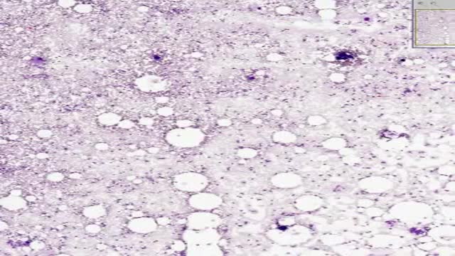
Histology of Bone Marrow Smear
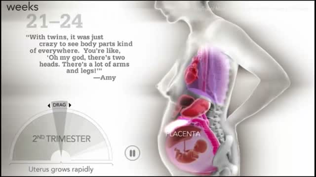
How a woman's body changes during Pregnancy

This could be caused by an infection, food poisoning, parasites, Crohn's disease, or reduced blood flow in the colon. Hemorrhoids are another common cause of GI or rectal bleeding. A hemorrhoid is an enlarged vein in your rectum or anus. These enlarged veins can rupture and bleed, causing rectal bleeding.
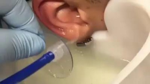
GIANT EAR WAX REMOVAL By using the elephant ear device.It's very useful video for medical students.Please share it!
Spermatogenesis is the process in which spermatozoa are produced from spermatogonial stem cells by way of mitosis and meiosis. The initial cells in this pathway are called spermatogonia, which yield primary spermatocytes by mitosis.
