トップ動画
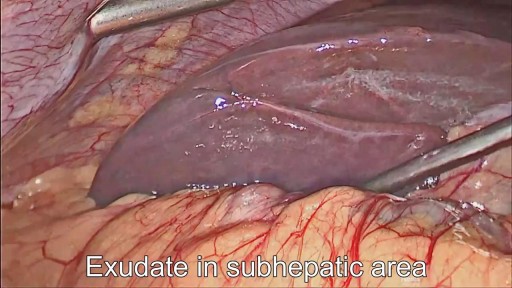
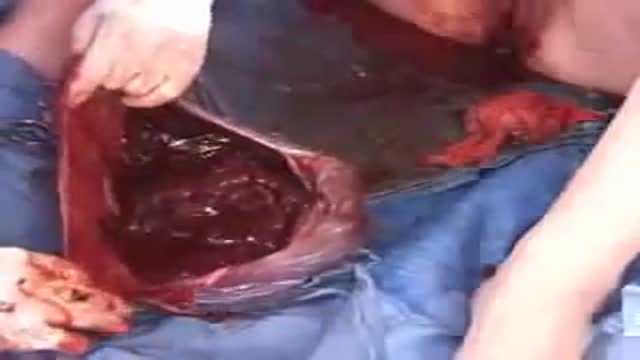
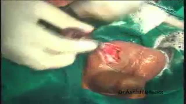
28 years old gentleman presented with huge liver abscess in the right lobe, with repeated attempts of percutaneous aspirations in the past. He was evaluated and subjected to Laparoscopic drainage. This video depicts feasibility of laparoscopy in deep seated liver abscesses. Video created by: Dr. Juneed M. Lanker Fellow Minimal Access Surgery Apollo Hospitals Chennai.
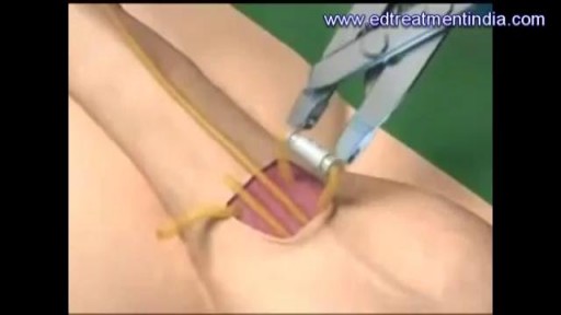
A penile prosthesis is another treatment option for men with erectile dysfunction. These devices are either malleable (bendable) or inflatable. The simplest type of prosthesis consists of a pair of malleable rods surgically implanted within the erection chambers of the penis. With this type of implant the penis is always semi-rigid and merely needs to be lifted or adjusted into the erect position to initiate sex. This type of implant is a good choice for men with spinal cord injuries and/or limited hand strength. Today, many men choose a hydraulic, inflatable prosthesis, which allows them to have an erection when they choose, and it's easier to conceal. It is also more natural. A penile implant is usually used when there is a clear medical cause for ED and when the problem is unlikely to resolve or improve naturally or with other medical treatments. Sometimes a penile prosthesis is implanted during surgery to reconstruct the penis when scarring has caused erections to curve (Peyronie's disease). Penile implant surgeries take about an hour and are typically done in an outpatient center. A man can resume sexual intercourse by 6 weeks after surgery.
The most common symptoms of infection from animal bites are redness, pain, swelling, and inflammation at the site of the bite. You should seek immediate medical treatment if any of these symptoms continue for more than 24 hours. Other symptoms of infection include: pus or fluid oozing from the wound
Most babies will move into delivery position a few weeks prior to birth, with the head moving closer to the birth canal. When this fails to happen, the baby’s buttocks and/or feet will be positioned to be delivered first. This is referred to as “breech presentation.” Breech births occur in approximately 1 out of 25 full-term births.

Intrauterine insemination (IUI) is a fertility treatment that involves placing sperm inside a woman's uterus to facilitate fertilization. The goal of IUI is to increase the number of sperm that reach the fallopian tubes and subsequently increase the chance of fertilization
Injection in buttocks
Mouth ulcers are sores that appear in the mouth, often on the inside of the cheeks. Mouth ulcers, also known as aphthous ulcers, can be painful when eating, drinking or brushing teeth. Occasional mouth ulcers are usually harmless and clear up on their own. Seek medical advice if they last longer than 3 weeks or keep coming back. Mouth ulcers cannot be caught from someone else. Up to 1 in 5 people get recurrent mouth ulcers.

Watch that video to know the Premature Ejaculation Causes and Cures
Loyola Full Male Exam Part 4 A video from Loyola medical school, Chicago showing the full examination of the male
This video shows the delivery of the placenta after delivery of the fetus

The typical radiograph is of a well-defined, rounded, retrocardiac opacity with an air-fluid level. In this image, the radiolucent gas is highlighted in blue, while the gastric contents are highlighted in the green. In many cases of hiatal hernia, there will not be an air bubble below the left hemidiaphragm. This is a relatively expected finding considering that the stomach is no longer in its usual position. The anatomical position of the herniated organ can be further elucidated on the lateral radiograph. Here we can see that the stomach is in the middle mediastinum posterior to the heart and above the diaphragm. Hiatal hernias can look similar to a retrocardiac lung abscess or another cavitary lesion, but it will change in size and shape between radiographs. Large hernias can shift the mediastinum to the right and result in a widening of the carinal angle. They can even give the appearance of cardiomegaly. In this radiograph, the cardiac silhouette is distinctly visible within the confines of the hiatal hernia. To review, a hiatal hernia on an AP chest radiograph typically appears as a round retrocardiac opacity with an air-fluid level.
🌐 Check out our website for more video lectures
https://www.med4vl.com
📺 Subscribe To My Channel and Get More Great Quizzes and Tutorials
https://www.youtube.com/channe....l/UC95TzSH1B_2EjaZMg
#FOAMrad #MedEd #radiology
Disclaimer: All the information provided by Medical Education for Visual Learners and associated videos are strictly for informational purposes only. It is not intended as a substitute for medical advice from your health care provider or physician. It should not be used to overrule the advice of a qualified healthcare provider, nor to provide advice for emergency medical treatment. If you think that you or someone that you know may be suffering from a medical condition, then please consult your physician or seek immediate medical attention.

Please remember that this video is to be used for educational purposes. You must follow your facility or colleges' policies and procedure checklists to ensure you are completing the skill satisfactorily. Thanks for watching!
Music from #Uppbeat (free for Creators!):
https://uppbeat.io/t/swoop/blue-sea
License code: W9DFUQ4II7YVHA59

This video demonstrates Laparoscopic Cholecystectomy Fully Explained Skin-to-Skin Video with Near Infrared Cholangiography performed by Dr R K Mishra at World Laparoscopy Hospital. A laparoscopic cholecystectomy is a minimally invasive surgical procedure that involves removing the gallbladder. It is typically performed using small incisions in the abdomen, through which a laparoscope (a thin tube with a camera and light) and surgical instruments are inserted. The surgeon uses the laparoscope to visualize the inside of the abdomen and to guide the instruments in removing the gallbladder.
Near-infrared cholangiography is a technique that uses a special camera and fluorescent dye to visualize the bile ducts during surgery. The dye is injected into the cystic duct (the tube that connects the gallbladder to the bile ducts) and the camera detects the fluorescence emitted by the dye, allowing the surgeon to see the bile ducts more clearly.
The combination of laparoscopic cholecystectomy and near-infrared cholangiography has become a standard of care in many hospitals and surgical centers. It allows for a more precise and efficient surgery, reducing the risk of complications such as bile duct injury.
The use of indocyanine green (ICG) with near-infrared imaging during laparoscopic cholecystectomy has several advantages. Here are some of them:
Better visualization of the biliary anatomy: ICG with near-infrared imaging allows for better visualization of the biliary anatomy during surgery. This helps the surgeon identify important structures, such as the cystic duct and the common bile duct, and avoid injuring them.
Reduced risk of bile duct injury: With better visualization of the biliary anatomy, the risk of bile duct injury during surgery is reduced. Bile duct injury is a serious complication that can occur during laparoscopic cholecystectomy and can lead to long-term health problems.
Improved surgical precision: ICG with near-infrared imaging also improves surgical precision. The surgeon can better see the tissues and structures being operated on, which can help reduce the risk of bleeding and other complications.
Shorter operating time: The use of ICG with near-infrared imaging can shorten the operating time for laparoscopic cholecystectomy. This is because the surgeon can more quickly and accurately identify the biliary anatomy, which can help streamline the surgery.
Overall, the use of ICG with near-infrared imaging is a valuable tool in laparoscopic cholecystectomy that can improve surgical outcomes and reduce the risk of complications.
Like any surgical procedure, laparoscopic cholecystectomy (gallbladder removal) has potential complications. Here are some of the most common ones:
Bleeding: Bleeding during or after the surgery is a possible complication of laparoscopic cholecystectomy. Most cases are minor and can be easily controlled, but in rare cases, significant bleeding may require a blood transfusion or even additional surgery.
Infection: Any surgical procedure carries a risk of infection. After laparoscopic cholecystectomy, there is a risk of infection at the site of the incisions or within the abdomen. Symptoms may include fever, pain, redness, or drainage from the incision sites.
Bile leakage: In some cases, a small amount of bile may leak from the bile ducts into the abdominal cavity after gallbladder removal. This can cause abdominal pain, fever, and sometimes requires further surgery or treatment.
Injury to nearby organs: During the surgery, there is a small risk of unintentional injury to nearby organs such as the liver, intestines, or bile ducts. This can cause additional complications and may require further treatment.
Adverse reactions to anesthesia: As with any surgery requiring general anesthesia, there is a small risk of adverse reactions to the anesthesia, such as an allergic reaction, respiratory problems, or heart complications.
Most patients recover without complications following a laparoscopic cholecystectomy, but it is important to discuss any concerns or questions with your surgeon beforehand.
Contact us
World Laparoscopy Hospital
Cyber City, Gurugram, NCR Delhi
INDIA : +919811416838
World Laparoscopy Training Institute
Bld.No: 27, DHCC, Dubai
UAE : +971525857874
World Laparoscopy Training Institute
8320 Inv Dr, Tallahassee, Florida
USA : +1 321 250 7653
Regenerate response
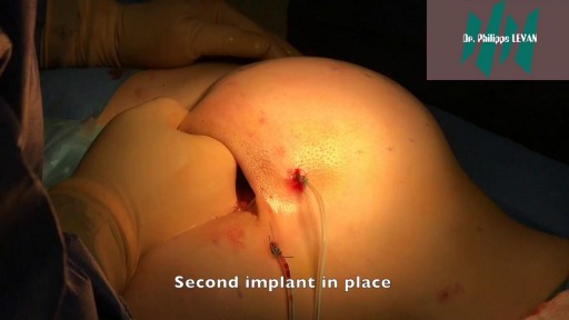
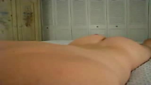
Butt implants are a popular plastic surgery procedure among those who wish to enhance the appearance, shape, and size of their rear ends. Buttock augmentation involves the surgical insertion of artificial body implants into a patient’s buttocks to create a larger, shapelier, and more sensuous rear end. Patients who have underdeveloped buttocks can achieve a more proportionate figure with butt implants. Women who wish to achieve an “hour glass” figure or are unhappy with the size of their buttocks can benefit from female butt implants. Men with flat or poorly developed buttocks can enhance the shape of the area to their liking with male butt implants. Many buttock augmentation patients say that their clothes fit better, they feel more attractive, and their confidence levels have improved.
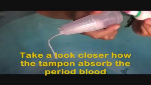
How to Use Tampons for The First Time Demo Video
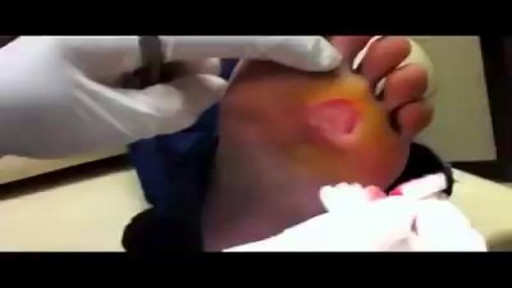
Remove a Plantar Wart from a foot Procedure
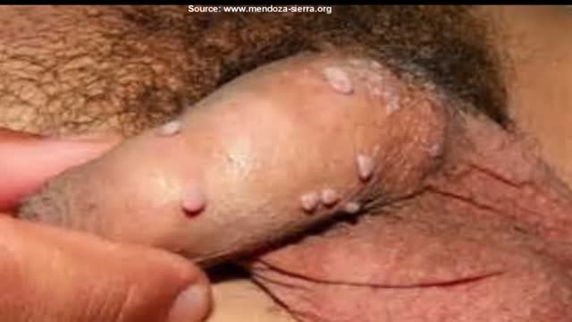
Genital warts are one of the most common types of sexually transmitted infections. At least half of all sexually active people will become infected with human papillomavirus (HPV), the virus that causes genital warts, at some point during their lives. Women are somewhat more likely than men to develop genital warts. As the name suggests, genital warts affect the moist tissues of the genital area. Genital warts may look like small, flesh-colored bumps or have a cauliflower-like appearance. In many cases, the warts are too small to be visible. Like warts that appear elsewhere on your body, genital warts are caused by the human papillomavirus (HPV). Some strains of genital HPV can cause genital warts, while others can cause cancer. Vaccines can help protect against certain strains of genital HPV
Brought to you by http://nursing-resource.com
Orchidectomy and Orchidopexy in Testicular Torsion

Traditional African treatment for infertility
