- Physical Examination
- Surgical Examination
- Ophthalmology
- Clinical Skills
- Orthopedics
- Surgery Videos
- Laparoscopy
- Pediatrics
- Funny Videos
- Cardiothoracic Surgery
- Nursing Videos
- Plastic Surgery
- Otorhinolaryngology
- Histology and Histopathology
- Neurosurgery
- Dermatology
- Pediatric Surgery
- Urology
- Dentistry
- Oncology and Cancers
- Anatomy Videos
- Health and Fitness
- Radiology
- Anaesthesia
- Physical Therapy
- Pharmacology
- Interventional Radiology
- Cardiology
- Endocrinology
- Gynecology
- Emergency Medicine
- Psychiatry and Psychology
- Childbirth Videos
- General Medical Videos
- Nephrology
- Physiology
- Diet and Food Health
- Diabetes Mellitus
- Neurology
- Women Health
- Osteoporosis
- Gastroenterology
- Pulmonology
- Hematology
- Rheumatology
- Toxicology
- Nuclear Medicine
- Infectious Diseases
- Vascular Disease
- Reproductive Health
- Burns and Wound Healing
- Other
Top videos
Factitious disorder is the term used to describe a pattern of behavior centered on the exaggeration or outright falsifications of one’s own health problems or the health problems of others. Some people with this disorder fake or exaggerate physical problems; others fake or exaggerate psychological problems or a combination of physical and psychological problems. Factitious disorder differs from a pattern of falsified or exaggerated behavior called malingering. While malingerers make their claims out of a motivation for personal gain, people with factitious disorder have no such motivation.

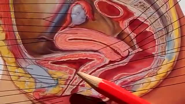
Watch that video to know if it is safe to have sex during period or not
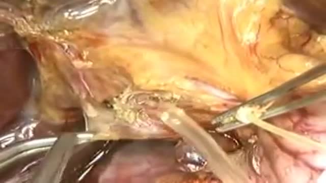
Liver transplantation is surgery to remove a diseased or injured liver and replace it with a healthy whole liver or a segment of a liver from another person, called a donor. People with either acute or chronic liver failure may need a liver transplant to survive.

DIRTIEST PARTS OF YOUR BODY
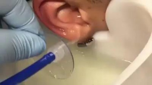
GIANT EAR WAX REMOVAL By using the elephant ear device.It's very useful video for medical students.Please share it!
Interventional Nephrology is a new and emerging subspecialty of Nephrology that mainly deals with ultrasonography of kidneys and ultrasound-guided renal biopsy, insertion of peritoneal dialysis catheters, tunneled dialysis catheters as a vascular access for patients undergoing hemodialysis as well as percutaneous ...
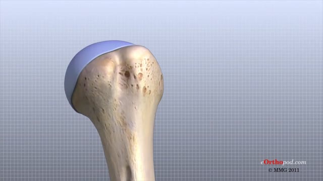
The shoulder joint is formed where the humerus (upper arm bone) fits into the scapula (shoulder blade), like a ball and socket. Other important bones in the shoulder include: The acromion is a bony projection off the scapula. The clavicle (collarbone) meets the acromion in the acromioclavicular joint.
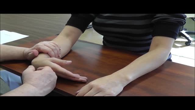
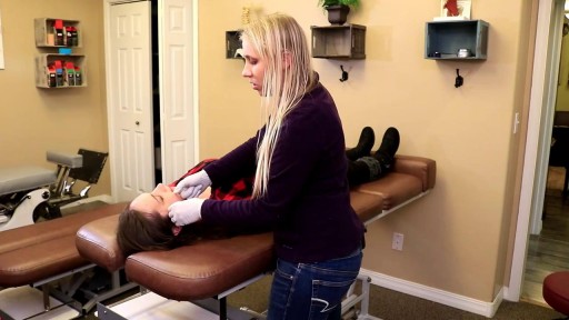
Hold your elbows at shoulder level and place the backs of your hands together with your wrists bent at 90 degrees. This position increases the pressure on the median nerve. If the test reproduces or worsens your symptoms (pain and tingling in your hands), you may have carpal tunnel syndrome.
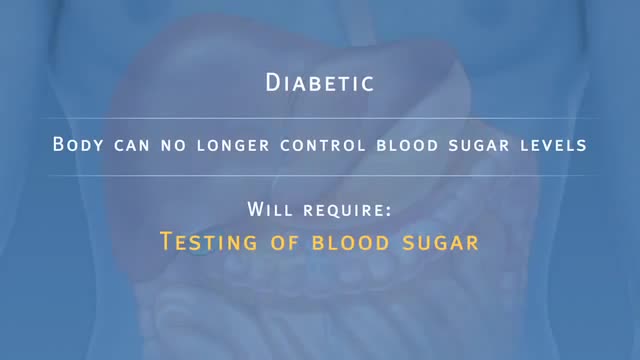
Pancreatic Auto Islet Transplantation is a procedure used to maintain insulin production and secretion in patients with chronic pancreatitis that are undergoing a total pancreatectomy, or removal of the entire pancreas. When all other medical therapies fail to control the pain, removal of the pancreas may be an option; however it can leave a person diabetic, which means that the body can no longer control blood sugar levels, and will require intensive testing of blood sugar and injections of insulin. The pancreas is an organ, located in the upper abdominal cavity, behind the stomach, liver and colon. Within the pancreas, specialized clusters of cells known as islets produce insulin, which maintain healthy blood sugar levels. The pancreas also produces enzymes to help digest food. In order to alleviate pain and maintain insulin production, the pancreas is removed from the body, processed and the islets are harvested. Once the pancreas is removed, it is placed in a solution and put into a machine where the pancreas is digested. The islets are then infused into the patient’s liver. Within a short time, the islets are expected to start producing insulin. In 80% of patients, the pain from pancreatitis is relieved by a total pancreatectomy. Over time, some patients may be diabetic and will need to take insulin to maintain healthy blood sugar levels. All patients will take pancreatic enzymes to help digest food after surgery.
The urinary bladder is a hollow muscular organ that collects urine from the kidneys before disposal by urination. A hollow muscular, and distensible (or elastic) organ, the bladder sits on the pelvic floor. Urine enters the bladder via the ureters and exits via the urethra.

Your egg usually lives for just 12 to 24 hours, but sperm will live inside you for anything from a few hours to seven days, with one to three days the optimum time. ... But because a small number of sperm are long-living, having sex up to six days before ovulation can also result in pregnancy.

This could be caused by an infection, food poisoning, parasites, Crohn's disease, or reduced blood flow in the colon. Hemorrhoids are another common cause of GI or rectal bleeding. A hemorrhoid is an enlarged vein in your rectum or anus. These enlarged veins can rupture and bleed, causing rectal bleeding.
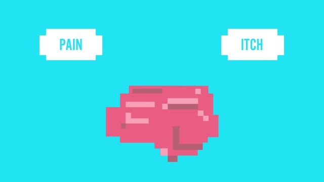
Pruritis is itchy skin that makes you want to scratch. It can be caused by many things. Normally, itchy skin isn't serious, but it can make you uncomfortable. Sometimes, itchy skin is caused by a serious medical condition. It can occur in association with a primary rash (e.g. dermatitis) or may occur because of hypersensitive nerves in the skin (neuropathic pruritus). ... Scratching a localised itch may lead to lichen simplex, prurigo or prurigo nodularis. Systemic causes of pruritus. Sytemic diseases may cause generalised pruritus.
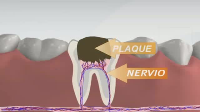
Root canals are common procedures and can help save your tooth from extraction. Dentists at Aspen Dental practices have been safely and expertly performing root canal procedures for over two decades.
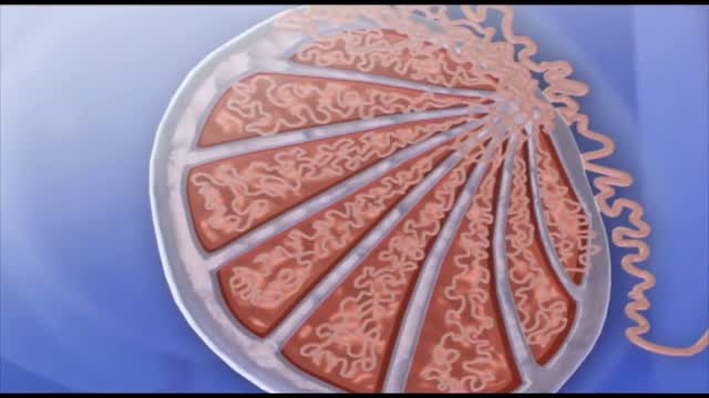
Spermatogenesis is the process in which spermatozoa are produced from spermatogonial stem cells by way of mitosis and meiosis. The initial cells in this pathway are called spermatogonia, which yield primary spermatocytes by mitosis.
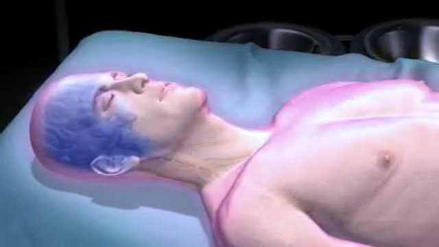
6 987 24 MORE How Does Anesthesia Work? Credit: itsmejust | Shutterstock If you’ve ever had surgery, unless you are super tough, you’ve gone through it with the benefit of anesthetics. But, how do these body-numbing elixirs work? Prior to the invention of anesthesia in the mid-1800s, surgeons had to hack off limbs, sew up wounds and remove mysterious growths with nothing to dull the patient's pain but opium or booze. While these drugs may have numbed the patient, they didn’t always completely block the pain, or erase the memory of it. Since then, doctors have gotten much better at putting us out with drug combinations that ease pain, relax muscles and, in some cases, put us in a deep state of hypnosis that gives us temporary amnesia. Today, there are two primary types of anesthesia drugs: those that knockout the whole body (general) and those that only numb things up locally.

What Really Happens When You Swallow Gum?
Rehydration Tips: Kids & Teens (Ages 1+) Give clear liquids (avoid milk and milk products) in small amounts every 15 minutes. ... If your child vomits, start over with a smaller amount of fluid (2 teaspoons, or about 10 milliliters) and continue as above. ... After no vomiting for about 8 hours, introduce solid foods slowly.
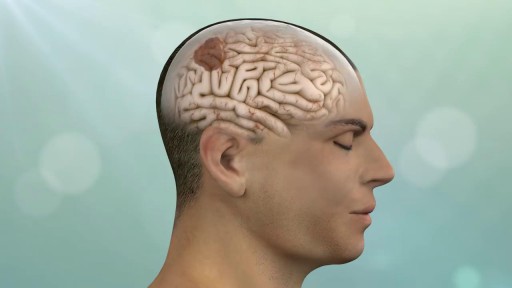
Keyhole surgery can be helpful for patients with brain, pituitary and skull base tumors. With cutting edge technology and a proven track record in keyhole and endonasal endoscopic approaches, we make surgery safer, less invasive and more effective.