Top videos

Psoriasis: treatment options related issues
We have just enhanced the smile of another wonderful patient! She just received 6 mini dental implants place by DR. Jue www.sugarlanddentalspa.com.

LIPO LASER PAPADA COSMETIC LASER CENTER

DR.JUE IS SUGAR LAND' S PREMIERE LUMINEERS AND SNAP ON SMILE DENTIST. DR. JUE HAS BEEN FEATURED ON FOX NEWS FOR LUMINEERS! Lumineers . The Den-Mat Corporation has patented a type of porcelain veneer called Lumineer, and we are proud to announce our full certification in Lumineer application. Lumineers work great for fixing gapped teeth, teeth that slope inward, or teeth that are to small. Because there is no drilling, Lumineers are nearly painless to apply.
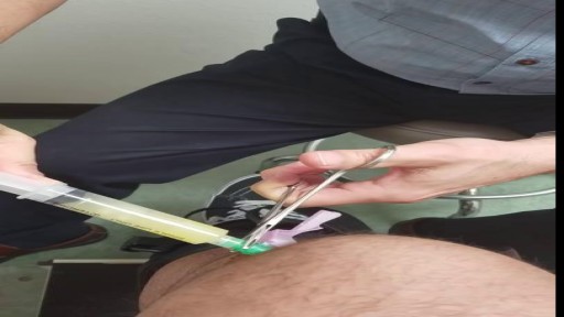
Cutting Championship Ring Stuck in Finger

What is hemodialysis, and why would someone need it? How does hemodialysis work? Can people perform hemodialysis at home? John Kevin Tucker, M.D., Nephrologist at Brigham and Women's Hospital and Vice President for Education at Mass General Brigham, discusses hemodialysis and how it helps people who have lost their kidney function to maintain normal lives.
Subscribe Link: https://www.youtube.com/channe....l/UCYrLjATd88gPwIKnt
0:00 - Intro
0:26 - The Condition
2:06 - Hemodialysis: How It Works
4:37 - In-Center Hemodialysis Care Team
About Mass General Brigham:
Mass General Brigham combines the strength of two world-class academic medical centers, five nationally ranked specialty hospitals, 11 community hospitals, and dozens of health centers. Our doctors and researchers accelerate medical breakthroughs and drive innovations in patient care. They are leaders in medical education, serving as Harvard Medical School faculty and training the next generation of physicians. Mass General Brigham’s mission is to deliver the best, affordable health care to patients everywhere. Together, we transform the health of our communities and beyond.
#MassGeneralBrigham #MGB #Hemodialysis
Visit Mass General Brigham: https://www.massgeneralbrigham.org/
Find us on social:
Twitter: https://twitter.com/MassGenBrigham
Instagram: https://www.instagram.com/massgeneralbrigham/
Facebook: https://www.facebook.com/MassGeneralBrigham/
LinkedIn: https://www.linkedin.com/compa....ny/mass-general-brig
Mass General Brigham:
https://www.youtube.com/massgeneralbrigham
Kidney Failure: Signs, Dialysis Options, and Hemodialysis Explained | Mass General Brigham
https://youtu.be/azy7yc19QYQ

![What does a fistula for dialysis look like? [CHT CERTIFICATION REVIEW] 2022](https://i.ytimg.com/vi/gjB5eKo4eh8/maxresdefault.jpg)
If this is the first time visiting us, make sure to subscribe to our channel here: https://bit.ly/2yXNBYp
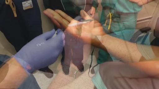
What does a fistula for dialysis look like?
A fistula for dialysis is a surgical connection between a vein and an artery.
In this video, I will show you a real fistula and how we should evaluate it before a dialysis connection.
Additional videos:
💉How to properly cannulate a fistula: https://youtu.be/IqoHnzFyhJQ
💉 What is a fistula for dialysis treatment: https://youtu.be/B5EEf-MklFk
💉 The 10-second assessment for fistulas: https://youtu.be/Uqo0LhjZSI8
💉 If you would like to be trained as a dialysis professional focused on offering quality of care to renal patients, visit our program details here: https://utopiahcc.com/hemodialysis-technician/
For nursing and technician schools😷 🩺 🎓, we can offer a special renal failure class to your students. For inquiries please contact us: info@utopiahcc.com
Where to find us:
Facebook: https://www.facebook.com/utopiahealth
Email: info@utopiahcc.com
Website: utopiahcc.com
🤔 Looking for renal and dialysis continuing education for your certification renewal? Check out our CE package where you will get a little over 40 contact hours for a small price and receive your certificates immediately.
Here's the link: https://bit.ly/3dbPvDZ
Want to watch *Free Dialysis Training Videos*?
https://utopiahcc.com/free-dia....lysis-video-training
__________________________________________________________
Additional resources:
What Does a Healthy AV Fistula Look Like? | Azura Vascular ...
www.azuravascularcare.com infodialysisaccess healt...
Jul 17, 2018 — An AV fistula is a surgically-created permanent access located under the skin, making a direct connection between a vein and an artery. An AV fistula is typically created in the non-dominant arm. If the veins in your arm are not large or healthy enough to support a fistula, it may be created in your leg.
Preparing for Dialysis (AV Fistula) Fact Sheets Yale ...
www.yalemedicine.org › conditions › preparing-dialysi...
To undergo dialysis, patients need a surgical procedure to create an access point for the dialysis machine. An AV fistula is the most common access point.
Vascular Access for Hemodialysis - Life Options
lifeoptions.org living-with-kidney-failure vascular-a...
Jump to How a Catheter Looks and Feels — This makes a pattern that looks a bit like a rope ladder. The next best way—for fistulas ONLY—is the “Buttonhole ...
Fistula or Graft Surgery · Needle Fear · How a Fistula or Graft Looks...
Taking Care of Your Fistula - DaVita
www.davita.com dialysis preparing-for-dialysis › ta...
An arteriovenous (AV) fistula is a type of access used for hemodialysis. ... access because it utilizes the patient's own vessels and does not require permanent placement of foreign materials such ... Look for redness or swelling around the fistula area. ... This sound may change from a whooshing noise to a whistle-like sound.
Vascular Access for Hemodialysis - Department of Surgery
surgery.ucsf.edu conditions--procedures vascular-ac...
The patient does not need anesthesia for this procedure. ... A vascular surgeon performs AV graft surgery, much like AV fistula surgery, in an outpatient center or ...
Frequently Asked Questions about Dialysis Access Surgery ...
www.bidmc.org transplant-institute frequently-aske...
Dialysis access surgery creates the vascular opening so a needle can be inserted for ... fluid and to correct electrolytes like potassium, sodium, phosphate and calcium, to name a few. ... Where are AV fistulas located and how long do they last?
Fistula and Graft Placement (Eric K. Peden, MD) - YouTube
www.youtube.com watch
Mar 28, 2016 — ... Bootcamp 2015 August 14 - 16, 2015 "Dialysis Access" Fistula and Graft Placement (Eric K. Peden, MD) DICET@Houstonmethodist.org.

What is hemodialysis and how does it work? Who needs it? How do you prepare for it? In the United States, over 30 million Americans have kidney disease, and sometimes, kidney disease progresses to kidney failure or end-stage renal disease. When this happens, you cannot survive unless you have a kidney transplant or some form of dialysis. So today we're going to talk about hemodialysis.
Your kidneys are the two kidney bean-shaped organs that are located in your lower back, or in your flanks. And the kidneys are responsible for filtering out or cleaning your blood. They get rid of excess waste, excess toxins, and excess fluids. If your kidneys stop functioning, then you develop renal failure or end-stage renal disease.
What is Hemodialysis?
Hemodialysis, or blood dialysis, is the filtering of your blood outside of your body. So, if your kidneys stop working properly, the hemodialysis acts as a substitute kidney. Now it's important to note that hemodialysis does not actually correct your own kidney function. It does not fix or treat your kidneys.
#hemodialysis #drfrita
What is The Dialyzer?
The dialyzer is actually the filter. It's the main powerhouse of the hemodialysis system, and it is what actually acts as the substitute kidney. In the dialyzer, you have these hollow fibers that run through it, and these fibers are bathed in something called dialysates, or dialysis fluid.
How Often Are Patients Treated With Hemodialysis?
Most patients who are on hemodialysis are on it between three and six hours, about three days a week, especially if they go to a center.
How Does Hemodialysis Work?
So when you are on dialysis, how does your blood get from your body to the hemodialysis machine and then back to your body? Well, it does so through tubes, and those tubes are connected to your access, and we'll talk about access in just a moment. But as far as the tubing, the tubing is connected to your body.
Types Of Hemodialysis Access
Arteriovenous Fistula or AV Fistula
The AV fistula is the gold standard as far as hemodialysis access is concerned because it gives you the most efficient hemodialysis and it is the least likely to be infected.
Arteriovenous Graft or AV Graft
The AV graft is very similar to the AV fistula in that you still have a surgically connected artery and a vein, usually in the arm, but in the case where if you have veins that are rather thin or arteries that are thin and maybe too weak in order to really give you a properly functioning, substantial AV fistula, then the vascular surgeon may opt to add an artificial material in order to make that shunt a little stronger, or little more durable. And so, an AV graft is another option for dialysis access.
Catheter
If you're in a situation where you need temporary dialysis, or if you have acute kidney injury, then you may have a temporary Vascath placed, and it's usually placed in a vein of the neck, the internal jugular vein, or it can be placed in the groin, or in the femoral vein.
Who Needs Hemodialysis Treatment?
How do you know if you need hemodialysis, and when is it time to prepare? Well, if you follow up with your kidney doctor (nephrologist) regularly, he or she will be watching your labs. They'll be able to see those signs of your kidneys not functioning properly.
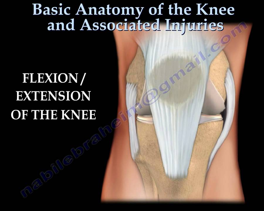
Dr. Ebraheim’s educational animated video describing the anatomy and associated injuries of the knee joint.
Disrupted quadriceps
•Patient is unable to actively extend the knee.
The most common cause of ACL ruptures:
•Traumatic force being applied during twisting motion.
•Side stepping or landing from a jump.
Patient complains of:
•Immediate pain
•Knee giving way
•Swelling
Aspiration of the knee
•If aspiration of the knee joint shows evidence of blood within the joint there is 75-80% chance of ACL and meniscal injury.
Lachamn’s test- ACL knee exam
•Knee is flexed at 30 degrees.
•ACL tear of the knee is identified by pulling on the tibia and examining the frontward motion of the lower leg in comparison to the upper leg.
Radiological exam – ACL
•MRI of the knee joint shows bone lesions or bruising associated with tears of the ACL. Injury is found in the typical location; middle of the femoral condyle and posterior part of the tibia laterally.
Posterior cruciate ligament tear (PCL)
•Common cause of injury is a bent knee hitting a dashboard in a car accident.
Tibial Sag Test –PCL knee exam
Quadriceps active test-PCL knee exam
•The examiner stabilizes the leg of the patient and then the patient is asked to actively contract the quadriceps muscle.
•The tibia is seen actively reduced from the posterior subluxed position.
Lachman’s test-PCL knee exam
•Knee is bent 20-30 degrees.
•The posterior drawer test is carried out while the patient is in a supine position and the knee is flexed to 90 degrees.
•The amount of translation of the tibia relative to the femur is observed.
The dial test is performed while the patient is in the supine or prone position and both knees are in 90 and 30 degrees of flexion. More than 10 degrees of external rotation indicates significant injury.
Common meniscal tears
Symptoms include
•Knee pain
•Pain with straightening the knee
•Swelling
•Locking
•Weakness

Common causes of the knee pain
Knee pain is very common and in this video we will present the most common problems that can cause pain in the knee. (Patella) itself, which is in front of the knee, or from the tendons that are attached to the kneecap (patellar tendon and quadricep tendon). One of the most common problems is patellar chondromalacia which is chronic pain due to the softening of the cartilage beneath the kneecap. The cartilage of the kneecap will have some erosions, defects, or holes from mild to complete inside the joint (exactly in the back of the kneecap).
• Pain in the front of the knee
• Occurs more in young people
• Becomes worse from climbing up stairs and going downstairs
Treatment is usually nonsteroidal anti-inflammatory medication, physical therapy, and surgery is very rare. Also in front of the kneecap, the patient may get pain due to prepatellar bursitis.
When there is prepatellar bursitis, the patient will see that the swelling, the inflammation, and the pain is located over the front of the kneecap. The bursa becomes inflamed and fills with fluid at the top of the knee, causing pain, swelling, tenderness and a lump in that area on top of the kneecap. If the pain is in front of the knee but below or above the patella, this may indicate that the patient has tendonitis. Patellar tendonitis is an overuse condition that often occurs in athletes who perform repetitive jumping activities. Patellar tendonitis is a knee pain that is associated with focal patellar tendon tenderness and it is usually activity related. It is located below the kneecap and is called "jumper's knee". Patellar tendonitis affects approximately 20% of jumping athletes. There will be tenderness to palpation at the distal pole of the patella in extension and not in flexion. Quadriceps inflexibility, atrophy and hamstring tightness are predisposing factors for this condition. Treatment is rest, anti-inflammatory medication, stretching and strengthening of the hamstrings and quadriceps. Use an eccentric exercise program. The early stages of patellar tendonitis will respond well to nonoperative treatment. Another important cause of knee pain is a meniscal tear. The meniscus is the cushion that protects the cartilage in the knee. Injury will cause pain on the medial or the lateral side of the knee exactly at the level of the joint. The patient will complain of a history of locking, instability and swelling of the knee. McMurray test will be positive. A painful pop or click is obtained as the knee is brought from flexion to extension with either internal or external rotation of the knee. Arthritis of the knee Knee arthritis is very common. The cartilage cells die with age and its repair response decreases in the joint collapses with increased breakdown of the framework of the cartilage. The patient will have progressive blurring away of the cartilage of the joint with decreased joint space as seen on x-rays. Another source of pain is the Baker's cyst. The cyst is in the back of the knee between the semimembranosus yes and the medial gastrocnemius muscles. Another important source of knee pain is a ligament injury. Here is a normal knee without a ligament injury. Here you can see from the front, you can see the lateral and medial collateral ligament. You can see the ACL and PCL from the side view. These ligaments are usually injured as a result of a sports activity. Here is an example of a sports knee injury. Here is an example of the medial collateral ligament injury. This is the most commonly injury knee ligament injury to this ligament is on the inner part of the knee. Here is an example of an injury of the anterior cruciate ligament. It involves a valgus stress to the knee. Lachman test is usually positive, and MRI is diagnostic. Another important cause of knee pain is iliotibial band syndrome of the knee. Inflammation of the thickening of the iliotibial band results from excessive friction as the iliotibial band slides over the lateral femoral condyle. The iliotibial band is a thick band of fascia that extends along the lateral thigh from the iliac crest to the knee. And as the knee moves, the IT band was repeatedly shifted forwards and backwards across the lateral femoral condyle. The patient will complain of swelling, tenderness, and crepitus over the lateral femoral condyle. The condition occurs in the ITB S occurs in runners, cyclist and athletes that require repeated knee flexion and extension. The pain may be reproduced by doing a single-leg squat. The Ober's test is used to at assess tightness of the iliotibial band. MRI may show edema in the area of the ITB. Treatment is usually nonoperative with rest and ice, physical therapy, with stretching, proprioception, and improvement in neuromuscular coordination. Training modification and injections may be helpful. Surgery is a last resort. Surgical excision of the scarred inflamed part of the iliotibial band.

![Kneecap [patella] button loosens after total #kneereplacement #kneeinjury #fracture](https://i.ytimg.com/vi/1pgqwZDgIPs/maxresdefault.jpg)

Dr. Carlos Benitez guides us through ultrasound images of the knee and how to identify knee injuries.
We are looking for 5 patients with knee pain who want to get significantly better in the next 30 days. Click this link to let me know you're interested and learn more.
https://www.drdavidgeier.com/work-with-me/contact/
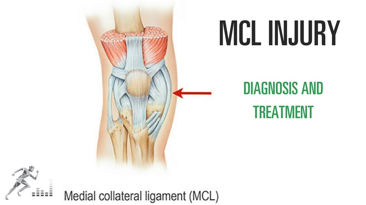
One of the most common knee injuries in contact and collision sports is a medial collateral ligament (MCL) injury. This is a ligament on the medial (side closest to the midline) side of your knee that provides stability against side-to-side stress to the knee. You might injure it by cutting maneuvers in sports like soccer or hockey. You can also suffer an MCL injury if another player hits you on the outside of your knee.
Please note: I don't respond to questions and requests for specific medical advice left in the comments to my videos. I receive too many to keep up (several hundred per week), and legally I can't offer specific medical advice to people who aren't my patients (see below). If you want to ask a question about a specific injury you have, leave it in the comments below, and I might answer it in an upcoming Ask Dr. Geier video. If you need more detailed information on your injury, go to my Resources page: https://www.drdavidgeier.com/resources/
The content of this YouTube Channel, https://www.youtube.com/user/drdavidgeier (“Channel”) is for INFORMATIONAL PURPOSES ONLY. The Channel may offer health, fitness, nutritional and other such information, but such information is intended for educational and informational purposes only. This content should not be used to self-diagnose or self-treat any health, medical, or physical condition. The content does not and is not intended to convey medical advice and does not constitute the practice of medicine. YOU SHOULD NOT RELY ON THIS INFORMATION AS A SUBSTITUTE FOR, NOR DOES IT REPLACE, PROFESSIONAL MEDICAL ADVICE, DIAGNOSIS, OR TREATMENT. You should consult with your healthcare professional before doing anything contained on this Channel. You agree that Dr. Geier is not responsible for any actions or inaction on your part based on the information that is presented on the Channel. Dr. David Geier Enterprises, LLC makes no representations about the accuracy or suitability of the content. USE OF THE CONTENT IS AT YOUR OWN RISK.
Unlike tears of the ACL, MCL injuries most often heal without surgery. You might need to wear a hinged knee brace for 2-6 weeks. The length of time you miss from sports or exercise varies depending on the location and severity of the injury.
In this video, I share my thoughts on the nature of an MCL injury, the diagnosis, the treatment options and return to sports.
Please remember, while I appreciate your questions, I cannot and will not offer specific medical advice by email, online, on my show, or in the comments at the end of these posts. My responses are meant to provide general medical information and education. Please consult your physician or health care provider for your specific medical concerns.

This 3D animation video explains airway clearance anatomy & physiology in the lungs.
Learn more about Baxter Respiratory Health products at www.hillrom.com/en/products-ca....tegory/non-invasive-
Rx Only. For safe and proper use of product mentioned herein, please refer to the Instructions for Use or Operator manual.
The information contained in these videos is provided for educational purposes only and is not intended nor implied to be a substitute for professional medical advice. You assume full responsibility for how you choose to use this information. Please speak with your healthcare provider about any questions you may have regarding a medical condition.
Baxter retains all right, title, and interest in and to the video, and retains the right to demand that you immediately cease use of the video and unembed the video. Baxter may discontinue or disable videos you have embedded at any time for any reason. You will not misrepresent the content contained in the video or use it in conjunction with price comparisons, in derogatory comparisons or in negative comparisons, with Baxter's competitor's products, or in derogatory or negative commentaries about Baxter's products - doing so may subject you to liability. Any and all claims made by you regarding the use, operation, quality, etc. of Baxter's products are your own, and you shall be responsible for ensuring that all such claims comply fully with all applicable federal, state and local laws.
US-FLC174-230024 v1
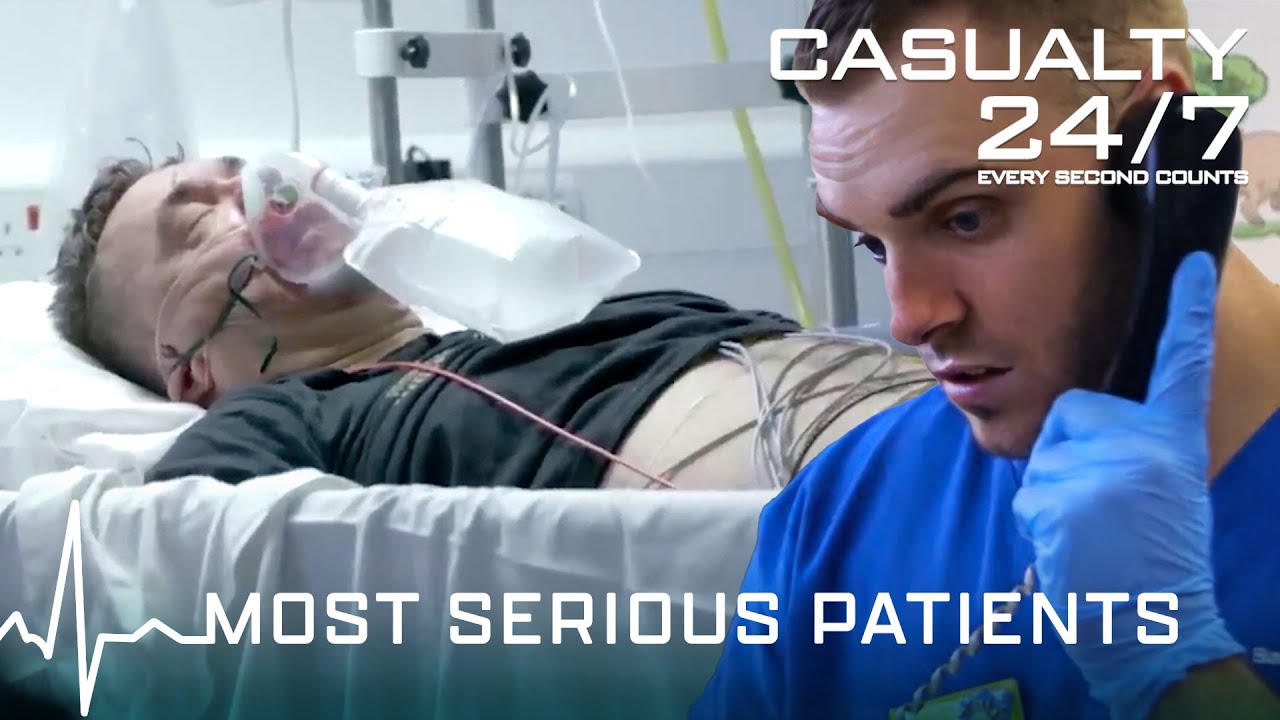
In this compilation, Barnsley Hospital is facing a very busy day with a high number of patients being treated, the doctors and nurses face some of their toughest shifts when they treat critical patients and rare illnesses as well as making tough decisions.
⌚️Timecodes:
00:00 Season 2 Episode 1
08:56 Season 4 Episode 1
16:53 Season 3 Episode 10
30:36 Season 3 Episode 13
37:45 Season 2 Episode 9
46:51 Season 1 Episode 2
52:52 Season 1 Episode 3
58:02 Season 2 Episode 2
01:09:39 Season 2 Episode 11
01:18:37 Season 2 episode 12
🟦 Click Link below to subscribe: https://www.youtube.com/channe....l/UCHPgATT2HtFrxmueq
About Casualty 24/7:
Casualty 24/7 shows how the doors of Barnsley A&E department are open every hour, of every day. They allow a peek inside their medical emergency teams, and how they deal with critical situations revolving around people's lives and illnesses. The team are close-knit and exchange typical Yorkshire humour to get them through their often long and tough days.
Watch our playlists:
🔵 Season 1 Full Episodes | Casualty 24/7:
https://www.youtube.com/playli....st?list=PLWrY8x74oDM
🔵 Season 2 Full Episodes | Casualty 24/7:
https://www.youtube.com/playli....st?list=PLWrY8x74oDM
🔵 Season 3 Full Episodes | Casualty 24/7:
https://www.youtube.com/playli....st?list=PLWrY8x74oDM
🔵 Season 4 Full Episodes | Casualty 24/7:
https://www.youtube.com/playli....st?list=PLWrY8x74oDM
🔵 Compilation Videos of Casualty 24/7:
https://www.youtube.com/playli....st?list=PLWrY8x74oDM
#SeriousIllness #Casualty247 #EmergencyServices #AandE #BHNFT #OurFutureSouthYorkshire

Dr. Nick demonstrates how to numb a toe for a patient who had a subungual hematoma “collection of blood under the nail”. This patient stubbed his toe and needed to have the nail removed.
#satisfying #reaction #amazing
MAKE SURE TO SUBSCRIBE FOR ALL THE NEW SURGICAL AND EDUCATIONAL VIDEOS COMING!!
👉🏻For more information visit :
https://drnickcampi.com
👉🏻Follow me on TikTok!!
https://vm.tiktok.com/ZMeXLbc5F/I’ll
👉🏻Connect with me!!
https://www.instagram.com/drnickcampitelli
👉🏻Check out this video of how we remove an ingrown toenail!
https://youtu.be/JyZo8aZDYds
👉🏻Dr. Nick Campitelli Performs latest Minimally Invasive Bunion Surgery! Watch this video!
https://youtu.be/eRpABMsCbOU
Dr. Nick Campitelli is a podiatrist who specializes in foot and ankle surgery in the Akron and Cleveland Ohio area. He is the Residency Director of the Western Reserve Hospital / University Hospital Podiatric Medicine and Surgery Residency Program.
*** All content found on the this YouTube video including: text, images, audio, or other formats were created for informational purposes only. The Content is not intended to be a substitute for professional medical advice, diagnosis, or treatment. Always seek the advice of your physician or other qualified health provider with any questions you may have regarding a medical condition. Never disregard professional medical advice or delay in seeking it because of something you heard on this video. ***
◦
Overview
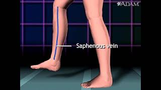
Heart bypass surgery creates a new route, called a bypass, for blood and oxygen to reach the heart.
Heart bypass surgery begins with an incision in the chest, and the breastbone is cut exposing the heart. Next, a portion of the saphenous vein, which is very large, is harvested from the inside of the leg. Pieces of this large vein are used to bypass the blocked coronary arteries, which are arteries that supply blood to the heart. The venous graft is sewn to the aorta, the main artery of the body, and to the affected coronary artery, to bypass the blocked site.
The internal mammary artery from the chest may also be used to bypass a clogged artery.
Several arteries may be bypassed depending on the condition of the heart. After the graft is created, the breastbone and chest are closed.
