- Physical Examination
- Surgical Examination
- Ophthalmology
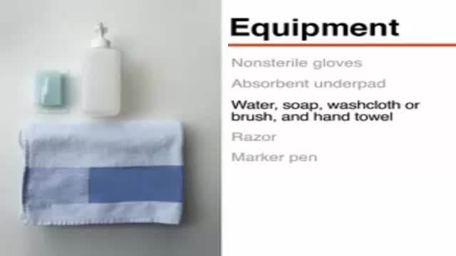
- Clinical Skills
- Orthopedics
- Surgery Videos
- Laparoscopy
- Pediatrics
- Funny Videos
- Cardiothoracic Surgery
- Nursing Videos
- Plastic Surgery
- Otorhinolaryngology
- Histology and Histopathology
- Neurosurgery
- Dermatology
- Pediatric Surgery
- Urology
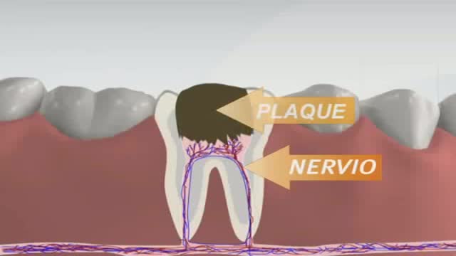
- Dentistry
- Oncology and Cancers
- Anatomy Videos
- Health and Fitness
- Radiology
- Anaesthesia
- Physical Therapy
- Pharmacology
- Interventional Radiology
- Cardiology
- Endocrinology
- Gynecology
- Emergency Medicine
- Psychiatry and Psychology
- Childbirth Videos
- General Medical Videos
- Nephrology
- Physiology
- Diet and Food Health
- Diabetes Mellitus
- Neurology
- Women Health
- Osteoporosis
- Gastroenterology
- Pulmonology
- Hematology
- Rheumatology
- Toxicology
- Nuclear Medicine
- Infectious Diseases
- Vascular Disease
- Reproductive Health
- Burns and Wound Healing
- Other
Top videos
Some of these advancements include the use of robots to perform the surgery and the use of computer mapping scanners and software and even 3D printers to make the artificial knee implant. We are also seeing a lot of different advances being made by the medical device manufacturers.Some of these changes are designed to make the devices more durable than the 10 to 20 years they are currently rated to last. This is important when younger patients who are years away from being considered elderly have a total knee replacement, they will typically need another artificial knee implanted at a later date since they are likely to outlive the implant, which doesn’t always happen with elderly patients.

Slicosis is caused by inhalation of unbound (free) crystalline silica dust and is characterized by nodular pulmonary fibrosis. Chronic silicosis initially causes no symptoms or only mild dyspnea but over years can advance to involve most of the lung and cause dyspnea, hypoxemia, pulmonary hypertension, and respiratory impairment. Diagnosis is based on history and chest x-ray findings. No effective treatment exists except supportive care and, for severe cases, lung transplantation.
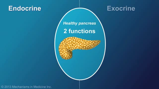
Chronic pancreatitis is a long-standing inflammation of the pancreas that alters the organ's normal structure and functions. It can present as episodes of acute inflammation in a previously injured pancreas, or as chronic damage with persistent pain or malabsorption.
Watch that Hemorrhoids Repairing Medical Video
This video: Polycythemia vera (pol-e-sigh-THEE-me-uh VEER-uh) is a slow-growing type of blood cancer in which your bone marrow makes too many red blood cells. Polycythemia vera may also result in production of too many of the other types of blood cells — white blood cells and platelets. These excess cells thicken your blood and cause complications, such as such as a risk of blood clots or bleeding. Polycythemia vera isn't common. It usually develops slowly, and you may have it for years without noticing signs or symptoms. Often, polycythemia vera is found during a blood test done for some other reason. Without treatment, polycythemia vera can be life-threatening. However, with proper medical care, many people experience few problems related to this disease. Over time, there's a risk of progressing to more-serious blood cancers, such as myelofibrosis or acute leukemia.
What are the symptoms of spinal meningitis in adults? Causes. The most common cause of viral meningitis is. ... Symptoms. Viral meningitis usually begins with symptoms of a viral infection, such as fever, a general feeling of illness (malaise), cough, muscle aches, vomiting, loss of appetite, and headache. ... Diagnosis. ... Treatment. ... Prognosis.
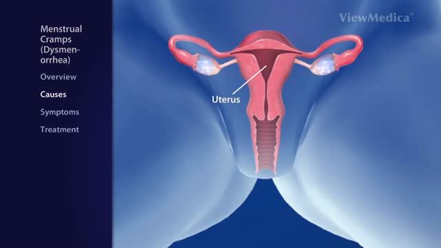
Menstrual cramps (dysmenorrhea) are throbbing or cramping pains in the lower abdomen. ... Menstrual cramps may be caused by identifiable problems, such as endometriosis or uterine fibroids. Treating any underlying cause is key to reducing the pain
Indications for intervention in patients with a renal artery aneurysm (RAA) include the following [20, 8, 13, 14] : Rupture Symptomatic RAA - Hypertension (from associated renal artery stenosis, refractory to medical management), pain, renal ischemia or infarction secondary to embolization from the aneurysm sac RAAs in females who are pregnant or are contemplating pregnancy Diameter greater than 2 cm Enlarging RAA RAA associated with acute dissection Currently, there is no consensus regarding the size at which an RAA should be repaired in an asymptomatic patient. Experts have recommended RAA repair at diameters ranging from 1.5 to 3 cm, [8] though most suggest 2 cm. Some reports have even suggest that larger asymptomatic saccular aneurysms may be managed expectantly. Note that aneurysm rupture at a diameter of 1.5 cm has been reported. Complete calcification of the wall of the aneurysm sac manifests in about 40% of patients. This was once believed to confer protection against rupture [21] ; however, this belief has since been questioned. [30] Asymptomatic, small (<2 cm in diameter) RAAs do not usually require treatment. One notable exception is an RAA in a woman who is pregnant or contemplating pregnancy. In view of the increased risk of rupture in such cases, even small asymptomatic aneurysms should be repaired in this population. For diagnosis and preinterventional planning, gadolinium-enhanced magnetic resonance angiography (MRA) and computed tomography (CT) angiography (CTA) with three-dimensional (3D) reconstruction have essentially replaced conventional arteriography. Regular follow-up examination with ultrasonography (US) or CT) is recommended in patients who are treated expectantly. Spontaneous cure by thrombosis of small aneurysms has been described. Further refinements in endovascular techniques may allow more RAAs to be treated in this manner. So far, excellent short- and intermediate-term results have been described in the literature [40] ; however, there remains a need for further long-term outcome data.
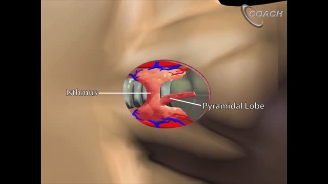
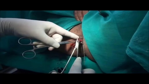
Minimally invasive open thyroidectomy (MIT) is similar to conventional thyroidectomy in its surgical approach. The major difference is the length of the neck incision. A smaller incision improves cosmesis and reduces discomfort. Typically, a skin incision less than 6 cm is considered minimally invasive. The remainder of the procedure is exactly the same as is used in conventional thyroidectomy. Adaptations to this technique include transection rather than lateral retraction of the strap muscles (the Sofferman technique). [1]
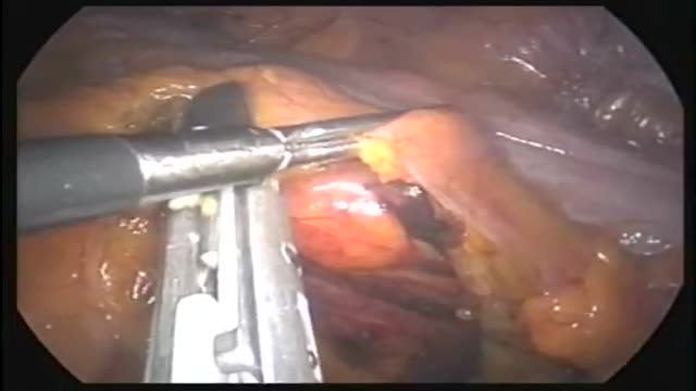
This operation can be performed as an open or laparoscopic (keyhole procedure). During the operation the sigmoid colon is removed. This involves taking away the blood vessels and lymph nodes to that part of the bowel. The surgeon then re-makes the join (anastomosis) between the remaining left side of the colon and the top of the rectum. The surgeon may use either sutures or special staples to make this join.
Invasive intracranial pressure monitoring. The most common surgically placed monitors for ICP measurement are intraventricular catheters (external ventricular drain [EVD] or a ventriculostomy drain) and fiberoptic ICP monitors implanted into the parenchyma of the brain.
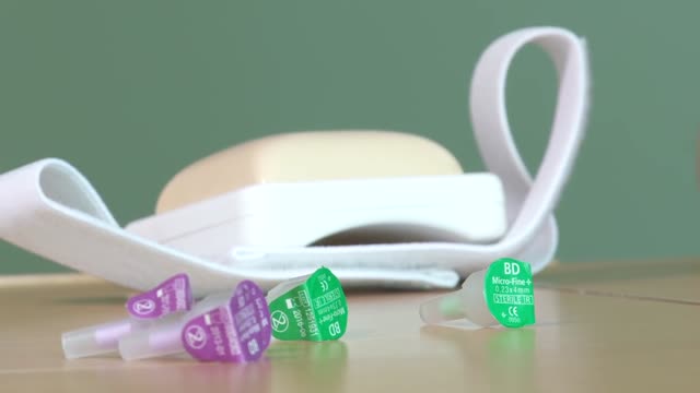
Insert the needle into the rubber stopper of the insulin bottle. Push the plunger down to inject air into the bottle (this allows the insulin to be drawn more easily). Leave the needle in the bottle. Turn the bottle and syringe upside-down.


Vitiligine Cura, Vitiligine Rimedi Naturali, Vitiligine Omeopatia, Rimedi Per La Vitiligine -- http://vitiligine-cura.good-info.co --- Vitiligine Cura, Vitiligine Rimedi Naturali, Vitiligine Omeopatia, Rimedi Per La Vitiligine. Soffri Di Uno Qualunque Dei Seguenti Sintomi Emotivi O Fisici? Qualsiasi tipo di vitiligine (qualsiasi livello di gravità) su viso, schiena, guance, palmi delle mani, gambe o piedi? Sei affetto da macchie o scoloramento della pelle? Provi ansia nel doverti togliere la maglia in pubblico? Provi costantemente insicurezza? Provi esasperazione per il disturbo della vitiligine? Spendi molti soldi in farmaci o parafarmaci che sembrano non funzionare? Vuoi curare la vitiligine ma non sai quale sia la giusta cura a causa di un sovraccarico di informazioni? "Una Presentazione Video Gratuita Spiega Un Singolare Consiglio Per Eliminare La Vitiligine Per Sempre In 45-60 Giorni - Garantito!" http://vitiligine-cura.good-info.co

Surgery to repair a torn rotator cuff most often involves re-attaching the tendon to the head of humerus (upper arm bone). A partial tear, however, may need only a trimming or smoothing procedure called a debridement. A complete tear is repaired by stitching the tendon back to its original site on the humerus.

Pruritis is itchy skin that makes you want to scratch. It can be caused by many things. Normally, itchy skin isn't serious, but it can make you uncomfortable. Sometimes, itchy skin is caused by a serious medical condition. It can occur in association with a primary rash (e.g. dermatitis) or may occur because of hypersensitive nerves in the skin (neuropathic pruritus). ... Scratching a localised itch may lead to lichen simplex, prurigo or prurigo nodularis. Systemic causes of pruritus. Sytemic diseases may cause generalised pruritus.
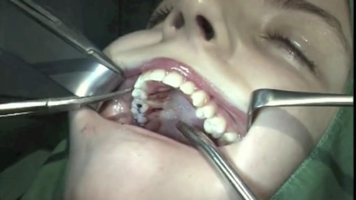
Simply put, there isn’t enough room for wisdom teeth because our jaws don’t grow to be big enough to have enough space for them to come in. Since there isn’t enough room for them to erupt properly, wisdom teeth tend to come in at an angle or they don’t fully emerge, which causes problems for the rest of the mouth. Third molars (the wisdom teeth) routinely damage the teeth right next door, called second molars. Dentists recommend removing wisdom teeth before they become a problem and to avoid a more complicated surgery. Read more at Ask the Dentist: https://askthedentist.com/wisdom-teeth-removal/
Root canals are common procedures and can help save your tooth from extraction. Dentists at Aspen Dental practices have been safely and expertly performing root canal procedures for over two decades.

Joint Replacement Surgery is part of Orthopedics hence surgical as well as non-surgical techniques are carried out by orthopedic surgeons. The burden of pain can be reduced with the help of minimally invasive orthopedic therapies prescribed by doctors. https://goo.gl/VhzaUr

Initially, lead poisoning can be hard to detect — even people who seem healthy can have high blood levels of lead. Signs and symptoms usually don't appear until dangerous amounts have accumulated. Lead poisoning symptoms in children Signs and symptoms of lead poisoning in children include: Developmental delay Learning difficulties Irritability Loss of appetite Weight loss Sluggishness and fatigue Abdominal pain Vomiting Constipation Hearing loss Seizures Eating things, such as paint chips, that aren't food (pica) Lead poisoning symptoms in newborns Babies exposed to lead before birth might: Be born prematurely Have lower birth weight Have slowed growth Lead poisoning symptoms in adults Although children are primarily at risk, lead poisoning is also dangerous for adults. Signs and symptoms in adults might include: High blood pressure Joint and muscle pain Difficulties with memory or concentration Headache Abdominal pain Mood disorders Reduced sperm count and abnormal sperm Miscarriage, stillbirth or premature birth in pregnant women Causes Lead is a metal that occurs naturally in the earth's crust, but human activity — mining, burning fossil fuels and manufacturing — has caused it to become more widespread. Lead was also once used in paint and gasoline and is still used in batteries, solder, pipes, pottery, roofing materials and some cosmetics. Lead in paint Lead-based paints for homes, children's toys and household furniture have been banned in the United States since 1978. But lead-based paint is still on walls and woodwork in many older homes and apartments. Most lead poisoning in children results from eating chips of deteriorating lead-based paint. Water pipes and imported canned goods Lead pipes, brass plumbing fixtures and copper pipes soldered with lead can release lead particles into tap water. Lead solder in food cans, banned in the United States, is still used in some countries. Other sources of lead exposure Lead sometimes can also be found in: Soil. Lead particles from leaded gasoline or paint settle on soil and can last years. Lead-contaminated soil is still a major problem around highways and in some urban settings. Some soil close to walls of older houses contains lead. Household dust. Household dust can contain lead from lead paint chips or from contaminated soil brought in from outside. Pottery. Glazes found on some ceramics, china and porcelain can contain lead that can leach into food served or stored in the pottery. Toys. Lead is sometimes found in toys and other products produced abroad. Cosmetics. Tiro, an eye cosmetic from Nigeria, has been linked to lead poisoning. Herbal or folk remedies. Lead poisoning has been linked to greta and azarcon, traditional Hispanic medicines, as well as some from India, China and other countries. Mexican candy. Tamarind, an ingredient used in some candies made in Mexico, might contain lead. Lead bullets. Time spent at firing ranges can lead to exposure. Occupations. People are exposed to lead and can bring it home on their clothes when they work in auto repair, mining, pipe fitting, battery manufacturing, painting, construction and certain other fields
