- Physical Examination
- Surgical Examination
- Ophthalmology
- Clinical Skills
- Orthopedics
- Surgery Videos
- Laparoscopy
- Pediatrics
- Funny Videos
- Cardiothoracic Surgery
- Nursing Videos
- Plastic Surgery
- Otorhinolaryngology
- Histology and Histopathology
- Neurosurgery
- Dermatology
- Pediatric Surgery
- Urology
- Dentistry
- Oncology and Cancers
- Anatomy Videos
- Health and Fitness
- Radiology
- Anaesthesia
- Physical Therapy
- Pharmacology
- Interventional Radiology
- Cardiology
- Endocrinology
- Gynecology
- Emergency Medicine
- Psychiatry and Psychology
- Childbirth Videos
- General Medical Videos
- Nephrology
- Physiology
- Diet and Food Health
- Diabetes Mellitus
- Neurology
- Women Health
- Osteoporosis
- Gastroenterology
- Pulmonology
- Hematology
- Rheumatology
- Toxicology
- Nuclear Medicine
- Infectious Diseases
- Vascular Disease
- Reproductive Health
- Burns and Wound Healing
- Other
Top videos
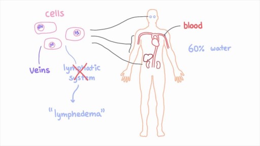
The lymphatic system is a network of specialized vessels (lymph vessels) throughout the body whose purpose is to collect excess lymph fluid with proteins, lipids, and waste products from the tissues. This fluid is then carried to the lymph nodes, which filter waste products and contain infection-fighting cells called lymphocytes. The excess fluid in the lymph vessels is eventually returned to the bloodstream. When the lymph vessels are blocked or unable to carry lymph fluid away from the tissues, localized swelling (lymphedema) is the result.

Dr. Lohith U, Consultant Surgical Gastroenterology, Bariatric and GI Oncology, Manipal Hospital Sarjapur, speaks about advanced laparoscopic surgery in this informative video. He shares details about the procedure,insights on its advantages, and how it can help treat gastrointestinal issues without making large incisions in a patient’s body. To know more watch this video.
Consult the experts here - https://bit.ly/3RiWqPr
#ManipalHospitalSarjapur #YourManipal #LifesOn #Healthcare #LaparoscopicSurgery

Mysterious massage from East Asia(CHINA).it can cure cure Erectile dysfunction,can let their life better.This video from mainland of China,so the language is Chinese mandarin.but you can see English show on the video too.Tiedang gong means kongfu of Iron penis&balls.

Synthol, otherwise known as site enhancement oil is used by some people (including bodybuilders) to increase the apparent size of their muscles by directly injecting the oil into their muscle tissue. Users treat it as a short cut of looking like a body builder, without the actual hard work of bodybuilding training. With repeated injections, a larger volume of synthol builds up inside the muscle, expanding its size like a balloon filling up with air. Side effects of synthol can cause nerve damage, stroke, ulcers, pulmonary embolisms, and much more. Injecting synthol is very dangerous and if that doesn’t deter potential users, there is also a problem from an aesthetic standpoint; synthol use makes ones body look deformed (just see for yourself in the pictures below).

The Hypertensive urgency must be distinguished from hypertensive emergency. Urgency is defined as severely elevated blood pressure (ie, systolic >220 mm Hg or diastolic >120 mm Hg) with no evidence of target organ damage.

Vaginal discharge serves an important housekeeping function in the female reproductive system. Fluid made by glands inside the vagina and cervix carries away dead cells and bacteria. This keeps the vagina clean and helps prevent infection. Most of the time, vaginal discharge is perfectly normal. The amount can vary, as can odor and hue (its color can range from clear to a milky white-ish), depending on the time in your menstrual cycle. For example, there will be more discharge if you are ovulating, breastfeeding, or are sexually aroused. The smell may be different if you are pregnant or you haven't been diligent about your personal hygiene. None of those changes is cause for alarm. However, if the color, smell, or consistency seems significantly unusual, especially if it accompanied by vaginal itching or burning, you could be noticing an infection or other condition. What causes abnormal discharge? Any change in the vagina's balance of normal bacteria can affect the smell, color, or discharge texture. These are a few of the things that can upset that balance:

Watch that video of a Snake bite causes girl’s leg to rot away with necrosis

Cisplatin is in a class of drugs known as platinum-containing compounds used to treat various types of cancers including metastatic testicular and ovarian tumors. The molecule was first discovered in 1845, but did not receive FDA approval until 1978. Today it is known as the "penicillin of cancer drugs," because it is so effective for many different cancers. There are three key players involved in Cisplatin's mechanism: (1) Cisplatin, (2) DNA (3) and an HMG Protein. Most Cisplatin enters the body through active transport, but some molecules are passively defused through the cell membrane. Once in the nucleus, Cisplatin can form an adduct with two consecutive guanine bases within a strand of DNA. The molecule loses its chlorine atoms in exchange for the nitrogen atoms of the target guanines. Cisplatin can bond more tightly with nitrogen because nitrogen balances the platinum charge more effectively than chlorine. It is this adduct-induced DNA bend that allows binding of proteins which contain the high mobility group, HMG domain. Once the protein is bound to the DNA, it inserts a wedge-like phenyl group of phenylalanine 37 into the widened minor groove created by the bend. The tightly bound HMG protein causes destacking of the nucleotide bases, resulting in the DNA helix becoming kinked. In this way, Cisplatin can be thought of as a monkey wrench in the DNA repair system. With the HMG protein bound to the DNA, the modified strand is not repaired properly and so the cell dies. The success of Cisplatin depends on its ratio of efficacy between cancerous and healthy cells.
Examination of the abdomen from the USMLE collection
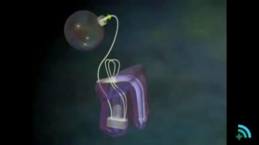
he inflatable penile prosthesis consists of two attached cylinders -- a reservoir and a pump -- which are placed surgically in the body. The two cylinders are inserted in the penis and connected by tubing to a separate reservoir of saline. The reservoir is implanted under the rectus muscles in the lower abdomen.

Shane Shapiro, M.D., orthopedic physician at Mayo Clinic in Florida, performs a bone marrow aspiration and concentration for BMAC/stem cell injection into arthritic knees. This procedure is part of a Mayo Clinic IRB approved, FDA monitored clinical research trial which can be searched on at http://ClinicalTrials.gov.
Mayo Clinic and the Mayo Center for Regenerative Biotherapeutics is studying biologically based non-surgical treatments for osteoarthritis. One such treatment is the harvesting of the patient's own stem cells from their bone marrow.
"In our procedure we draw cellular rich bone marrow from both sides of the pelvis. We then filter the resulting product and concentrate the stem cells and their corresponding growth factors. Using an ultrasound to image the knee joint, we are then able to precisely inject the cells into the arthritic knee. We are currently demonstrating that this procedure is safe and can relieve pain. We also hope to be able to slow the progression of the degenerative joint disease and perhaps one day regrow cartilage in the arthritic joint."
------
Hear Dr. Shapiro discus this procedure in detail here: http://youtu.be/8Djpsc66hKI
Learn more about the Mayo Clinic Center for Regenerative Biotherapeutics here: http://goo.gl/rnRdtU
------
Mayo Clinic...
On Facebook: http://Facebook.com/MayoClinic
On Twitter: http://twitter.com/MayoClinic
On Google+: http://google.com/+MayoClinic
On Instagram: http://instagram.com/MayoClinic
On Pinterest: http://Pinterest.com/MayoClinic
On YouTube: http://YouTube.com/MayoClinic
On the blogs: http://connect.MayoClinic.org
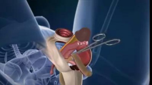
A walk through of an interactive about male to female sex reassignment surgery.
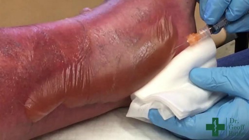
his patient had spilled boiling water on his lower leg a couple days before. This isn't complicated but the teaching points should focus on draining the large blistered areas and attempting to maintain moisture as long as we can so the skin doesn't contract down on itself.

Labia minoraplasty is an elective procedure that can reduce the size and reshape the inner vaginal lips. Large or asymmetrical labia minora can leave you feeling self-conscience in tight clothing or during intimacy. Long labia may result in rubbing, irritation or discomfort during intercourse and exercise. Certain skin conditions can cause increased sensitivity or tearing of the labia minora. In some cases, the labia minora may be fused with tissue in the labia majora and require medical correction.
Ankylosing spondylitis is an inflammatory disease that, over time, can cause some of the vertebrae in your spine to fuse. This fusing makes the spine less flexible and can result in a hunched-forward posture. If ribs are affected, it can be difficult to breathe deeply. Ankylosing spondylitis affects men more often than women. Signs and symptoms typically begin in early adulthood. Inflammation also can occur in other parts of your body — most commonly, your eyes. There is no cure for ankylosing spondylitis, but treatments can lessen your symptoms and possibly slow progression of the disease.
A breech birth occurs when a baby is born bottom first instead of head first. Around 3-5% of pregnant women at term (37–40 weeks pregnant) will have a breech baby. Most babies in the breech position are born by a caesarean section because it is seen as safer than being born vaginally.

An abscess is a collection of pus. Pus is a thick fluid that usually contains white blood cells, dead tissue and germs (bacteria). The usual cause of an abscess is an infection with bacteria. Certain bacteria are more likely to be 'pus-forming' as they make chemicals (toxins) that can damage the body's tissues.
The majority of fetuses are in a breech presentation early in pregnancy. By week 38th week of gestation, however, the fetus normally turns to a cephalic presentation. Although the fetal head is the widest single diameter, the fetus’s buttocks [ breech], plus the lower extremities, actually takes up more space. The fundus, being the largest part of the uterus, probably accounts for the fact that in approximately 97% of all pregnancies, the fetus turns so that the buttocks and lower extremities are in the fundus. Vaginal delivery of a breech presentation requires great skill if the fetus is not to be damaged. With the low rate of vaginal breech deliveries in the developed world, experience is being lost. 6% of women with breech presentation still have a vaginal breech delivery as they present too late - so units need to retain a high level of preparedness. Types of breech presentation: I. Complete breech [ flexed breech]: The fetal attitude is one of complete flexion, with hips and knees both flexed and the feet tucked in beside the buttocks. The presenting part consists of two buttocks, external genitalia and two feet. It is commonly present in multiparae. II. Incomplete breech: This is due to varying degrees of extension of thighs or legs at podalic pole. Three varieties are possible; - Breech with extended legs [ frank breech ]: The breech presents with the hips flexed and legs extended on the abdomen. 70% of breech presentations are of this type and it is particularly common in primigravidae whose good uterine muscle tone inhibits flexion of the legs and free turning of the fetus. - Footling breech: This is rare. One or both feet present because neither hips nor knees are fully flexed. The feet are lower than the buttocks, which distinguishes it from the complete breech. - Knee presentation: This is very rare. Thighs are extended but the knees are flexed, bringing the knees down to present at the brim.

Diastasis recti often occurs during pregnancy and can persist after pregnancy. It affects core strength and the appearance of the abdominal muscles.
Dr. Erick Sanchez repairs the abdominal muscles with every tummy tuck. This short video shows the muscle repair portion of the surgery with a bonus after photo at the end!
To request a consultation with Dr. Sanchez, visit sanchezplasticsurgery.com and click Request a Consultation. Fill out the form and someone will get in touch with you to answer all your questions.
Expected cost can be found at the bottom of each procedure page on our website.
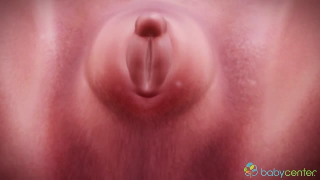
Your baby's sex is set at conception. At around 7 weeks, your baby's internal sex organs – such as ovaries and testes – begin to form in the abdomen. Male and female sex organs and genitalia look the same at this stage because they're derived from the same structures. At around 9 weeks, boys and girls begin to develop differently. In girls, a tiny bud emerges between the tissue of the legs. This bud will become the clitoris. The membrane that forms a groove below the bud separates to become the labia minora and the vaginal opening. By 22 weeks, the ovaries are completely formed and move from the abdomen to the pelvis. They already contain a lifetime supply of 6 million eggs. In boys, the bud develops into the penis and starts to elongate at around 12 weeks. The outer membrane grows into the scrotal sac that will later house the testicles. By 22 weeks, the testes have formed in the abdomen. They already contain immature sperm. Soon they'll begin their descent to the scrotum, but it's a long journey. They'll reach their destination late in pregnancy, or for some boys, after birth. If you're eager to find out whether you're having a girl or a boy, you'll have to wait until you're at least 17 weeks pregnant. That's when the genitals have developed enough to be seen on an ultrasound.
