- Physical Examination
- Surgical Examination
- Ophthalmology
- Clinical Skills
- Orthopedics
- Surgery Videos
- Laparoscopy
- Pediatrics
- Funny Videos
- Cardiothoracic Surgery
- Nursing Videos
- Plastic Surgery
- Otorhinolaryngology
- Histology and Histopathology
- Neurosurgery
- Dermatology
- Pediatric Surgery
- Urology
- Dentistry
- Oncology and Cancers
- Anatomy Videos
- Health and Fitness
- Radiology
- Anaesthesia
- Physical Therapy
- Pharmacology
- Interventional Radiology
- Cardiology
- Endocrinology
- Gynecology
- Emergency Medicine
- Psychiatry and Psychology
- Childbirth Videos
- General Medical Videos
- Nephrology
- Physiology
- Diet and Food Health
- Diabetes Mellitus
- Neurology
- Women Health
- Osteoporosis
- Gastroenterology
- Pulmonology
- Hematology
- Rheumatology
- Toxicology
- Nuclear Medicine
- Infectious Diseases
- Vascular Disease
- Reproductive Health
- Burns and Wound Healing
- Other
Top videos
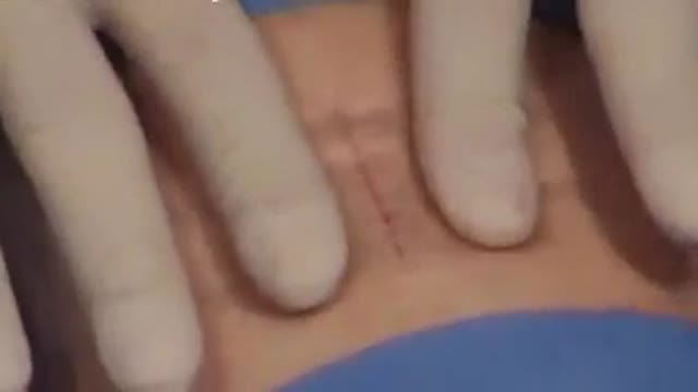
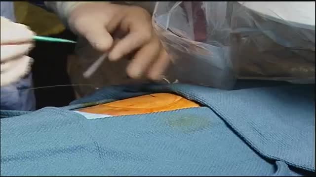
Wound-closure technologies are becoming less painful and more efficient at closing wounds
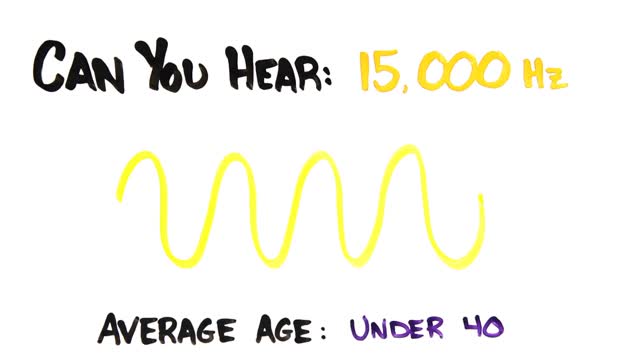
Hearing loss can affect anyone at any age, due to heredity, medical conditions or loud noise exposure. However, as we get older, we naturally become more susceptible to hearing loss because of changes to the delicate mechanics of our ears.

A simple continuous stitch can be a useful technique for skin closure when speed is important, e.g. closing a scalp laceration on a screaming child. The simple running, or continuous suture, is begun in the same way as a simple interrupted suture.
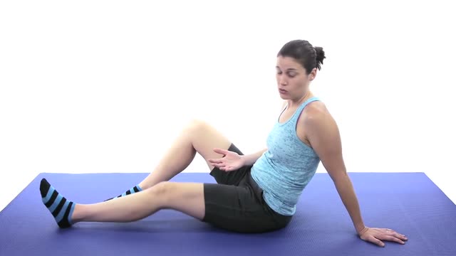
The ACL is one of the four main ligaments within the knee that connect the femur to the tibia. The knee is essentially a hinged joint that is held together by the medial collateral (MCL), lateral collateral (LCL), anterior cruciate (ACL) and posterior cruciate (PCL) ligaments.

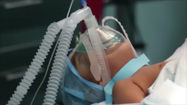
The majority of epileptic seizures are controlled by medication, particularly anticonvulsant drugs. The type of treatment prescribed will depend on several factors, including the frequency and severity of the seizures and the person's age, overall health, and medical history. An accurate diagnosis of the type of epilepsy is also critical to choosing the best treatment. Drug Therapy Many drugs are available to treat epilepsy. Although generic drugs are safely used for most medications, anticonvulsants are one category where doctors proceed with caution. Most doctors prefer to use brand-name anticonvulsants, but realize that many insurance companies will not cover the cost. As a result, it is acceptable to start taking a generic anticonvulsant medication, but if the desired control is not achieved, the patient should be switched to the brand-name drug.
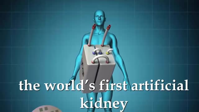
Artificial kidney is often a synonym for hemodialysis, but may also, more generally, refer to renal replacement therapies (with exclusion of kidney transplantation) that are in use and/or in development.
An intra-aortic balloon pump (IABP) is a mechanical device that helps the heart pump blood. This device is inserted into the aorta, the body's largest artery. It is a long, thin tube called a catheter with a balloon on the end of it. If you are hospitalized, your doctor may insert an IABP.
Myasthenia gravis is a chronic autoimmune neuromuscular disease characterized by varying degrees of weakness of the skeletal (voluntary) muscles of the body. The name myasthenia gravis, which is Latin and Greek in origin, literally means "grave muscle weakness."

The arm and leg muscles are affected later. Myasthenia gravis (MG) is an autoimmune disease — a disease that occurs when the immune system attacks the body's own tissues. In MG, that attack interrupts the connection between nerve and muscle — the neuromuscular junction.

Wash your hands thoroughly with soap and water before and after treating the wound. Wash the area with mild soap and running water to reduce the risk of infection. Pat dry. Apply antibiotic ointment and cover with a clean bandage or sterile dressing. Antibiotic prophylaxis should be considered, especially if there is a high risk of infection, such as with cat bites, with puncture wounds, with wounds to the hand, and in persons who are immunosuppressed. Amoxicillin/clavulanate is the first-line prophylactic antibiotic.

If you’ve suffered a sporting knee injury, how do you know when it’s serious? In this short video, Yorkshire Knee Clinic’s Dave Duffy reveals the two key tests that tell you whether your knee needs urgent, specialist attention.
𝗡𝗼𝘁𝗲𝘀 𝗳𝗼𝗿 𝘁𝗵𝗲 𝘀𝗾𝘂𝗲𝗮𝗺𝗶𝘀𝗵: This video features only features a model of the knee. There is no live footage from operations.
Discover more about sports knee injuries: https://yorkshirekneeclinic.com/sports-injuries/
Discover more about Dave Duffy: https://yorkshirekneeclinic.com/about/dave-duffy/

Classical PKU is an autosomal recessive disorder, caused by mutations in both alleles of the gene for phenylalanine hydroxylase (PAH), found on chromosome 12. In the body, phenylalanine hydroxylase converts the amino acid phenylalanine to tyrosine, another amino acid.

Johns Hopkins orthopaedic hip and knee surgeon, Savyasachi "Savya" Thakkar, explains how to prepare for knee replacement surgery, and what to expect before and after surgery. To learn more about our hip and knee replacement division, visit https://www.hopkinsmedicine.org/ortho. #KneeReplacement #JohnsHopkins
Q&A's
0:15 What causes someone to need a knee replacement?
0:29 What should patients do in advance of surgery?
1:10 Do you recommend physical therapy BEFORE surgery?
1:43 Will joint implants set off metal detectors at airports?
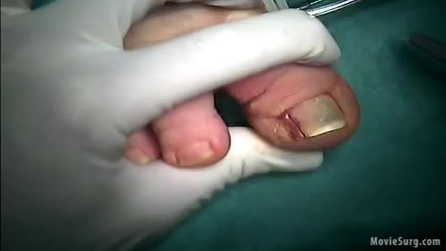
irregular, curved toenails. footwear that places a lot of pressure on the big toes, such as socks and stockings that are too tight or shoes that are too tight, narrow, or flat for your feet. toenail injury, including stubbing your toe, dropping something heavy on your foot, or kicking a ball repeatedly. poor posture.
Empyema can develop after you have pneumonia. Many different types of bacteria may cause pneumonia, but the two most common are Streptococcus pneumoniae and Staphylococcus aureus. Occasionally, empyema may happen after you've had surgery on your chest. Medical instruments can transfer bacteria into your pleural cavity

Como Eliminar Verrugas, Tratamiento Para Verrugas Genitales, Como Sacar Verrugas, Verrugas Planas --- http://sinverrugasylunares.plus101.com -- Sabías Que Con Vinagre De Manzana Puedes Eliminas Tus Verrugas? No es un secreto que el uso de vinagre de manzana trae enormes beneficios a la piel, es capaz de activar la regeneración de la piel sin importar la lesión que se haya sufrido. A continuación voy a enseñarte cómo puedes utilizar el vinagre de manzana para eliminar las verrugas y evitar que estas vuelvan a brotar. Voy a enseñarte 2 formas para eliminar las verrugas con vinagre de manzana. Método 1: Lávate las manos (o cualquier otra parte del cuerpo donde las verruga se ha formado). …bien antes de empezar con este tratamiento. Toma una taza de vinagre de manzana y dilúyela con agua. Sumerge el área afectada en esta solución durante 20 minutos todos los días. Usa este tratamiento regularmente. Con el tiempo la verruga se desprenderá sin dejar rastro en tu piel. Método 2: Lávate las manos (o la parte afectada) antes de comenzar. Utiliza una bola de algodón y sumérgela en vinagre de manzana. A continuación, escurre el exceso de vinagre y coloca el algodón sobre la verruga. Conoce el verdadero método natural para eliminar las verrugas y lunares ahora: click aqui: http://sinverrugasylunares.plus101.com
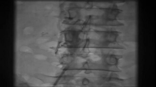
IVC filter is placed to prevent fatal Pulmonary Embolism due to Deep Venous Thrombosis (DVT). This particular patient had extensive DVT of Ilio-Femoral veins leading to massive swelling of left lower limb. The IVC filter was inserted via the Right Femoral Vein. This case was the first IVC filter placement of North Bengal and adjoining areas.

Waardenburg syndrome is a group of genetic conditions that can cause hearing loss and changes in coloring (pigmentation) of the hair, skin, and eyes. Although most people with Waardenburg syndrome have normal hearing, moderate to profound hearing loss can occur in one or both ears. The hearing loss is present from birth (congenital). People with this condition often have very pale blue eyes or different colored eyes, such as one blue eye and one brown eye. Sometimes one eye has segments of two different colors. Distinctive hair coloring (such as a patch of white hair or hair that prematurely turns gray) is another common sign of the condition. The features of Waardenburg syndrome vary among affected individuals, even among people in the same family.
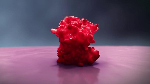
The pathobiology of MM is complex and the root underlying cause of myeloma is the multistep genetic changes in the postgerminal center B cell. In addition, the bone marrow microenvironment plays a crucial role.[2] The interaction between myeloma cells and the microenvironment is mediated through adhesive interactions via cell-surface receptors, paracrine loops involving several cytokines, such as IL-6, VEGF and IL-10, and suppression of cell-mediated immunity.[2–4] IMiDs modulate many of these interactions leading to decreased myeloma cell growth and survival. Thalidomide was the first IMiD introduced to treat MM. It was initially synthesized in Germany in the late 1950s to treat insomnia and morning sickness. It was withdrawn from the market in 1961 because of its teratogenic effects. Its immunomodulatory properties were realized when it was observed to improve erythema nodosum leprosum, a painful immunologic reaction of leprosy, leading to its approval by the FDA in 1998 with tight prescribing and marketing regulations. Subsequent research showed the diverse mechanism of action of thalidomide including its immunomodulatory effect by inhibition of de novo IgM antibody synthesis,[5] modulation of the T-cell subset by increasing the T-helper cells, inhibitory effects on the TNF-α and antiangiogenic activity leading to its use in MM. Significantly higher response rates in combination with dexamethasone led to its approval in the treatment of newly diagnosed MM in 2006. Lenalidomide, a second-generation IMiD, was developed from the structural backbone of the thalidomide molecule by the addition of an amino group (NH2-) at position 4 of the phthaloyl ring and removal of the carbonyl group (C = O) of the 4-amino-substituted phthaloyl ring (Table 1).[6] In addition to immunomodulatory effects, other mechanisms of action have been described such as direct cytotoxicity via induction of apoptosis, inhibition of cell adhesion molecules and inhibition of growth signals that promote bone marrow angiogenesis
The brain is the most complex organ in our body. It controls everything we do, from simple things such as breathing, to complex things such as co-ordinating our movements. The brain stores our memories, allows us to think and speak, and controls how we behave
