- Physical Examination
- Surgical Examination
- Ophthalmology
- Clinical Skills
- Orthopedics
- Surgery Videos
- Laparoscopy
- Pediatrics
- Funny Videos
- Cardiothoracic Surgery
- Nursing Videos
- Plastic Surgery
- Otorhinolaryngology
- Histology and Histopathology
- Neurosurgery
- Dermatology
- Pediatric Surgery
- Urology
- Dentistry
- Oncology and Cancers
- Anatomy Videos
- Health and Fitness
- Radiology
- Anaesthesia
- Physical Therapy
- Pharmacology
- Interventional Radiology
- Cardiology
- Endocrinology
- Gynecology
- Emergency Medicine
- Psychiatry and Psychology
- Childbirth Videos
- General Medical Videos
- Nephrology
- Physiology
- Diet and Food Health
- Diabetes Mellitus
- Neurology
- Women Health
- Osteoporosis
- Gastroenterology
- Pulmonology
- Hematology
- Rheumatology
- Toxicology
- Nuclear Medicine
- Infectious Diseases
- Vascular Disease
- Reproductive Health
- Burns and Wound Healing
- Other
Top videos

How To Stop Hair Loss Naturally, Hair Regrowth Shampoo, Tips For Hair Regrowth, Hair Loss Stop--- http://how-to-regrow-your-hair.info-pro.co/ --- Natural Hair Regrowth Treatment, Looking for ideas on natural hair regrowth treatment? There are actually a lot of safe and effective natural methods that you can try in order to reverse hair loss. So what are those natural methods? Here are some of them: Eat your way to better hair: Hard to believe isn’t it? To think that what you’re actually eating can affect your hair in so many ways, positively and negatively. Want to slow down the process of hair loss and get your hair back to the healthy original state you always remembered it in? Then you might want to start transitioning over to a low fat high fiber diet. Aside from this, you’ll also want to concentrate on foods that contain biotins, which play an essential role in maintaining healthy hair. Fish, cooked eggs, whole milk, and various nuts and fruits – all of these are good sources of biotins so it’s best that you take note of them. Drink plenty of water: It’s not only that gets thirsty, as even your hair requires the moisture that water provides. Dehydration can lead to symptoms like constipation, eczema, thick dandruff, wrinkly skin, foul breath and hair loss. Remember that your body is made up of 98% water and you need to maintain it at optimum levels if you want to keep your hair in place. An easy way to quickly replenish and establish enough water in your body is to routinely drink at least 8 ounces of water immediately after you urinate. You’ll know you’re getting enough water when you observe that you are urinating more frequently. Supplements: The fact that you are experiencing hair loss is a surefire indication that something is amiss in the nutrition department. If you are however looking for a definitive guide on the products that will be available, one useful source you can use will be found at http://how-to-regrow-your-hair.info-pro.co/

Wash your hands thoroughly with soap and water before and after treating the wound. Wash the area with mild soap and running water to reduce the risk of infection. Pat dry. Apply antibiotic ointment and cover with a clean bandage or sterile dressing. Antibiotic prophylaxis should be considered, especially if there is a high risk of infection, such as with cat bites, with puncture wounds, with wounds to the hand, and in persons who are immunosuppressed. Amoxicillin/clavulanate is the first-line prophylactic antibiotic.

» A kidney stone, also known as a renal calculus (from the Latin rēnēs, “kidneys,” and calculus, “pebble”), is a solid concretion or crystal aggregation formed in the kidneys from dietary minerals in the urine. » Urolithiasis (UL) is one of the most common diseases, with approximately 7.6% incidence in Western India. Although most patients have only one stone episode, 25% of patients experience recurrent stone formation. UL therefore has a significant impact on quality of life and socioeconomic factors.

Watch that video to know if oral sex is safe or not
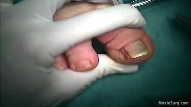
irregular, curved toenails. footwear that places a lot of pressure on the big toes, such as socks and stockings that are too tight or shoes that are too tight, narrow, or flat for your feet. toenail injury, including stubbing your toe, dropping something heavy on your foot, or kicking a ball repeatedly. poor posture.
Temporal arteritis is a condition in which the temporal arteries, which supply blood to the head and brain, become inflamed or damaged. It is also known as cranial arteritis or giant cell arteritis. Although this condition usually occurs in the temporal arteries, it can occur in almost any medium to large artery in the body. The journal Arthritis & Rheumatology states that approximately 228,000 people in the United States are affected by temporal arteritis. According to the American College of Rheumatology, people over the age of 50 are more likely than younger people to develop the condition. Women are also more likely than men to have temporal arteritis. It is most prevalent in people of northern European or Scandinavian descent. Although the exact cause of the condition is unknown, it may be linked to the body’s autoimmune response. Also, excessive doses of antibiotics and certain severe infections have been linked to temporal arteritis. There’s no known prevention. However, once diagnosed, temporal arteritis can be treated to minimize complications.

Waardenburg syndrome is a group of genetic conditions that can cause hearing loss and changes in coloring (pigmentation) of the hair, skin, and eyes. Although most people with Waardenburg syndrome have normal hearing, moderate to profound hearing loss can occur in one or both ears. The hearing loss is present from birth (congenital). People with this condition often have very pale blue eyes or different colored eyes, such as one blue eye and one brown eye. Sometimes one eye has segments of two different colors. Distinctive hair coloring (such as a patch of white hair or hair that prematurely turns gray) is another common sign of the condition. The features of Waardenburg syndrome vary among affected individuals, even among people in the same family.

You can now test your knowledge with a free lesson quiz on NURSING.com!
Click here for your free quiz: https://bit.ly/3Jcl93Z
Tracheostomy Suctioning- Nursing Skills
FREE Nursing School Cheat Sheets at: http://www.NURSING.com
Get the full lesson on Trach Suctioning here:
https://nursing.com/lesson/ski....lls-03-03-trach-suct
Get Access to Thousands of Lessons here:
https://nursing.com/courses/
Welcome to the NURSING Family, we call it the most supportive nursing cohort on the planet.
At NURSING.com, we want to help you remove the stress and overwhelm of nursing school so that you can focus on becoming an amazing nurse.
Check out our freebies and learn more at: (http://www.nursing.com)
Tracheostomy Suctioning- Nursing Skills:
In this video we’re going to talk about suctioning a tracheostomy. You may need to do this before you do trach care or just because the patient requires suctioning. Make sure that you assess the patient before you start so that you know what their one sounds are, and what their oxygen saturation is. We love you guys! Go out and be your best selves today! And, as always, happy nursing!
Bookmarks:
0.05 Introduction to trach suctioning
0:21 Suction setup
0:42 Opening suction kit
1:55 Sterile water
2:13 Starting trach suctioning
2:00 Catheter insertion
3:00 Catheter pass #2
3:26 Listen to lungs
3:31 Outro
Visit us at https://nursing.com/medical-disclaimer/ for disclaimer information.
NCLEX®, NCLEX-RN® are registered trademarks of the National Council of State Boards of Nursing, INC. and hold no affiliation with NURSING.com.
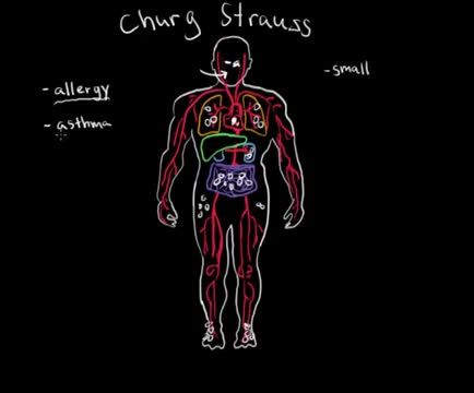
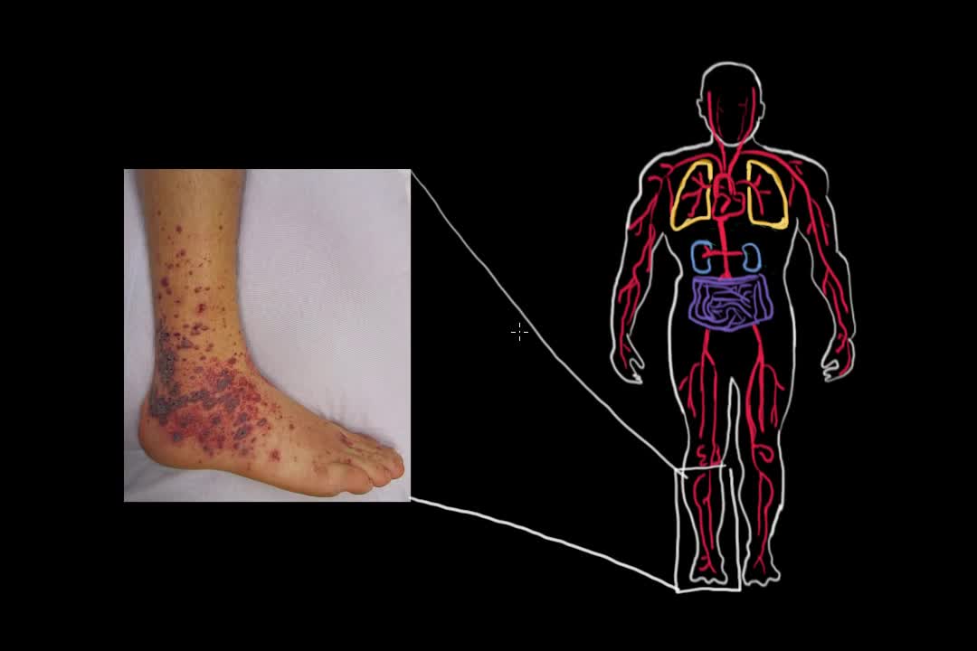
Eosinophilic granulomatosis with polyangiitis (EGPA)—or, as it was traditionally termed, Churg-Strauss syndrome—is a rare systemic necrotizing vasculitis that affects small-to-medium-sized vessels and is associated with severe asthma and blood and tissue eosinophilia. [1] Like granulomatosis with polyangiitis (Wegener granulomatosis), and the microscopic form of periarteritis (ie, microscopic polyangiitis), EGPA is an antineutrophil cytoplasmic antibody (ANCA)–associated vasculitide. [2, 3, 4, 5] In 1951, Churg and Strauss first described the syndrome in 13 patients who had asthma, eosinophilia, granulomatous inflammation, necrotizing systemic vasculitis, and necrotizing glomerulonephritis. [3] In 1990, the American College of Rheumatology (ACR) proposed the following six criteria for the diagnosis of Churg-Strauss syndrome [6] : Asthma (wheezing, expiratory rhonchi) Eosinophilia of more than 10% in peripheral blood Paranasal sinusitis Pulmonary infiltrates (may be transient) Histological proof of vasculitis with extravascular eosinophils Mononeuritis multiplex or polyneuropathy

Early Signs that Cancer is Growing in Your Body

This is an introduction to ventilator settings like FIO2, PEEP, Flow rate,trigger,TV, and RR. I also discuss how these settings relate to CO2 and O2 control and to complications like oxygen toxicity and barotrauma with an emphasis on physiology.
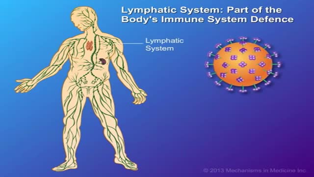
CD4 T-cells (a type of white blood cell) are important to your body's defence against infections. This animation describes how your immune system is weakened by the HIV virus, which targets CD4 T-cells and leads to their gradual decline in number. Low to very low levels of CD4 cells put you at risk for 'opportunistic infections' that take advantage of the body's weakened immune system.
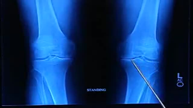
total knee joint replacement surgery
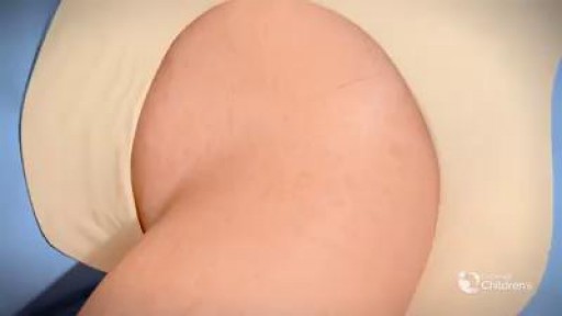
The definition of DDH is not universally agreed upon. Typically, the term DDH is used in referring to patients who are born with dislocation or instability of the hip, which may then result in hip dysplasia. More broadly, DDH may be defined simply as abnormal growth of the hip. Abnormal development of the hip includes the osseous structures, such as the acetabulum and the proximal femur, as well as the labrum, capsule, and other soft tissues. This condition may occur at any time, from conception to skeletal maturity. The author prefers to use the term hip dysplasia, considering it both simpler and more accurate. Internationally, this disorder is still referred to as congenital dislocation of the hip.

Obesity is one of the most pervasive, chronic diseases in need of new strategies for medical treatment and prevention. As a leading cause of United States mortality, morbidity, disability, healthcare utilization and healthcare costs, the high prevalence of obesity continues to strain the United States healthcare system. Obesity is defined as excess adipose tissue. There are several different methods for determining excess adipose (fat) tissue; the most common being the Body Mass Index (BMI) (see below). A fat cell is an endocrine cell and adipose tissue is an endocrine organ. As such, adipose tissue secretes a number of products, including metabolites, cytokines, lipids, and coagulation factors among others. Significantly, excess adiposity or obesity causes increased levels of circulating fatty acids and inflammation. This can lead to insulin resistance, which in turn can lead to type 2 diabetes.

Therapeutic anticoagulation is recommended for all women with acute VTE; prophylactic anticoagulation is recommended for women at risk, such as those with a past history of thrombosis or thrombophilia or with a mechanical heart valve. The preferred anticoagulants during pregnancy are the heparin compounds.
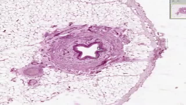
Histology of Ureter

Histology of Secretory Endometrium
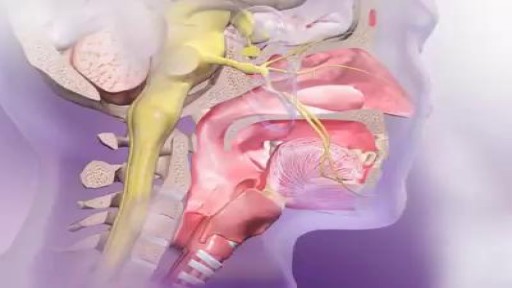
A sneeze, or sternutation, is a semi-autonomous, convulsive expulsion of air from the lungs through the nose and mouth, usually caused by foreign particles irritating the nasal mucosa

Adrenoleukodystrophy, or ALD, is a deadly genetic disease that affects 1 in 18 000 people. It most severely affects boys and men. This brain disorder destroys myelin, the protective sheath that surrounds the brain's neurons -- the nerve cells that allow us to think and to control our muscles.
