- Physical Examination
- Surgical Examination
- Ophthalmology
- Clinical Skills
- Orthopedics
- Surgery Videos
- Laparoscopy
- Pediatrics
- Funny Videos
- Cardiothoracic Surgery
- Nursing Videos
- Plastic Surgery
- Otorhinolaryngology
- Histology and Histopathology
- Neurosurgery
- Dermatology
- Pediatric Surgery
- Urology
- Dentistry
- Oncology and Cancers
- Anatomy Videos
- Health and Fitness
- Radiology
- Anaesthesia
- Physical Therapy
- Pharmacology
- Interventional Radiology
- Cardiology
- Endocrinology
- Gynecology
- Emergency Medicine
- Psychiatry and Psychology
- Childbirth Videos
- General Medical Videos
- Nephrology
- Physiology
- Diet and Food Health
- Diabetes Mellitus
- Neurology
- Women Health
- Osteoporosis
- Gastroenterology
- Pulmonology
- Hematology
- Rheumatology
- Toxicology
- Nuclear Medicine
- Infectious Diseases
- Vascular Disease
- Reproductive Health
- Burns and Wound Healing
- Other
Top videos

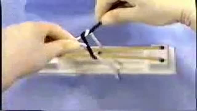
Nose Packing Application & Removal
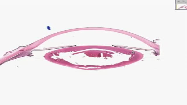
Histology of Eye
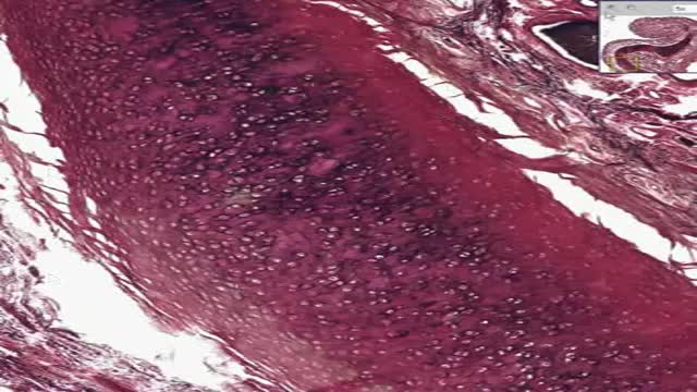
Histology of Elastic Cartilage

A video from the American Academy of Family Physicians
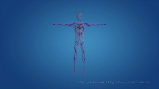
Human Circulatory System and heart video
Lupus is an autoimmune disease that can affect almost any part of your body, most often your joints, skin, kidneys, heart, lungs, blood, or brain. Your two kidneys are part of your renal system, which also includes two ureters, the bladder, and the urethra. As the primary organs of the renal system, your kidneys are responsible for: Maintaining the correct amount and type of body fluids Removing waste products and toxic substances Regulating the hormones (chemical messengers) that help control blood pressure and blood volume
Squared Notch-1
The G-SHOT® (clinical description: G-Spot Amplification™ or GSA™), is a simple, nonsurgical, physician-administered treatment that can temporarily augment the Grafenburg spot (G-Spot) in sexually active women with normal sexual function.
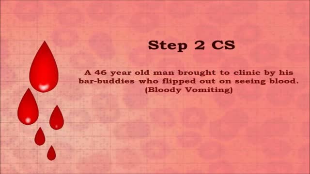
USMLE Step 2 CS - Hemetemesis This is just preview video. To get full access please visit our website : www.usmletutoring.com
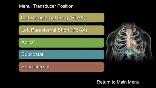
"How to Perform a Transthoracic Echocardiographic Study Volume 1: Transducer Position and Anatomy" is an instructional video, offered by ASE, and can be used for professional lectures and offers an interactive section for flexible presentations. The video includes an overview of relevant cardiac anatomy, a step by step presentation of all Transducer Positions, and the sequential transducer movements to acquire standard echo images needed to complete a Transthoracic Echocardiographic Study.
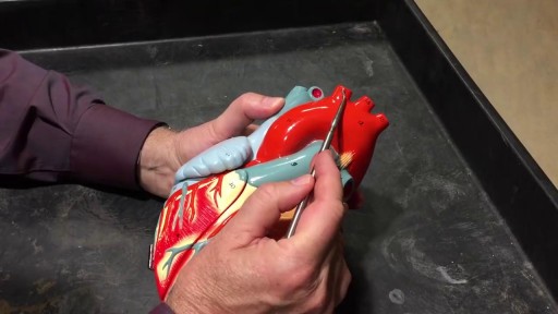
The cardiovascular system is a closed system if the heart and blood vessels. The heart pumps blood through a closed system of blood vessels. Blood vessels allow blood to circulate to all parts of the body. Arteries usually colored red because oxygen rich, carry blood away from the heart to capillaries within the tissues. Veins usually colored blue because oxygen poor, carry blood to the heart from the capillaries.

Histology of Medium Artery and Vein

Taking the guesswork out of insulin management with new advancements.
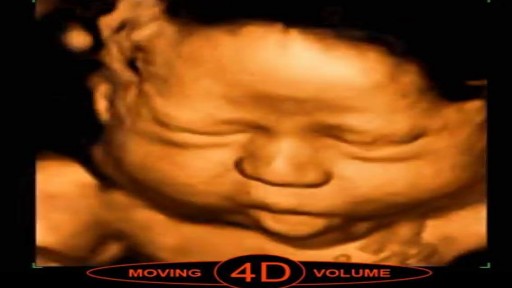
3D scans show still pictures of your baby in three dimensions. 4D scans show moving 3D images of your baby, with time being the fourth dimension. It's natural to be really excited by the prospect of your first scan. But some mums find the standard 2D scans disappointing when all they see is a grey, blurry outline.
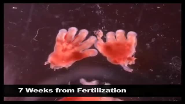
Abortion real ghraphics
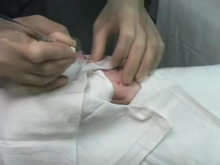
Arterial line placement is a common procedure in various critical care settings. Intra-arterial blood pressure (BP) measurement is more accurate than measurement of BP by noninvasive means, especially in the critically ill. [1] Intra-arterial BP management permits the rapid recognition of BP changes that is vital for patients on continuous infusions of vasoactive drugs. Arterial cannulation also allows repeated arterial blood gas samples to be drawn without injury to the patient.
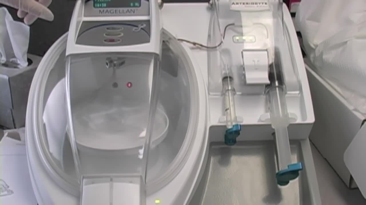
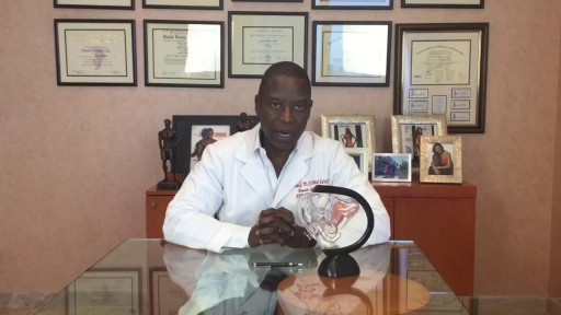
Stem Cell Injection Treatment - Stem Cell Therapy
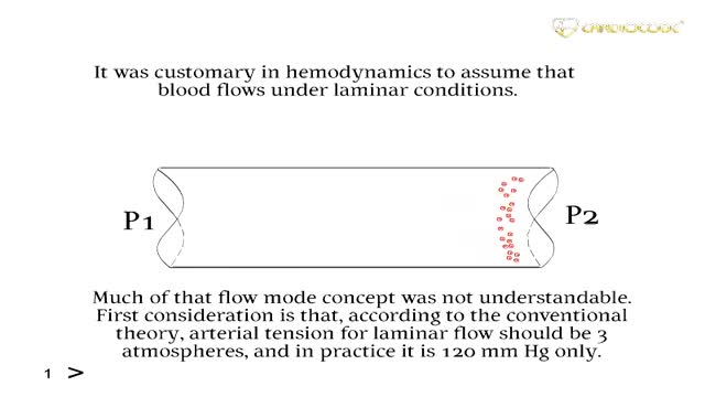
New methods in heart diseases diagnostics and imaging
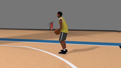
ACL tears are treatable using arthroscopy and minimally-invasive surgical techniques. The surgical success rates for ACL reconstruction exceed 95%. The anterior cruciate ligament is one of the major supportive ligaments in the knee
