- Physical Examination
- Surgical Examination
- Ophthalmology
- Clinical Skills
- Orthopedics
- Surgery Videos
- Laparoscopy
- Pediatrics
- Funny Videos
- Cardiothoracic Surgery
- Nursing Videos
- Plastic Surgery
- Otorhinolaryngology
- Histology and Histopathology
- Neurosurgery
- Dermatology
- Pediatric Surgery
- Urology
- Dentistry
- Oncology and Cancers
- Anatomy Videos
- Health and Fitness
- Radiology
- Anaesthesia
- Physical Therapy
- Pharmacology
- Interventional Radiology
- Cardiology
- Endocrinology
- Gynecology
- Emergency Medicine
- Psychiatry and Psychology
- Childbirth Videos
- General Medical Videos
- Nephrology
- Physiology
- Diet and Food Health
- Diabetes Mellitus
- Neurology
- Women Health
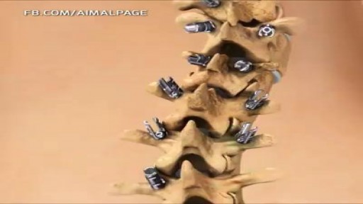
- Osteoporosis
- Gastroenterology
- Pulmonology
- Hematology
- Rheumatology
- Toxicology
- Nuclear Medicine
- Infectious Diseases
- Vascular Disease
- Reproductive Health
- Burns and Wound Healing
- Other
Top videos
What could cause a blockage in the stomach? Mechanical causes of intestinal obstruction may include: Adhesions or scar tissue that forms after surgery. Foreign bodies (objects that are swallowed and block the intestines) Gallstones (rare) Hernias. Impacted stool. Intussusception (telescoping of one segment of bowel into another) Tumors blocking the intestines. Less common radiologic signs are seen in specific circumstances. Most closed-loop obstructions (75%) are caused by adhesions. A closed-loop obstruction occurs when a loop of bowel is not decompressed by the caudal passage of gas and fluid.
If it is not removed, tooth decay will begin. The acids in plaque damage the enamel covering your teeth. It also creates holes in the tooth called cavities. Cavities usually do not hurt, unless they grow very large and affect nerves or cause a tooth fracture.
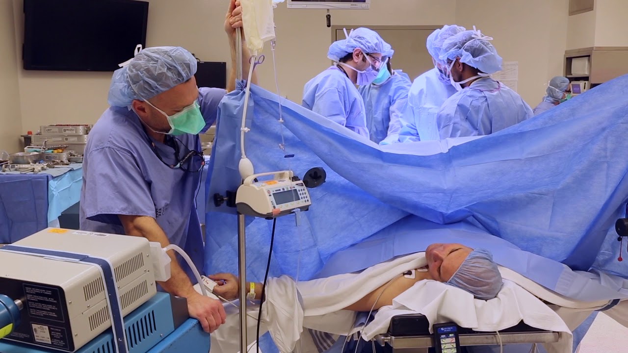
Women's College Hospital is revolutionizing the way knee-replacement surgery is done. It is starting to provide the procedure as an ambulatory service. Patients can go home from hospital four hours after having the surgery. In some other hospitals knee replacement surgery patients have to stay as long as 4 days.
Read an excerpt from Theresa Boyle's story:
It’s been less than four hours since Greg Nemez underwent knee-replacement surgery and the 56-year-old Mississauga man is already on his way home from hospital.
This past Monday, he became the fifth patient at Toronto’s Women’s College Hospital to undergo the outpatient procedure, which normally requires a hospital stay of two or three days.
“I’m happy ... You have that freedom of movement from before. It’s like wow,” he said on the elevator as he was leaving the hospital.
After years of being unable to hold his leg straight, the real-estate agent can finally do so. A 20-year-old football injury had left him with severe arthritis and pain.
Read the full story:
https://www.thestar.com/news/g....ta/2018/04/11/he-got
Follow the Toronto Star on social media:
Facebook: https://www.facebook.com/torontostar/
Twitter: https://twitter.com/TorontoStar
Instagram: https://www.instagram.com/thetorontostar/
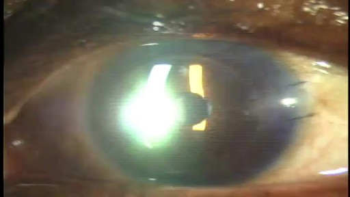
Stephen Slade shows a 1 week post op patient after DSAEK. DSAEK is an excellent option for many patients with corneal disease. In DSAEK, only the thin, inner layer is replaced, so the healing is typically much faster than a full thickness cornea graft.
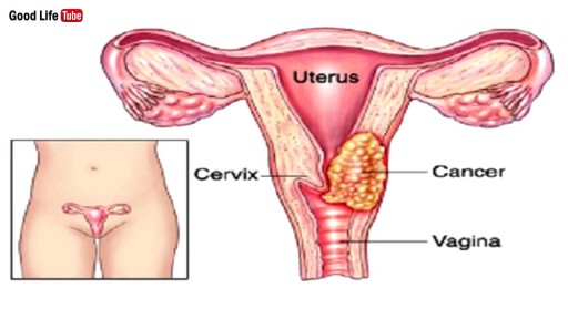
Watch that video to know the Signs of Genital Cervical Cancer You Should Know
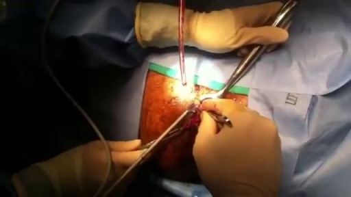
Watch that Hemorrhoids Surgical Repairing Medical Operation
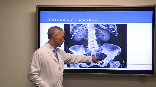
Treatment for kidney stones varies, depending on the type of stone and the cause. Small stones with minimal symptoms Most kidney stones won't require invasive treatment. You may be able to pass a small stone by: Drinking water. Drinking as much as 2 to 3 quarts (1.9 to 2.8 liters) a day may help flush out your urinary system. Unless your doctor tells you otherwise, drink enough fluid — mostly water — to produce clear or nearly clear urine. Pain relievers. Passing a small stone can cause some discomfort. To relieve mild pain, your doctor may recommend pain relievers such as ibuprofen (Advil, Motrin IB, others), acetaminophen (Tylenol, others) or naproxen sodium (Aleve). Medical therapy. Your doctor may give you a medication to help pass your kidney stone. This type of medication, known as an alpha blocker, relaxes the muscles in your ureter, helping you pass the kidney stone more quickly and with less pain. Large stones and those that cause symptoms Kidney stones that can't be treated with conservative measures — either because they're too large to pass on their own or because they cause bleeding, kidney damage or ongoing urinary tract infections — may require more extensive treatment. Procedures may include: Using sound waves to break up stones. For certain kidney stones — depending on size and location — your doctor may recommend a procedure called extracorporeal shock wave lithotripsy (ESWL). ESWL uses sound waves to create strong vibrations (shock waves) that break the stones into tiny pieces that can be passed in your urine. The procedure lasts about 45 to 60 minutes and can cause moderate pain, so you may be under sedation or light anesthesia to make you comfortable. ESWL can cause blood in the urine, bruising on the back or abdomen, bleeding around the kidney and other adjacent organs, and discomfort as the stone fragments pass through the urinary tract. Surgery to remove very large stones in the kidney. A procedure called percutaneous nephrolithotomy (nef-row-lih-THOT-uh-me) involves surgically removing a kidney stone using small telescopes and instruments inserted through a small incision in your back. You will receive general anesthesia during the surgery and be in the hospital for one to two days while you recover. Your doctor may recommend this surgery if ESWL was unsuccessful. Using a scope to remove stones. To remove a smaller stone in your ureter or kidney, your doctor may pass a thin lighted tube (ureteroscope) equipped with a camera through your urethra and bladder to your ureter. Once the stone is located, special tools can snare the stone or break it into pieces that will pass in your urine. Your doctor may then place a small tube (stent) in the ureter to relieve swelling and promote healing. You may need general or local anesthesia during this procedure. Parathyroid gland surgery. Some calcium phosphate stones are caused by overactive parathyroid glands, which are located on the four corners of your thyroid gland, just below your Adam's apple. When these glands produce too much parathyroid hormone (hyperparathyroidism), your calcium levels can become too high and kidney stones may form as a result. Hyperparathyroidism sometimes occurs when a small, benign tumor forms in one of your parathyroid glands or you develop another condition that leads these glands to produce more parathyroid hormone. Removing the growth from the gland stops the formation of kidney stones. Or your doctor may recommend treatment of the condition that's causing your parathyroid gland to overproduce the hormone.
A pneumothorax is usually caused by an injury to the chest, such as a broken rib or puncture wound. It may also occur suddenly without an injury. A pneumothorax can result from damage to the lungs caused by conditions such as chronic obstructive pulmonary disease (COPD), asthma, cystic fibrosis, and pneumonia.
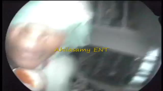
Results Sinusitis was characterized as acute in 26 patients, subacute in 5 (including 1 pyocele), and chronic in 8 (including 2 fungal infections). No tumors were found. Isolated sinus cysts were excluded from the study. Headache, the main symptom in 32 patients (82%), was localized most commonly on the vertex. Other common complaints were rhinitis, dizziness, eye symptoms, and fever. In 2 patients, the finding was occult. Eight patients (21%) presented with cranial nerve deficits, and 1 patient had an intracranial complication. Sinus irrigation was performed in 16 patients (41%) and sphenoidotomy was performed in 10 (26%). Fifteen patients (38%) were treated with antibiotic drugs alone. Within 3 months, 31 (84%) of 37 patients had recovered from the illness; 5 still experienced headaches despite having normalized radiographic findings; and 1 had permanent unilateral visual loss. Two patients were lost to follow-up.
Rheumatic heart disease (RHD) is the most common acquired heart disease in children in many countries of the world, especially in developing countries. The global burden of disease caused by rheumatic fever currently falls disproportionately on children living in the developing world, especially where poverty is widespread. RHD is a chronic heart condition caused by rheumatic fever that can be prevented and controlled. Rheumatic fever is caused by a preceding group A streptococcal (strep) infection. Treating strep throat with antibiotics can prevent rheumatic fever. Moreover, regular antibiotics (usually monthly injections) can prevent patients with rheumatic fever from contracting further strep infections and causing progression of valve damage. Consequences of rheumatic heart disease Acute rheumatic fever primarily affects the heart, joints and central nervous system. The major importance of acute rheumatic fever is its ability to cause fibrosis of heart valves, leading to crippling valvular heart disease, heart failure and death. The decline of rheumatic fever in developed countries is believed to be the result of improved living conditions and availability of antibiotics for treatment of group A streptococcal infection. Overcrowding, poor housing conditions, undernutrition and lack of access to healthcare play a role in the persistence of this disease in developing countries.
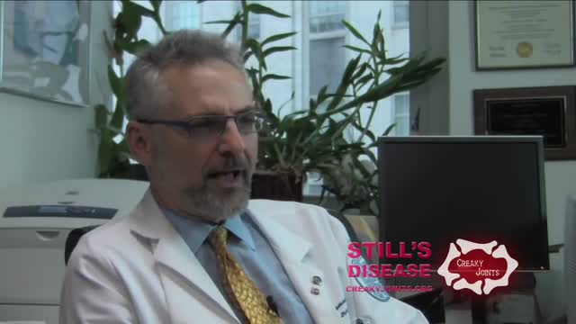
Adult-onset Still's disease (AOSD) is a rare systemic inflammatory disease characterized by the classic triad of persistent high spiking fevers, joint pain, and a distinctive salmon-colored bumpy rash. The disease is considered a diagnosis of exclusion.
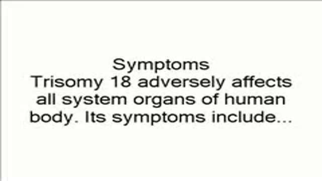
Trisomy 18, also called Edwards syndrome, is a chromosomal condition associated with abnormalities in many parts of the body. Individuals with trisomy 18 often have slow growth before birth (intrauterine growth retardation) and a low birth weight. Affected individuals may have heart defects and abnormalities of other organs that develop before birth. Other features of trisomy 18 include a small, abnormally shaped head; a small jaw and mouth; and clenched fists with overlapping fingers. Due to the presence of several life-threatening medical problems, many individuals with trisomy 18 die before birth or within their first month. Five to 10 percent of children with this condition live past their first year, and these children often have severe intellectual disability.
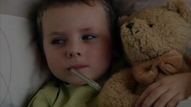
Diarrhea in Children: Common Causes and Treatments Diarrhea is the body's way of ridding itself of germs, and most episodes last a few days to a week. Diarrhea often occurs with fever, nausea, vomiting, cramps, and dehydration. Some of the most common reasons kids get diarrhea include: Infection from viruses like rotavirus, bacteria like salmonella and, rarely, parasites like giardia. Viruses are the most common cause of a child's diarrhea. Along with loose or watery stools, symptoms of a viral gastroenteritis infection often include vomiting, stomachache, headache, and fever. When treating viral gastroenteritis -- which can last 5-14 days -- it's important to prevent fluid loss. Offer additional breast milk or an oral rehydration solution (ORS) to infants and young children. Water alone doesn't have enough sodium, potassium, and other nutrients to safely rehydrate very young children. Be sure to talk to your doctor about the amount of fluids your child needs, how to make sure he or she gets them, when to give them, and how to watch for dehydration. Older children with diarrhea can drink anything they like to stay hydrated, including ORS and brand-name products (their names usually end in "lyte"). Popsicles can also be a good way to get fluids into a child who's been vomiting and needs to rehydrate slowly.
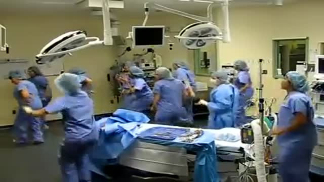
Zumba in Operation room
Scoliosis is a sideways curvature of the spine that occurs most often during the growth spurt just before puberty. While scoliosis can be caused by conditions such as cerebral palsy and muscular dystrophy, the cause of most scoliosis is unknown. Most cases of scoliosis are mild, but some children develop spine deformities that continue to get more severe as they grow. Severe scoliosis can be disabling. An especially severe spinal curve can reduce the amount of space within the chest, making it difficult for the lungs to function properly. Children who have mild scoliosis are monitored closely, usually with X-rays, to see if the curve is getting worse. In many cases, no treatment is necessary. Some children will need to wear a brace to stop the curve from worsening. Others may need surgery to keep the scoliosis from worsening and to straighten severe cases of scoliosis.

Twins in the Womb - Human Development

Major signs and symptoms include enlargement of the liver and spleen (hepatosplenomegaly), a low number of red blood cells (anemia), easy bruising caused by a decrease in blood platelets (thrombocytopenia), lung disease, and bone abnormalities such as bone pain, fractures, and arthritis.
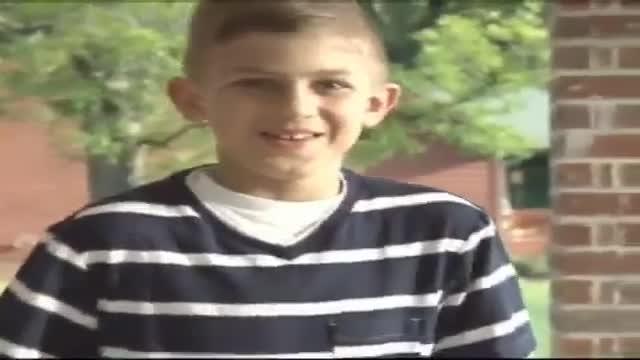
Adrenoleukodystrophy, or ALD, is a deadly genetic disease that affects 1 in 18 000 people. It most severely affects boys and men. This brain disorder destroys myelin, the protective sheath that surrounds the brain's neurons -- the nerve cells that allow us to think and to control our muscles.
