- Physical Examination
- Surgical Examination
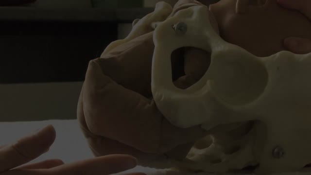
- Ophthalmology
- Clinical Skills
- Orthopedics
- Surgery Videos
- Laparoscopy
- Pediatrics
- Funny Videos
- Cardiothoracic Surgery
- Nursing Videos
- Plastic Surgery
- Otorhinolaryngology
- Histology and Histopathology
- Neurosurgery
- Dermatology
- Pediatric Surgery
- Urology
- Dentistry
- Oncology and Cancers
- Anatomy Videos
- Health and Fitness
- Radiology
- Anaesthesia
- Physical Therapy
- Pharmacology
- Interventional Radiology
- Cardiology
- Endocrinology
- Gynecology
- Emergency Medicine
- Psychiatry and Psychology
- Childbirth Videos
- General Medical Videos
- Nephrology
- Physiology
- Diet and Food Health
- Diabetes Mellitus
- Neurology
- Women Health
- Osteoporosis
- Gastroenterology
- Pulmonology
- Hematology
- Rheumatology
- Toxicology
- Nuclear Medicine
- Infectious Diseases
- Vascular Disease
- Reproductive Health
- Burns and Wound Healing
- Other
Top videos
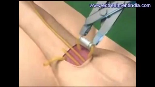
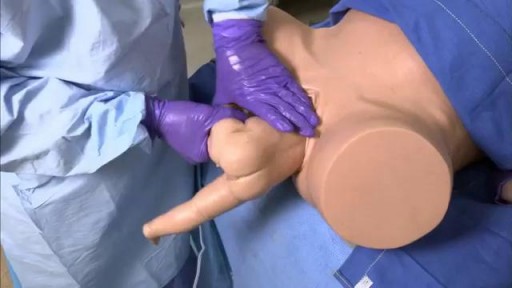
A penile prosthesis is another treatment option for men with erectile dysfunction. These devices are either malleable (bendable) or inflatable. The simplest type of prosthesis consists of a pair of malleable rods surgically implanted within the erection chambers of the penis. With this type of implant the penis is always semi-rigid and merely needs to be lifted or adjusted into the erect position to initiate sex. This type of implant is a good choice for men with spinal cord injuries and/or limited hand strength. Today, many men choose a hydraulic, inflatable prosthesis, which allows them to have an erection when they choose, and it's easier to conceal. It is also more natural. A penile implant is usually used when there is a clear medical cause for ED and when the problem is unlikely to resolve or improve naturally or with other medical treatments. Sometimes a penile prosthesis is implanted during surgery to reconstruct the penis when scarring has caused erections to curve (Peyronie's disease). Penile implant surgeries take about an hour and are typically done in an outpatient center. A man can resume sexual intercourse by 6 weeks after surgery.
A breech birth occurs when a baby is born bottom first instead of head first. Around 3-5% of pregnant women at term (37–40 weeks pregnant) will have a breech baby. Most babies in the breech position are born by a caesarean section because it is seen as safer than being born vaginally.

This minimally invasive technique allows surgeons to remove skull base tumors as large as softballs through the nose, with less trauma to the brain and critical nerves than with a traditional craniotomy.
To learn more, please visit https://www.upmc.com/
Pulmonary edema is usually caused by a heart condition. Other causes include pneumonia, exposure to certain toxins and drugs, and being at high elevations. Depending on the cause, pulmonary edema symptoms may appear suddenly or develop over time. Mild to extreme breathing difficulty can occur. Cough, chest pain, and fatigue are other symptoms. Treatment generally includes supplemental oxygen and medications.
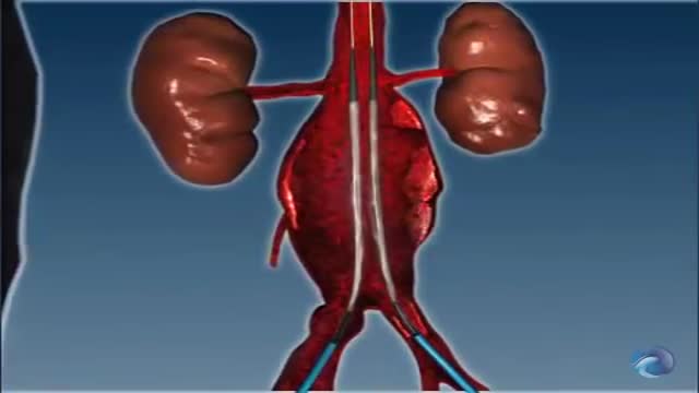
Endovascular Aneurysm Repair Endovascular aneurysm repair (or endovascular aortic repair) (EVAR) is a type of endovascular surgery used to treat pathology of the aorta, most commonly an abdominal aortic aneurysm (AAA).
The majority of fetuses are in a breech presentation early in pregnancy. By week 38th week of gestation, however, the fetus normally turns to a cephalic presentation. Although the fetal head is the widest single diameter, the fetus’s buttocks [ breech], plus the lower extremities, actually takes up more space. The fundus, being the largest part of the uterus, probably accounts for the fact that in approximately 97% of all pregnancies, the fetus turns so that the buttocks and lower extremities are in the fundus. Vaginal delivery of a breech presentation requires great skill if the fetus is not to be damaged. With the low rate of vaginal breech deliveries in the developed world, experience is being lost. 6% of women with breech presentation still have a vaginal breech delivery as they present too late - so units need to retain a high level of preparedness. Types of breech presentation: I. Complete breech [ flexed breech]: The fetal attitude is one of complete flexion, with hips and knees both flexed and the feet tucked in beside the buttocks. The presenting part consists of two buttocks, external genitalia and two feet. It is commonly present in multiparae. II. Incomplete breech: This is due to varying degrees of extension of thighs or legs at podalic pole. Three varieties are possible; - Breech with extended legs [ frank breech ]: The breech presents with the hips flexed and legs extended on the abdomen. 70% of breech presentations are of this type and it is particularly common in primigravidae whose good uterine muscle tone inhibits flexion of the legs and free turning of the fetus. - Footling breech: This is rare. One or both feet present because neither hips nor knees are fully flexed. The feet are lower than the buttocks, which distinguishes it from the complete breech. - Knee presentation: This is very rare. Thighs are extended but the knees are flexed, bringing the knees down to present at the brim.
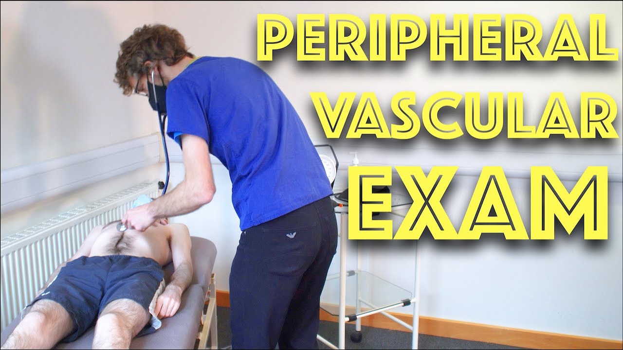
Peripheral Vascular Examination OSCE - Clinical Skills - Dr Gill
In the cardiovascular examination, particularly in the case of an OSCE station, we conclude the examination often by stating that the examiner would want to perform:
- An ECG
- Check full blood count
- and "do a peripheral vascular examination
In this video, we demonstrate that oft-talked about, but comparatively less common examination.
Starting off, with the examination of the hands, the radial, brachial and carotid pulses. before moving down to assess for a AAA, checking the femoral and popliteal pulses, before wrapping up around the ankle with the posterior tibial and dorsalis pedis pulses
For completeness, the cardiovascular examination is demonstrated here
https://www.youtube.com/watch?v=ECs9O5zl6XQ&t=2s
#PeripheralVascular #ClinicalSkills #DrGill

This video demonstrate Laparoscopic Cholecystectomy Full Length Skin to Skin Video with Infrared Cholangiography performed by Dr R K Mishra at World Laparoscopy Hospital. Infrared Cholegiography is performed by using Indocyanine Green during laparoscopic cholecystectomy surgery for gallbladder removal. Bile duct injury remains the most feared complication of laparoscopic cholecystectomy. Intraoperative cholangiography (IOC) is the current gold standard for biliary imaging and may reduce injury, but is not widely used because of the difficulties of doing it. Near-Infrared Fluorescence Cholangiography (NIRF-C) is a novel non-invasive method for real-time, radiation-free, intra-operative biliary mapping during laparoscopic cholecystectomy. We have experienced that NIRF-C is a safe and effective method for identifying biliary anatomy during laparoscopic cholecystectomy. Indocyanine green is a cyanine dye is very popular and used for many years in medical diagnostics. It is used for determining cardiac output, hepatic function, liver, and gastric blood flow, and for ophthalmic angiography. Now the use of this dye in lap chole has improved the safety of this surgery by NEAR INFRARED FLUORESCENT CHOLANGIOGRAPHY.
For more information please contact:
World Laparoscopy Hospital
Cyber City, Gurugram, NCR DELHI
INDIA 122002
Phone & WhatsApp: +919811416838, + 91 9999677788
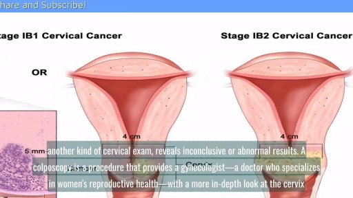
A cervical biopsy is a procedure that is sometimes done on women during an exam called a colposcopy to remove cervical tissue for examination. It is also called a punch biopsy. It is usually performed when a Pap smear result is either inconclusive or abnormal and a doctor wants to screen further for any cervical dysplasia or cervical cancer.
Suprapubic Catheterization / Cystostomy

While in residency, Marc Pelletier, MD, helped in a bypass surgery and knew it was the field in which he would excel. Watch as the Chief of Cardiac Surgery for University Hospitals Harrington Heart & Vascular Institute in Cleveland, Ohio explains, in detail, what happens in preparation for heart surgery, in the operating room and the feeling he experiences after surgery.
How does a heart-lung machine work? What is 'efficiency of motion'? These questions and more are answered in this compelling, dramatic look at heart surgery.
To learn more about heart surgery at University Hospitals: https://www.uhhospitals.org/fo....r-clinicians/special
University Hospitals is one of the nation’s leading health care systems, providing patient-centered care that meets the highest standards for quality and patient safety and have received numerous awards and recognitions from some of the most prestigious institutions in the country for our leadership and exceptional patient outcomes. As an accountable care organization, we foster long-term patient-provider relationships that help promote preventive care, increase wellness and healthy behaviors, decrease emergency episodes, and prevent hospitalizations. To learn more: https://www.uhhospitals.org
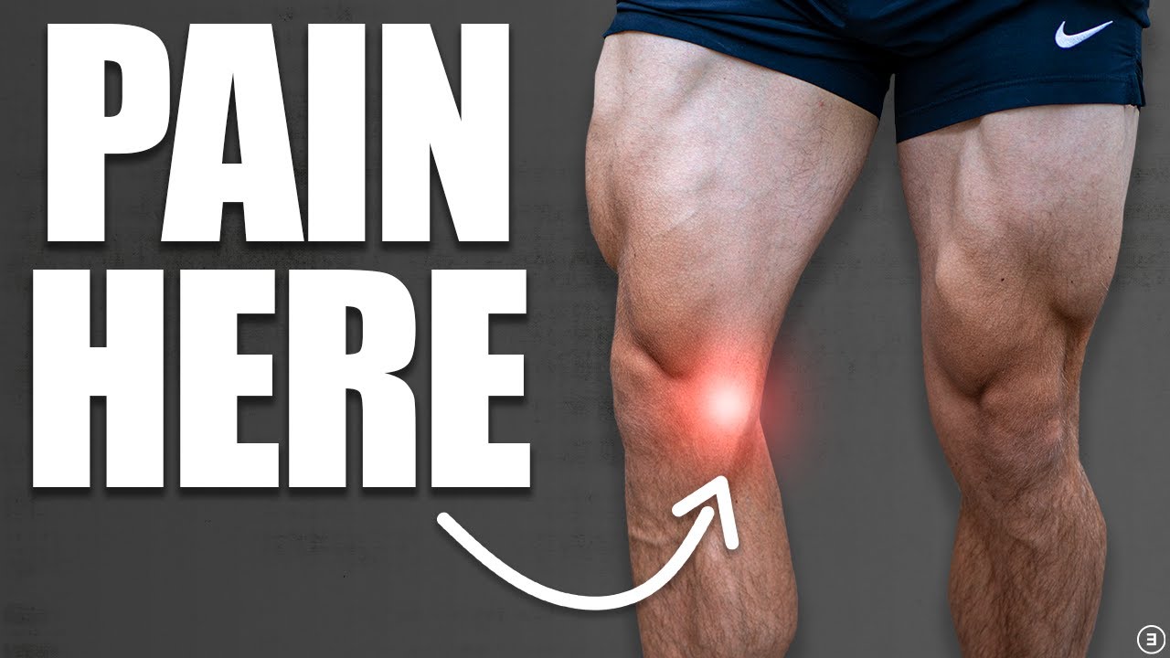
💪 Get our Knee Resilience program here: https://e3rehab.com/programs/r....esilience/knee-resil
In this video, I will walk you through a comprehensive rehab program for the most commonly injured knee ligament - the MCL.
💪 PROGRAMS: https://e3rehab.com/programs/
📩 MAILING LIST (exclusive deals, offers, and information): https://e3rehab.com/newsletter/
🏆 COACHING: https://e3rehab.com/coaching/
📝 ARTICLES: https://e3rehab.com/articles/
👕 APPAREL: https://e3rehab.com/clothing/
🎧 PODCAST: https://open.spotify.com/show/....5ZbaI145Bk94Guq7olMJ
AFFILIATES:
👟 Vivo Barefoot: Get 15% off all shoes! - https://www.vivobarefoot.com/e3rehab
📓 MASS (Monthly Research Review): http://bit.ly/E3MASS
📚 CSMi: https://humacnorm.com/e3rehab
🏋️ GYM EQUIPMENT: https://e3rehab.com/affiliates/
Follow Us:
YOUTUBE: https://www.youtube.com/@e3reh....ab?sub_confirmation=
INSTAGRAM: https://www.instagram.com/e3rehab
TWITTER: https://twitter.com/E3Rehab
FACEBOOK: https://www.facebook.com/e3rehab
TIKTOK: https://www.tiktok.com/@e3rehab
Intro (0:00)
Anatomy & Function (0:08)
Classification (1:11)
Treatment Options (1:46)
Bracing (3:30)
Rehab Overview (4:28)
Early Stage (5:27)
Mid-Stage(8:50)
Late Stage/Return to Sport (21:14)
Programming (22:13)
Summary (23:47)
---
Disclaimer: The information presented is not intended as medical advice or to be a substitute for medical counseling but intended for entertainment purposes only. If you are experiencing pain, please seek the appropriate healthcare professional.

Diastasis recti often occurs during pregnancy and can persist after pregnancy. It affects core strength and the appearance of the abdominal muscles.
Dr. Erick Sanchez repairs the abdominal muscles with every tummy tuck. This short video shows the muscle repair portion of the surgery with a bonus after photo at the end!
To request a consultation with Dr. Sanchez, visit sanchezplasticsurgery.com and click Request a Consultation. Fill out the form and someone will get in touch with you to answer all your questions.
Expected cost can be found at the bottom of each procedure page on our website.
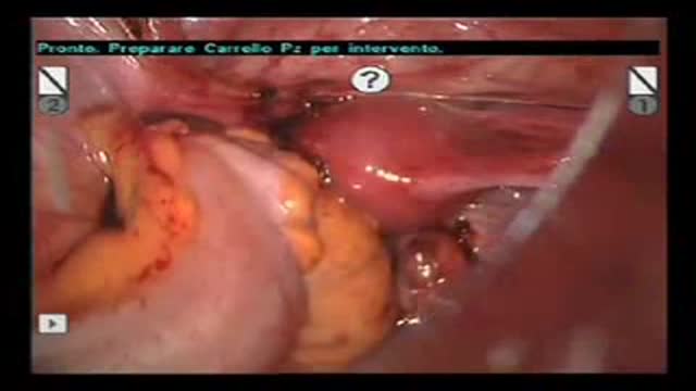
Robot-Assisted Laparoscopic Rectal resection for Endometriosis.Operation performed by D.Vitobello, director of divisione of Gynaecology, and G.Baldazzi,director of Surgical department. Abano Terme Hospital Padova (Italy)
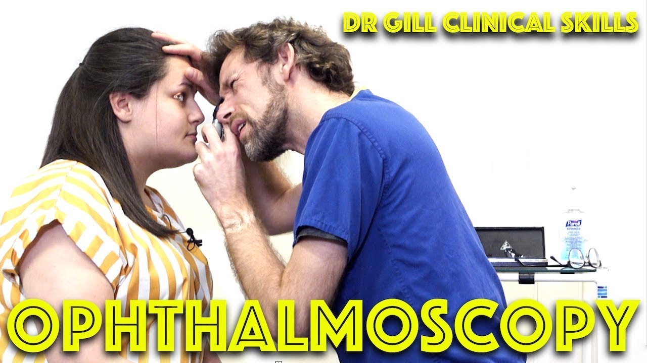
Ophthalmoscopy - Eye Clinical Examination - OSCE - Dr Gill
Direct Ophthalmoscopy use of the eyes is a very challenging clinical skill, incorporating both the examiner's knowledge of the retina, but also understanding the use of the ophthalmoscope
In this clinical skills tutorial, we look at the use of the direct ophthalmoscope as part of an ophthalmic examination
it should be noted that in the ideal circumstances, the room lights will be dimmed during the examination, and dilating eye drops used to improve the visualisation of the fundus
Some people may notice an ASMR effect from this clinical examination
#DrGill #Ophthalmoscopy #ClinicalSkills #EyeExam

Labia minoraplasty is an elective procedure that can reduce the size and reshape the inner vaginal lips. Large or asymmetrical labia minora can leave you feeling self-conscience in tight clothing or during intimacy. Long labia may result in rubbing, irritation or discomfort during intercourse and exercise. Certain skin conditions can cause increased sensitivity or tearing of the labia minora. In some cases, the labia minora may be fused with tissue in the labia majora and require medical correction.

An abscess is a collection of pus. Pus is a thick fluid that usually contains white blood cells, dead tissue and germs (bacteria). The usual cause of an abscess is an infection with bacteria. Certain bacteria are more likely to be 'pus-forming' as they make chemicals (toxins) that can damage the body's tissues.

Colorectal cancer (also known as colon cancer, rectal cancer or bowel cancer) is the development of cancer in the colon or rectum (parts of the large intestine). It is due to the abnormal growth of cells that have the ability to invade or spread to other parts of the body. People with HNPCC tend to develop colon cancer before age 50. Familial adenomatous polyposis (FAP). FAP is a rare disorder that causes you to develop thousands of polyps in the lining of your colon and rectum. People with untreated FAP have a greatly increased risk of developing colon cancer before age 40.
You may have heard that some positions, such as your partner on top (missionary position), are better than others for getting pregnant. In fact, there's no evidence to back these theories up. Experts just haven't done the research yet. What experts have done, though, is use scanning to show what's going on inside when you're doing the deed. The research looked at two positions: the missionary position and doggy style. (Doggy style being when you're on all fours, and your partner enters you from behind). Common sense tells us that these positions allow for deep penetration. This means that they're more likely to place sperm right next to your cervix (the opening of your uterus). The scans confirm that the tip of the penis reaches the areas between the cervix and vaginal walls in both of these positions. The missionary position allows the penis to reach the area at the front of the cervix. The rear entry position reaches the area at back of the cervix. It's amazing what some experts spend their time doing, isn't it! Other positions, such as standing up, or woman on top, may be just as good for getting sperm right next to the cervix. We just don't know yet. So, in the meantime, enjoy some variety in your sex life and keep it fun while you're trying for a baby. And talk to others who are hoping to get pregnant by joining our Actively trying group. Do I have to have an orgasm to conceive? Obviously, it's very important for your partner to reach orgasm if you are trying for a baby. There is no evidence, however, that you need to orgasm to conceive. The female orgasm is all about pleasure and satisfaction. It doesn't really help to get the sperm to the egg. Gentle contractions in your uterus can help the sperm along, but these happen without you having an orgasm. So, it's really not vital for you to reach orgasm after your partner, or even to reach orgasm at all, for you to conceive.
Ankylosing spondylitis is an inflammatory disease that, over time, can cause some of the vertebrae in your spine to fuse. This fusing makes the spine less flexible and can result in a hunched-forward posture. If ribs are affected, it can be difficult to breathe deeply. Ankylosing spondylitis affects men more often than women. Signs and symptoms typically begin in early adulthood. Inflammation also can occur in other parts of your body — most commonly, your eyes. There is no cure for ankylosing spondylitis, but treatments can lessen your symptoms and possibly slow progression of the disease.
