- Physical Examination
- Surgical Examination
- Ophthalmology
- Clinical Skills
- Orthopedics
- Surgery Videos
- Laparoscopy
- Pediatrics
- Funny Videos
- Cardiothoracic Surgery
- Nursing Videos
- Plastic Surgery
- Otorhinolaryngology
- Histology and Histopathology
- Neurosurgery
- Dermatology
- Pediatric Surgery
- Urology
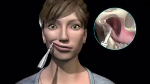
- Dentistry
- Oncology and Cancers
- Anatomy Videos
- Health and Fitness
- Radiology
- Anaesthesia
- Physical Therapy
- Pharmacology
- Interventional Radiology
- Cardiology
- Endocrinology
- Gynecology
- Emergency Medicine
- Psychiatry and Psychology
- Childbirth Videos
- General Medical Videos
- Nephrology
- Physiology
- Diet and Food Health
- Diabetes Mellitus
- Neurology
- Women Health
- Osteoporosis
- Gastroenterology
- Pulmonology
- Hematology
- Rheumatology
- Toxicology
- Nuclear Medicine
- Infectious Diseases
- Vascular Disease
- Reproductive Health
- Burns and Wound Healing
- Other
Top videos
A hematoma is a collection of blood outside of a blood vessel Some causes of hematomas are as pelvic bone fractures, fingernail injuries (subungual), bumps, passing blood clots, blood clot in the leg (DVT), blood cancers, and excessive alcohol use.

ROTIGS medical device by Honolulu inventor Dr. Brad NaPier makes airway intubations easier for medical professionals.
Watch that Cutting Inside Human Fat Body video
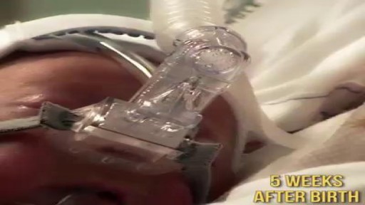
Meet Toby, the baby who was born premature at 24 weeks. He may be small, but he's definitely a fighter! Share his story.
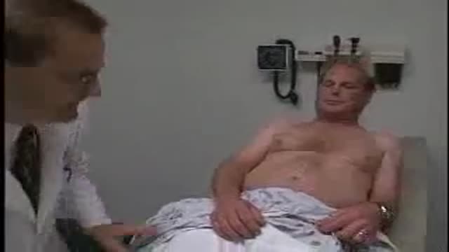
Loyola Full Male Exam Part 3 A video from Loyola medical school, Chicago showing the full examination of the male
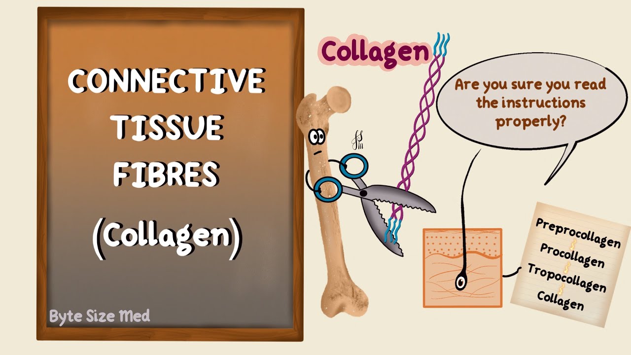
✨This video is on the protein fibres of connective tissue, the types, structure and synthesis of collagen and elastin. I hope it helps! ☀️
🌟What's in this video?
0:00 - Intro
0:07 - Connective Tissue Recap
0:39 - Connective Tissue Fibres
1:22 - Collagen
1:46 - Types of Collagen
3:40 - Structure of Collagen
4:40 - Collagen Synthesis
8:50 - Elastin
✨ Other videos you may need:
🔅 Connective Tissue : https://youtu.be/xw_ALdt5n-A
🔅 Cartilage : https://youtu.be/4inWF4H6pKE
🔅Epithelial Tissue: https://youtu.be/Gw5fC0zXaeU
🔅Structure of Blood Vessels: https://youtu.be/BAo2UqqyL3g
🔅Histology: https://www.youtube.com/playli....st?list=PL1rG930trF2
💫 For more videos like this, subscribe to my channel!
Byte Size Med: https://youtube.com/channel/UC....ZghvlgylH3r_CWfA18eF
📚Factual References & for Further Reading:
- DiFiore's Atlas of Histology
- Junqueira's Basic Histology
- Harper's Biochemistry
- Gartner's Concise Histology
- Openstax Anatomy and Physiology
https://openstax.org/details/b....ooks/anatomy-and-phy
- Openstax Biology
https://openstax.org/details/books/biology-2e
(The last two are links to open-source references. They are NOT affiliate links)
🌤 Note:
These are just a collection of my notes. So use them the way you would use borrowed notes from a friend. 📝
The images in this video are hand-drawn for illustration and explanation only.✍️ Hence, they may not be anatomically accurate. I am just one person making these videos. If there are any errors, that is unintentional. I try super hard to avoid them. Please let me know if you find any, so it gets clarified for other viewers. Science constantly evolves and changes. New discoveries are made everyday. So some of the information in these videos may become outdated. If you notice that, please let me know so I can update them.
⚡️Disclaimer:
These videos are NOT a substitute for a medical textbook. Textbooks are written by experts (which I do not claim to be), edited, proofread and referenced. Please use them.
The information has been sourced from multiple references as mentioned above. I draw all the pictures myself. But if I have inadvertently infringed on any copyright, that is completely unintentional. I only make these videos to impart education. If I have accidentally violated copyright in any way, do let me know so I can make the necessary changes or give credit to anyone who is owed the same.
These videos are NOT intended for patient education. They are NOT a substitute for diagnosis and treatment by a licensed medical professional. Always seek the advice of a qualified health care provider for any questions you may have regarding any medical condition, so that they can address your individual needs.
🔅They are ONLY meant to help students of medicine and health sciences with studying, and should be used for just that purpose and absolutely nothing else.
Byte Size Med. All Rights Reserved.

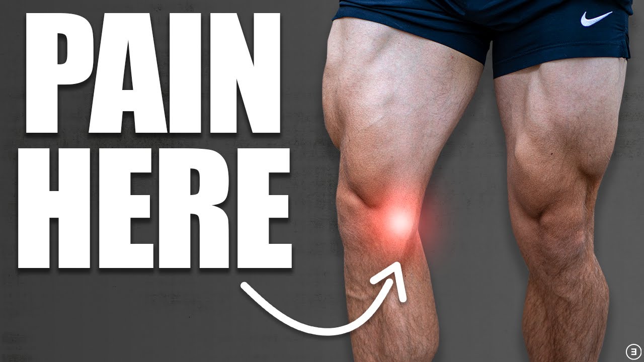
It depends upon which ligament is injured. If it is medial collateral ligament you feel pain when you walk ,sit and stand and you will be liming as well. If it is anterior cruciate ligament you feel pain when you walk on uneven ground.
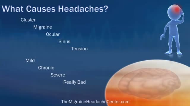
Your headache symptoms can help your doctor determine its cause and the appropriate treatment. Most headaches aren't the result of a serious illness, but some may result from a life-threatening condition requiring emergency care. Headaches are generally classified by cause: Primary headaches A primary headache is caused by overactivity of or problems with pain-sensitive structures in your head. A primary headache isn't a symptom of an underlying disease. Chemical activity in your brain, the nerves or blood vessels surrounding your skull, or the muscles of your head and neck (or some combination of these factors) can play a role in primary headaches. Some people may also carry genes that make them more likely to develop such headaches. The most common primary headaches are: Cluster headache Migraine (with and without aura) Tension headache (also known as tension-type headache) Trigeminal autonomic cephalalgia (TAC), such as cluster headache and paroxysmal hemicrania A few headache patterns also are generally considered types of primary headache, but are less common. These headaches have distinct features, such as an unusual duration or pain associated with a certain activity. Although generally considered primary, each could be a symptom of an underlying disease. They include: Chronic daily headaches (for example, chronic migraine, chronic tension-type headache, or hemicranias continua) Cough headaches Exercise headaches Sex headaches Some primary headaches can be triggered by lifestyle factors, including: Alcohol, particularly red wine Certain foods, such as processed meats that contain nitrates Changes in sleep or lack of sleep Poor posture Skipped meals Stress Secondary headaches A secondary headache is a symptom of a disease that can activate the pain-sensitive nerves of the head. Any number of conditions — varying greatly in severity — may cause secondary headaches. Possible causes of secondary headaches include: Acute sinusitis Arterial tears (carotid or vertebral dissections) Blood clot (venous thrombosis) within the brain — separate from stroke Brain aneurysm (a bulge in an artery in your brain) Brain AVM (brain arteriovenous malformation) — an abnormal formation of brain blood vessels Brain tumor Carbon monoxide poisoning Chiari malformation (structural problem at the base of your skull) Concussion Dehydration Dental problems Ear infection (middle ear) Encephalitis (brain inflammation) Giant cell arteritis (inflammation of the lining of the arteries) Glaucoma (acute angle closure glaucoma) Hangovers

With a portable pump controlled by a wireless handheld device that automatically delivers insulin.
💪 Get our Knee Resilience program here: https://e3rehab.com/programs/r....esilience/knee-resil
In this video, I will walk you through a comprehensive rehab program for the most commonly injured knee ligament - the MCL.
💪 PROGRAMS: https://e3rehab.com/programs/
📩 MAILING LIST (exclusive deals, offers, and information): https://e3rehab.com/newsletter/
🏆 COACHING: https://e3rehab.com/coaching/
📝 ARTICLES: https://e3rehab.com/articles/
👕 APPAREL: https://e3rehab.com/clothing/
🎧 PODCAST: https://open.spotify.com/show/....5ZbaI145Bk94Guq7olMJ
AFFILIATES:
👟 Vivo Barefoot: Get 15% off all shoes! - https://www.vivobarefoot.com/e3rehab
📓 MASS (Monthly Research Review): http://bit.ly/E3MASS
📚 CSMi: https://humacnorm.com/e3rehab
🏋️ GYM EQUIPMENT: https://e3rehab.com/affiliates/
Follow Us:
YOUTUBE: https://www.youtube.com/@e3reh....ab?sub_confirmation=
INSTAGRAM: https://www.instagram.com/e3rehab
TWITTER: https://twitter.com/E3Rehab
FACEBOOK: https://www.facebook.com/e3rehab
TIKTOK: https://www.tiktok.com/@e3rehab
Intro (0:00)
Anatomy & Function (0:08)
Classification (1:11)
Treatment Options (1:46)
Bracing (3:30)
Rehab Overview (4:28)
Early Stage (5:27)
Mid-Stage(8:50)
Late Stage/Return to Sport (21:14)
Programming (22:13)
Summary (23:47)
---
Disclaimer: The information presented is not intended as medical advice or to be a substitute for medical counseling but intended for entertainment purposes only. If you are experiencing pain, please seek the appropriate healthcare professional.

http://www.landging.com/accident-animation-workers-compensation-lifting-box.html
This lifting box accident animation demonstrates the injury covered by workers compensation program.

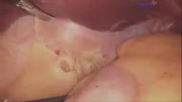
Soft simple painless surgical repair of umbilical hernia video
Total abdominal hysterectomy

Learn about electromagnetic navigation diagnostic bronchoscopy, a new technology used to diagnose small lung cancer tumors as small as a pencil eraser before they have the chance to spread. Cleveland Clinic physician Dr. Thomas Gildea demonstrates how this endobronchial ultrasound procedure, which involves using a small camera probe inserted thru the nose into the lungs, allows doctors to reach possible cancer in the lungs that they could never reliably get to before
Liver transplantation is surgery to remove a diseased or injured liver and replace it with a healthy whole liver or a segment of a liver from another person, called a donor. People with either acute or chronic liver failure may need a liver transplant to survive.
Sinus infections caused by viruses can use home (over-the-counter, OTC) treatments such as pain and fever medications (acetaminophen [Tylenol]), decongestants, and mucolytics. In addition, some health-care professionals suggest nasal irrigation or a sinus rinse solution to help relieve symptoms of sinus infections, even chronic sinusitis symptoms.
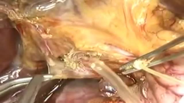
Dr. Jawad has been performing Bariatric Surgery in Central Florida since 1984, and Laparoscopic Bariatric Surgery since 1999, having completed over 2000 Bariatric Surgical Cases safely, and with great success. Here you can watch Dr. Jawad performing a Laparoscopic Adjustable Gastric Band procedure, with audio commentary describing the procedure.

An excerpt from the award-winning documentary “Exposure: Environmental Links to Breast Cancer” about the effects of radiation. Featuring Olivia Newton-John, Dr. Rosalie Bertell and Dr. Susan Love.
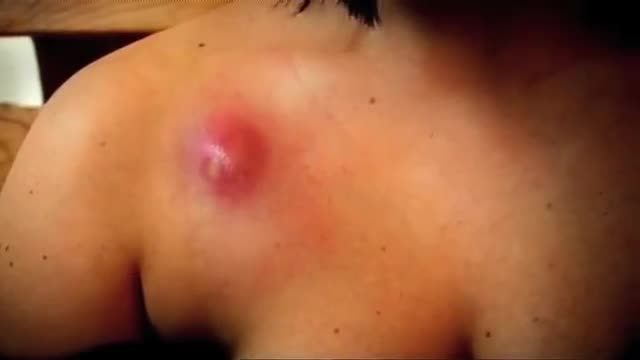
An abscess is a tender mass generally surrounded by a colored area from pink to deep red. Abscesses are often easy to feel by touching. The middle of an abscess is full of pus and debris. Painful and warm to touch, abscesses can show up any place on your body. The most common sites are in your armpits (axillae), areas around your anus and vagina(Bartholin gland abscess), the base of your spine (pilonidal abscess), around a tooth (dental abscess), and in your groin. Inflammation around a hair follicle can also lead to the formation of an abscess, which is called a boil (furuncle). Unlike other infections, antibiotics alone will not usually cure an abscess. In general an abscess must open and drain in order for it to improve. Sometimes draining occurs on its own, but generally it must be opened by a doctor in a procedure called incision and drainage (I&D).

How a simple conversation about colon cancer screening can save your life.
