- Physical Examination
- Surgical Examination
- Ophthalmology
- Clinical Skills
- Orthopedics
- Surgery Videos
- Laparoscopy
- Pediatrics
- Funny Videos
- Cardiothoracic Surgery
- Nursing Videos
- Plastic Surgery
- Otorhinolaryngology
- Histology and Histopathology
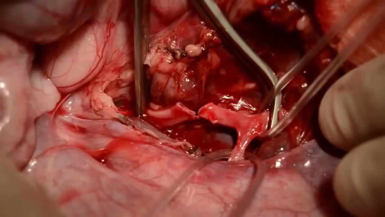
- Neurosurgery
- Dermatology
- Pediatric Surgery
- Urology
- Dentistry
- Oncology and Cancers
- Anatomy Videos
- Health and Fitness
- Radiology
- Anaesthesia
- Physical Therapy
- Pharmacology
- Interventional Radiology
- Cardiology
- Endocrinology
- Gynecology
- Emergency Medicine
- Psychiatry and Psychology
- Childbirth Videos
- General Medical Videos
- Nephrology
- Physiology
- Diet and Food Health
- Diabetes Mellitus
- Neurology
- Women Health
- Osteoporosis
- Gastroenterology
- Pulmonology
- Hematology
- Rheumatology
- Toxicology
- Nuclear Medicine
- Infectious Diseases
- Vascular Disease
- Reproductive Health
- Burns and Wound Healing
- Other
Top videos
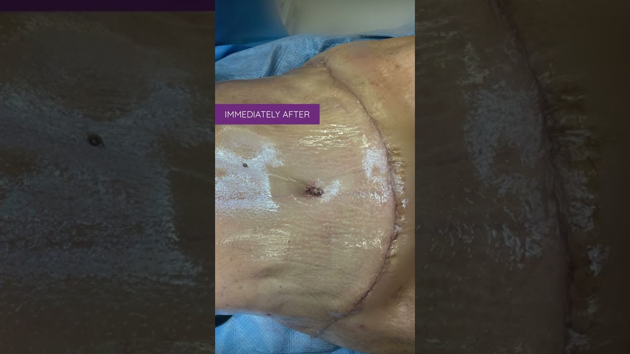
Liposuction in tummy tuck requires special planning and technique. I need to ensure that the blood circulation is well maintained for good healing. Yet proper liposuction is important to have a nice flat and contoured tummy.
#hdliposuction #tummytuck #lipoabdominoplasty #surgicalplanning #skinremovalsurgery #imeediatelyafter #plasticsurgeondubai #cocoonaclinic #drsanjayparashar #dubai
For more information visit www.drsanjayparashar.com
For more content, follow me on my social media
Instagram : https://www.instagram.com/drsanjayparashar/
Facebook : https://www.facebook.com/drsanjayparashar

Watch that Full Real Human Body Decomposition Process

In order to be able to look at tissues under a microscope, we need to first stain them with the right technique. Learn the main staining techniques used in histology today on our full video: https://khub.me/aux9w
Oh, are you struggling with learning anatomy? We created the ★ Ultimate Anatomy Study Guide ★ to help you kick some gluteus maximus in any topic. Completely free. Download yours today: https://khub.me/e0th1
As you probably know, histology is the study of the microscopic anatomy of cells and tissues. So we use staining methods to visualize and distinguish the different parts of cells and tissues since cells and their structures are usually transparent or colorless. The types of dyes used to color cells and their components can either be specific to particular structures, chemical groups or even molecules, and it can also be non-specific in which case most of the cell is stained in the same way.
When staining tissue samples, dyes that are used are either acidic or basic or a combination of the two. And why is that, you might be asking. Well, cellular structures such as nucleic acids or proteins have charged groups which are known as phosphate groups or carboxyl groups, just to name a couple. The dyes used in histology are colored organic compounds which also have a charge. Acidic dyes carry a negative charge and so they bind to positively-charged cell structures.
In the full version of this tutorial, we will cover some of the most common types of dyes used in histological staining of cells and their structures:
- basic dyes vs acidic dyes vs neutral dyes;
- hematoxylin and eosin;
- PAS - staining;
- Golgi method;
- Toluidine blue;
- Masson's trichrome;
- Osmium tetroxide;
To master this topic, click on the link and carry on watching the full video (available to Premium members): https://khub.me/aux9w !
Want to test your knowledge on the different types of cells and tissues? Take this quiz: https://khub.me/3g19f
Read more on how to interpret different histological sections on this complete article which goes through the different stains used in histology https://khub.me/saimh
For more engaging video tutorials, interactive quizzes, articles and an atlas of Human anatomy and histology, go to https://khub.me/pkvz2
Loyola Full Male Exam Part 1 A video from Loyola medical school, Chicago showing the full examination of the male
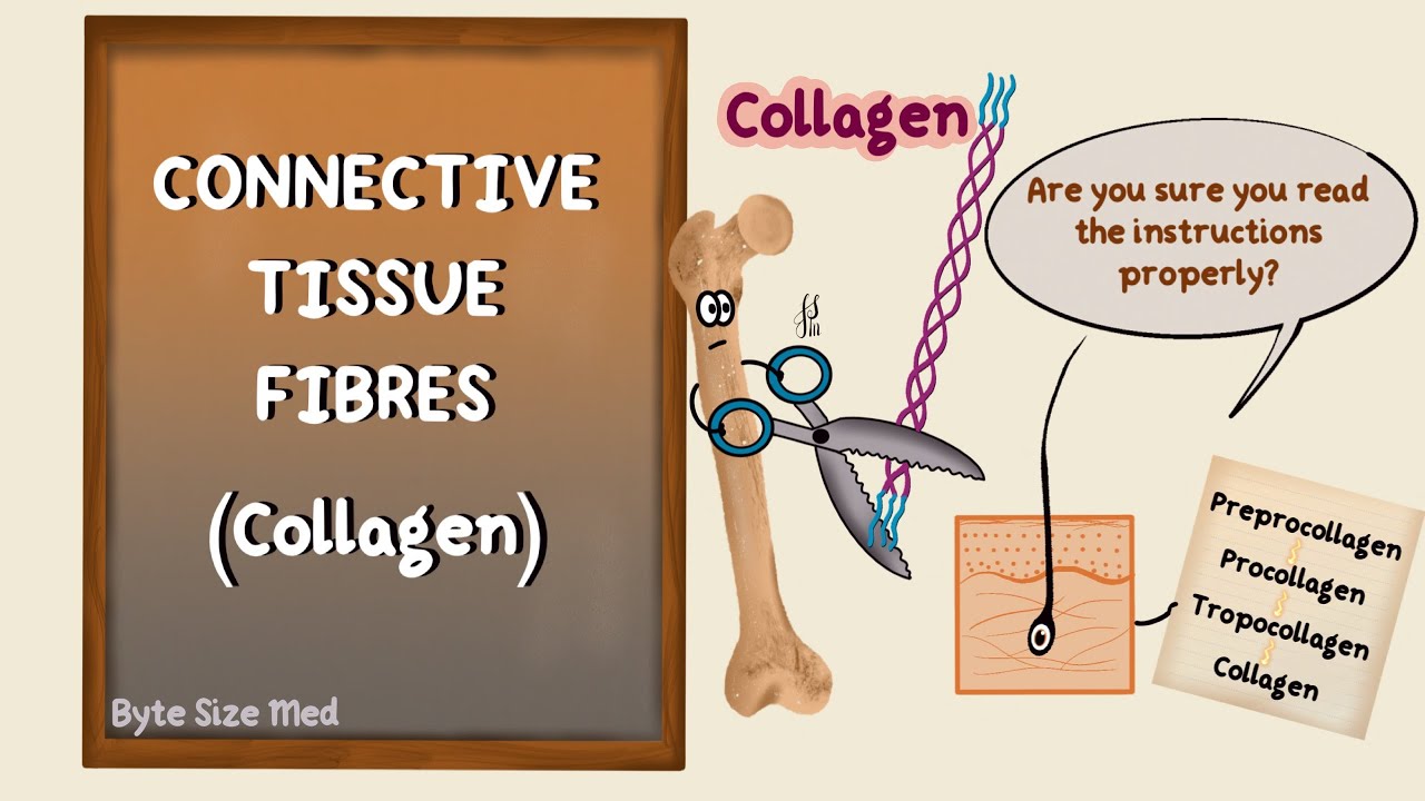
✨This video is on the protein fibres of connective tissue, the types, structure and synthesis of collagen and elastin. I hope it helps! ☀️
🌟What's in this video?
0:00 - Intro
0:07 - Connective Tissue Recap
0:39 - Connective Tissue Fibres
1:22 - Collagen
1:46 - Types of Collagen
3:40 - Structure of Collagen
4:40 - Collagen Synthesis
8:50 - Elastin
✨ Other videos you may need:
🔅 Connective Tissue : https://youtu.be/xw_ALdt5n-A
🔅 Cartilage : https://youtu.be/4inWF4H6pKE
🔅Epithelial Tissue: https://youtu.be/Gw5fC0zXaeU
🔅Structure of Blood Vessels: https://youtu.be/BAo2UqqyL3g
🔅Histology: https://www.youtube.com/playli....st?list=PL1rG930trF2
💫 For more videos like this, subscribe to my channel!
Byte Size Med: https://youtube.com/channel/UC....ZghvlgylH3r_CWfA18eF
📚Factual References & for Further Reading:
- DiFiore's Atlas of Histology
- Junqueira's Basic Histology
- Harper's Biochemistry
- Gartner's Concise Histology
- Openstax Anatomy and Physiology
https://openstax.org/details/b....ooks/anatomy-and-phy
- Openstax Biology
https://openstax.org/details/books/biology-2e
(The last two are links to open-source references. They are NOT affiliate links)
🌤 Note:
These are just a collection of my notes. So use them the way you would use borrowed notes from a friend. 📝
The images in this video are hand-drawn for illustration and explanation only.✍️ Hence, they may not be anatomically accurate. I am just one person making these videos. If there are any errors, that is unintentional. I try super hard to avoid them. Please let me know if you find any, so it gets clarified for other viewers. Science constantly evolves and changes. New discoveries are made everyday. So some of the information in these videos may become outdated. If you notice that, please let me know so I can update them.
⚡️Disclaimer:
These videos are NOT a substitute for a medical textbook. Textbooks are written by experts (which I do not claim to be), edited, proofread and referenced. Please use them.
The information has been sourced from multiple references as mentioned above. I draw all the pictures myself. But if I have inadvertently infringed on any copyright, that is completely unintentional. I only make these videos to impart education. If I have accidentally violated copyright in any way, do let me know so I can make the necessary changes or give credit to anyone who is owed the same.
These videos are NOT intended for patient education. They are NOT a substitute for diagnosis and treatment by a licensed medical professional. Always seek the advice of a qualified health care provider for any questions you may have regarding any medical condition, so that they can address your individual needs.
🔅They are ONLY meant to help students of medicine and health sciences with studying, and should be used for just that purpose and absolutely nothing else.
Byte Size Med. All Rights Reserved.
Medical Examination of the Lower Limbs
Throughout the body, there are several points at which blood vessels unite. The junctions are termed anastomoses. In the simplest sense, an anastomosis is any connection (made surgically or occurring naturally) between tube-like structures. Naturally occurring arterial anastomoses provide an alternative blood supply to target areas in cases where the primary arterial pathway is obstructed. They are most abundant in regions of the body where the blood supply may can be easily damaged or blocked (such as the joints or intestines). This article focuses on the arterial anastomotic networks of the upper limb.

For a full Surgical Airway Techniques resource: https://bit.ly/2rb9Nud
Video courtesy of Gauri Mankekar, MBBS, MS, PhD
Watch that video to learn How to Know When You Are Ovulating
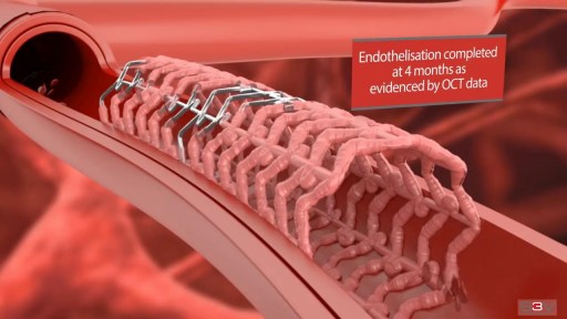
Angioplasty is a procedure to restore blood flow through the artery. You have angioplasty in a hospital. The doctor threads a thin tube through a blood vessel in the arm or groin up to the involved site in the artery. The tube has a tiny balloon on the end.
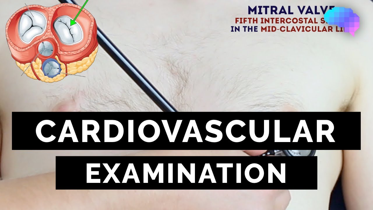
This video demonstrates how to perform a cardiovascular examination in an OSCE station.
You can access our step-by-step OSCE guide to accompany this video here: https://geekymedics.com/cardio....vascular-examination
Check out our other awesome clinical skills resources including:
• 🔥 Geeky Medics Bundles (discounted products): https://app.geekymedics.com/purchase/bundles/
• ✨ 1000+ OSCE Stations: https://app.geekymedics.com/pu....rchase/osce-stations
• 🏥 Geeky Medics OSCE Revision Book: https://app.geekymedics.com/purchase/book/
• 📝 150+ PDF OSCE Checklists: https://geekymedics.com/pdf-osce-checklists/
• 🗂️ 3000+ OSCE Flashcards: https://app.geekymedics.com/pu....rchase/flashcard-col
• 📱 Geeky Medics OSCE App: https://geekymedics.com/geeky-medics-app/
• 🩺 Medical Finals SBA Question Pack: https://app.geekymedics.com/pu....rchase/medical-stude
• 💊 PSA Question Pack: https://app.geekymedics.com/pu....rchase/prescribing-s
Chapters:
- Introduction 00:00
- General inspection 00:35
- Hands 00:46
- Schamroth's window test 01:07
- Capillary refill 01:27
- Pulses 01:35
- Carotid auscultation 02:21
- Carotid pulse 02:43
- Jugular venous pressure 02:55
- Hepatojugular reflux 03:09
- Inspection of the face 03:21
- Inspection of the chest 03:49
- Apex beat 04:12
- Heaves and thrills 04:28
- Heart valve ausculation 04:49
- Accentuation manoeuvres 05:45
- Lung base auscultation 06:23
- Sacral and pedal oedema 06:43
- Summary 07:10
Subscribe to our newsletter to be the first to know about our latest content: https://geekymedics.com/newsletter/ ✉️
Join the Geeky Medics community: 👩👩👧👧
Twitter: http://www.twitter.com/geekymedics
Instagram: https://instagram.com/geekymedics
Facebook: http://www.facebook.com/geekymedics
Always adhere to your medical school/local hospital guidelines when performing examinations or clinical procedures. DO NOT perform any examination or procedure on patients based purely upon the content of these videos. Geeky Medics accepts no liability for loss of any kind incurred as a result of reliance upon the information provided in this video.
Normal heart sounds and aortic regurgitation/stenosis sounds
Recorded on a Thinklabs Digital Stethoscope (https://www.thinklabs.com)
Some people have found this video useful for ASMR purposes.
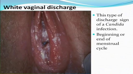
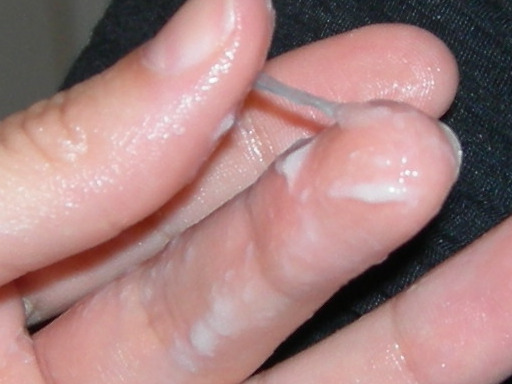
All you need to know about the Types of Female Genital Discharge

This video demonstrates how to perform an abdominal examination in an OSCE station.
You can access our step-by-step OSCE guide to accompany this video here: https://geekymedics.com/abdominal-examination/
Check out our other awesome clinical skills resources including:
• 🔥 Geeky Medics Bundles (discounted products): https://app.geekymedics.com/purchase/bundles/
• ✨ 1000+ OSCE Stations: https://app.geekymedics.com/pu....rchase/osce-stations
• 🏥 Geeky Medics OSCE Revision Book: https://app.geekymedics.com/purchase/book/
• 📝 150+ PDF OSCE Checklists: https://geekymedics.com/pdf-osce-checklists/
• 🗂️ 3000+ OSCE Flashcards: https://app.geekymedics.com/pu....rchase/flashcard-col
• 📱 Geeky Medics OSCE App: https://geekymedics.com/geeky-medics-app/
• 🩺 Medical Finals SBA Question Pack: https://app.geekymedics.com/pu....rchase/medical-stude
• 💊 PSA Question Pack: https://app.geekymedics.com/pu....rchase/prescribing-s
Chapters:
- Introduction 00:00
- General inspection 00:35
- Hands 00:47
- Asterixis 01:20
- Arms and axilla 01:32
- Face, eyes & mouth 01:45
- Lymph node palpation 02:19
- Chest inspection 02:50
- Inspection of abdomen 03:02
- Palpation of abdomen 03:34
- Percussion of abdomen 05:36
- Shifting dullness 06:30
- Auscultation of abdomen 06:55
- Summary 07:29
Subscribe to our newsletter to be the first to know about our latest content: https://geekymedics.com/newsletter/ ✉️
Join the Geeky Medics community: 👩👩👧👧
Twitter: http://www.twitter.com/geekymedics
Instagram: https://instagram.com/geekymedics
Facebook: http://www.facebook.com/geekymedics
Always adhere to your medical school/local hospital guidelines when performing examinations or clinical procedures. DO NOT perform any examination or procedure on patients based purely upon the content of these videos. Geeky Medics accepts no liability for loss of any kind incurred as a result of reliance upon the information provided in this video.
Some people have found this video useful for ASMR purposes.
Watch that video of Dissection Inside Fat Body

Surgical site infections (SSIs) remain a prevalent threat to patient safety. Proper surgical hand scrub or rub techniques are essential to decreasing the incidence of SSIs. This video provides instructions on the anatomical surgical hand scrub procedure using the brushstroke method. Learn more from the Department of Hospital Epidemiology and Infection Control (HEIC) at The Johns Hopkins Hospital: http://www.hopkinsmedicine.org/heic
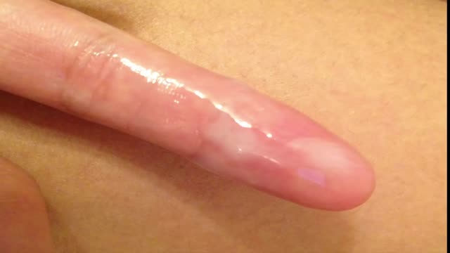
Most women have vaginal discharge at many different times throughout their cycle. During ovulation, white and watery discharge is common and accepted as normal. But, discharge after ovulation is widely believed to be a sign of pregnancy.

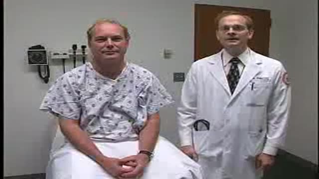
Inguinal or groin hernias are the most common type of hernias and most of the time occur in men. We talked with CU Medicine surgeon, Dr. Sam Phinney, about groin hernias and how they are treated. https://www.cumedicine.us/abou....t-cu-medicine/health

Cardiovascular Examination Clinical skills - Medical School Revision - Dr Gill
The cardiac exam is one of the clinical skills that medical students learn completely, as more often than not, patients will consult regularly about chest pain, and it is important to be able to identify key cardiovascular signs
As a junior doctor, the examination of the cardiovascular system can be almost a dreaded examination, as cardiac murmurs can literally take years of exposure in order to gain confidence with their identification through cardiac auscultation.
This video demonstrates not merely the examination of the heart, but the complete cardiovascular system including its peripheries.
I hope these clinical skill revision videos are helpful, please like and subscribe and join the community so that we can create more effective videos to help with your journey through medical school
#ClinicalExamination #ASMR #drgill
Some people have found this video useful for ASMR
Vasectomy is a minor surgical procedure wherein the vasa deferentia of a man are severed, and then tied or sealed in a manner such to prevent sperm from entering the seminal stream (ejaculate). Typically done in an outpatient setting, a traditional vasectomy involves numbing (local anesthetic) of the scrotum after which 1 (or 2) small incisions are made, allowing a surgeon to gain access to the vas deferens.
