- Physical Examination
- Surgical Examination
- Ophthalmology
- Clinical Skills
- Orthopedics
- Surgery Videos
- Laparoscopy
- Pediatrics
- Funny Videos
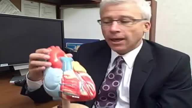
- Cardiothoracic Surgery
- Nursing Videos
- Plastic Surgery
- Otorhinolaryngology
- Histology and Histopathology
- Neurosurgery
- Dermatology
- Pediatric Surgery
- Urology
- Dentistry
- Oncology and Cancers
- Anatomy Videos
- Health and Fitness
- Radiology
- Anaesthesia
- Physical Therapy
- Pharmacology
- Interventional Radiology
- Cardiology
- Endocrinology
- Gynecology
- Emergency Medicine
- Psychiatry and Psychology
- Childbirth Videos
- General Medical Videos
- Nephrology
- Physiology
- Diet and Food Health
- Diabetes Mellitus
- Neurology
- Women Health
- Osteoporosis
- Gastroenterology
- Pulmonology
- Hematology
- Rheumatology
- Toxicology
- Nuclear Medicine
- Infectious Diseases
- Vascular Disease
- Reproductive Health
- Burns and Wound Healing
- Other
Top videos

There's only one group of people who really know what happens when you die: the dead. And since the dead won't be revealing their secrets anytime soon, it's up to scientists to explain what happens when a person dies. Death, just like life, is a process, scientists say. The first stage of this process is known as clinical death. It lasts from four to six minutes, beginning when a person stops breathing and the heart stops pumping blood. During this time, there may be enough oxygen in the brain that no permanent brain damage occurs. Other organs, such as the kidneys and eyes, also remain alive throughout clinical death.
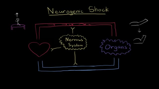
Neurogenic shock is a distributive type of shock resulting in low blood pressure, occasionally with a slowed heart rate, that is attributed to the disruption of the autonomic pathways within the spinal cord. It can occur after damage to the central nervous system such as spinal cord injury.
The protein inside red blood cells (a) that carries oxygen to cells and carbon dioxide to the lungs is hemoglobin (b). Hemoglobin is made up of four symmetrical subunits and four heme groups. Iron associated with the heme binds oxygen.
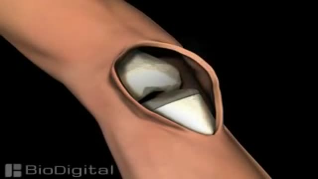
Knee replacement, also called arthroplasty, is a surgical procedure to resurface a knee damaged by arthritis. Metal and plastic parts are used to cap the ends of the bones that form the knee joint, along with the kneecap. This surgery may be considered for someone who has severe arthritis or a severe knee injury.
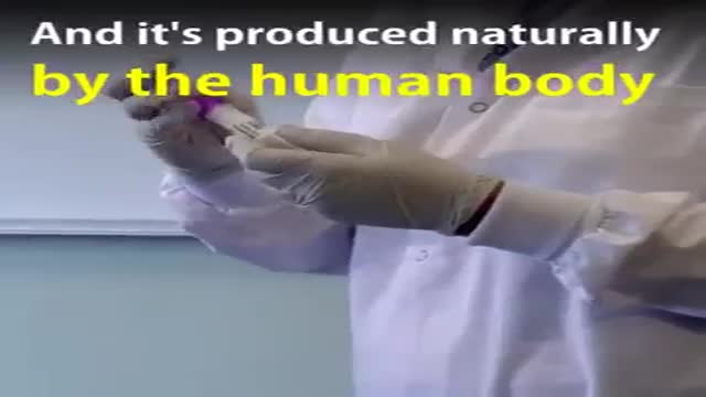
Natural painkiller found in human spit. Compound in saliva could be more powerful than morphine. A new painkilling substance has been discovered that is up to six times more potent than morphine when tested in rats — and it's produced naturally by the human body.

Why Does Thinking Hard Make You Tired?
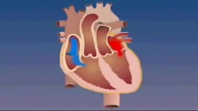
Transposition of the great arteries is a serious but rare heart defect present at birth (congenital), in which the two main arteries leaving the heart are reversed (transposed). Transposition of the great arteries changes the way blood circulates through the body, leaving a shortage of oxygen in blood flowing from the heart to the rest of the body. Without an adequate supply of oxygen-rich blood, the body can't function properly and your child faces serious complications or death without treatment.
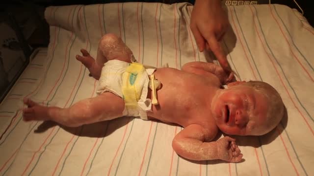
Although the Apgar score was developed in 1952 by an anesthesiologist named Virginia Apgar, you also might hear it referred to as an acronym for: Appearance, Pulse, Grimace, Activity, and Respiration. The Apgar test is usually given to a baby twice: once at 1 minute after birth, and again at 5 minutes after birth.
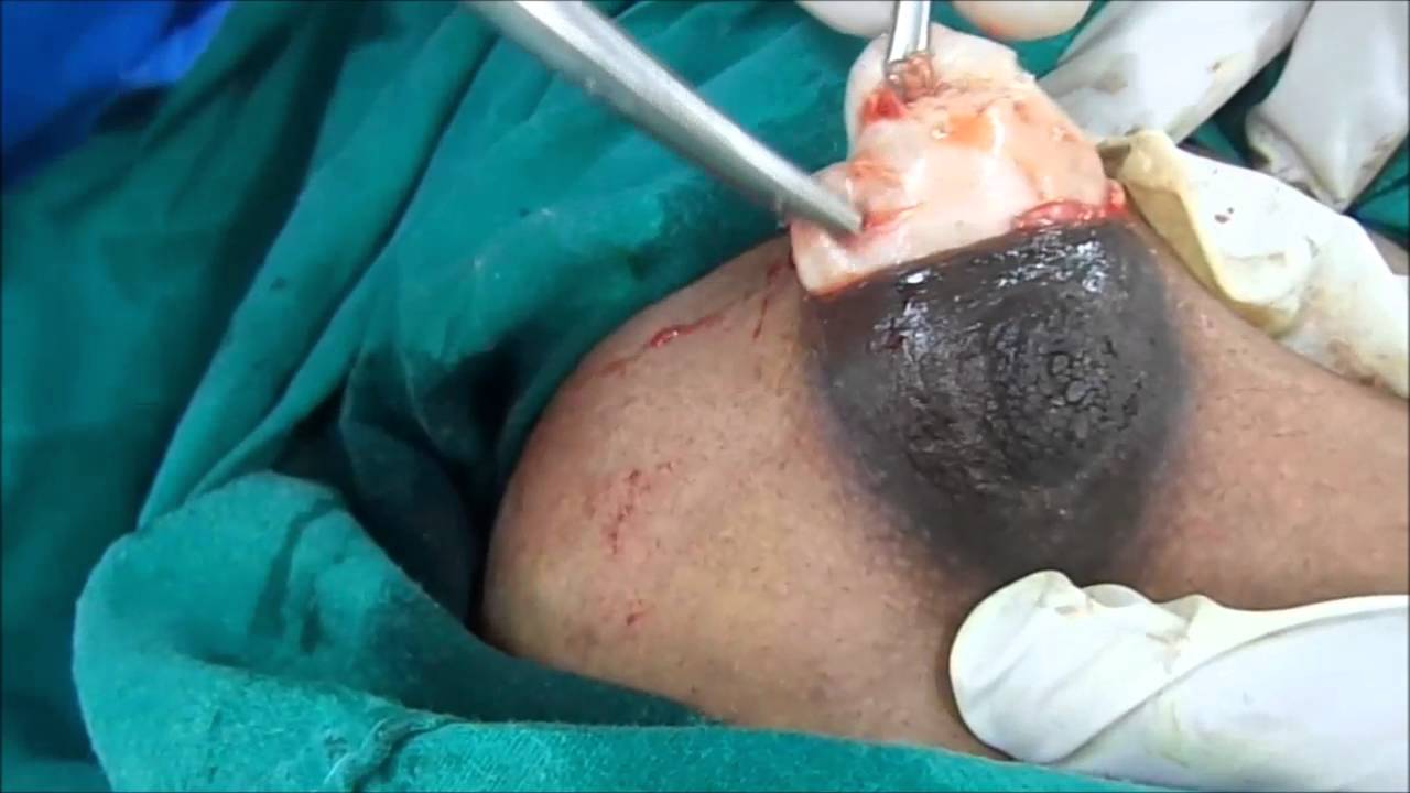
A Big Size Fibrodenoma Removal Under Local Anesthesia
Spine Examination

What's helping me become a better doctor
Marfan syndrome is a genetic disorder that affects the body’s connective tissue. Connective tissue holds all the body’s cells, organs and tissue together. It also plays an important role in helping the body grow and develop properly. marfan_general_2.jpg What is Marfan Syndrome?Connective tissue is made up of proteins. The protein that plays a role in Marfan syndrome is called fibrillin-1. Marfan syndrome is caused by a defect (or mutation) in the gene that tells the body how to make fibrillin-1. This mutation results in an increase in a protein called transforming growth factor beta, or TGF-β. The increase in TGF-β causes problems in connective tissues throughout the body, which in turn creates the features and medical problems associated with Marfan syndrome and some related disorders. Because connective tissue is found throughout the body, Marfan syndrome can affect many different parts of the body, as well. Features of the disorder are most often found in the heart, blood vessels, bones, joints, and eyes. Some Marfan features – for example, aortic enlargement (expansion of the main blood vessel that carries blood away from the heart to the rest of the body) – can be life-threatening. The lungs, skin and nervous system may also be affected. Marfan syndrome does not affect intelligence.

Coronary artery vasospasm, or smooth muscle constriction of the coronary artery, is an important cause of chest pain syndromes that can lead to myocardial infarction (MI), ventricular arrhythmias, and sudden death. It also plays a key role in the development of atherosclerotic lesions.Nov 22, 2016
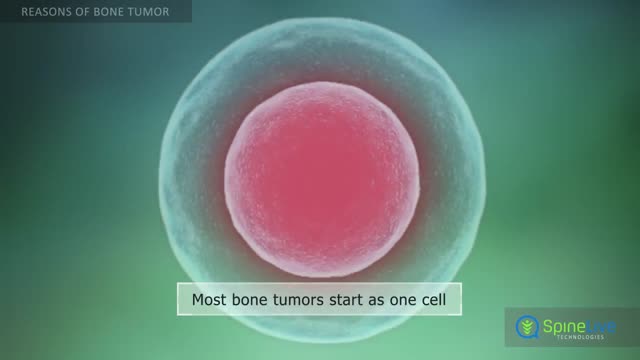
Bone tumors develop when cells in the bone divide without control, forming a mass of tissue. Most bone tumors are benign, which means they are not cancer and cannot spread. However, they may still weaken bone and lead to fractures or cause other problems. Bone cancer destroys normal bone tissue and may spread to other parts of the body (called metastasis). Benign Bone Tumors Benign tumors are more common than malignant tumors of the bones. These are a few common types of benign bone tumors: Osteochondroma is the most common benign bone tumor. It is more common in people under age 20. Giant cell tumor is a benign tumor, typically affecting the leg (malignant types of this tumor are uncommon). Osteoid osteoma is a bone tumor, often occurring in long bones, that occurs commonly in the early 20s. Osteoblastoma is a single tumor that occurs in the spine and long bones, mostly in young adults. Enchondroma usually appears in bones of the hand and feet. It often has no symptoms. It is the most common type of hand tumor.

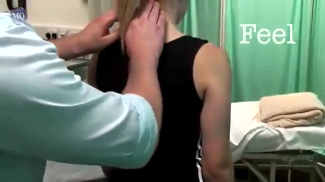
This video shows you how to conduct a clinical examination of the shoulder and to identify common causes of pain.
This video clip is part of the FIFA Diploma in Football Medicine and the FIFA Medical Network. To enrol or to find our more click on the following link http://www.fifamedicalnetwork.com
The Diploma is a free online course designed to help clinicians learn how to diagnose and manage common football-related injuries and illnesses. There are a total of 42 modules created by football medicine experts. Visit a single page, complete individual modules or finish the entire course.
The network provides the opportunity for clinicians around the world to meet and share ideas relating to football medicine. Ask about an interesting case, debate current practice and discuss treatment strategies. Create a profile and log on to interact with other health professionals from around the globe.
This is not medical advice. The content is intended as educational content for health care professionals and students. If you are a patient, seek care of a health care professional.
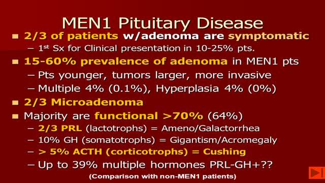
-MEN1 syndrome is composed of hyperparathyroidism, gastrinoma (pancreatic tumor) and pituitary tum or(remember the 3 Ps). Hyperparathyroidism in MEN1 is caused by hyperplasia of the parathyroid glands. Removal of 3 1/2 glands or total parathyroidectomy with autotransplantation is necessary.

Cervical Mucus
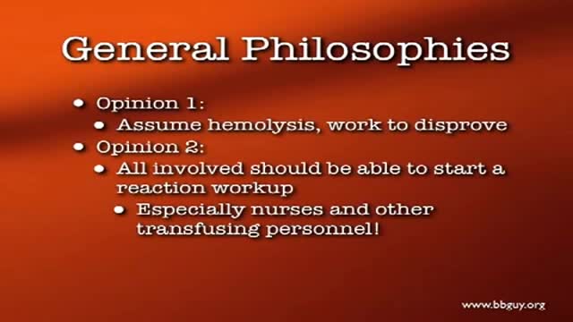
A hemolytic transfusion reaction is a serious complication that can occur after a transfusion of blood. The red blood cells that were given in the transfusion are destroyed by the patient's immune system. There are other types of allergic transfusion reactions that do not cause hemolysis.
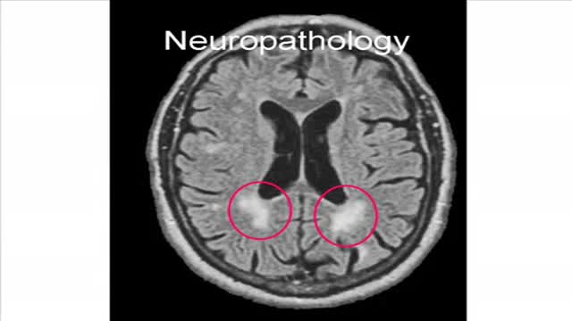
Binswanger's disease is a type of vascular dementia that involves white matter infarcts. Patients with this disease usually present with apathy, agitation, and bilateral corticospinal or bulbar signs
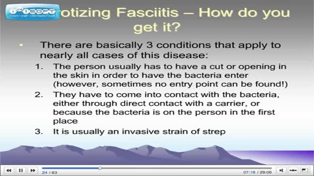
patient has fever, hypotension, swelling, and erythema of his left leg. Notably, his leg is more painful to palpation than might be expected after visual inspection. These symptoms are very concerning for necrotizing fasciitis. Necrotizing fasciitis is a fulminant infection of the subcutaneous tissue that spreads rapidly along the fascial planes and leads to extensive tissue necrosis and shock. Treatment • Requires surgical debridement & broad-spectrum antibiotics
