
- Physical Examination
- Surgical Examination
- Ophthalmology
- Clinical Skills
- Orthopedics
- Surgery Videos
- Laparoscopy
- Pediatrics
- Funny Videos
- Cardiothoracic Surgery
- Nursing Videos
- Plastic Surgery
- Otorhinolaryngology
- Histology and Histopathology
- Neurosurgery
- Dermatology
- Pediatric Surgery
- Urology
- Dentistry
- Oncology and Cancers
- Anatomy Videos
- Health and Fitness
- Radiology
- Anaesthesia
- Physical Therapy
- Pharmacology
- Interventional Radiology
- Cardiology
- Endocrinology
- Gynecology
- Emergency Medicine
- Psychiatry and Psychology
- Childbirth Videos
- General Medical Videos
- Nephrology
- Physiology
- Diet and Food Health
- Diabetes Mellitus
- Neurology
- Women Health
- Osteoporosis
- Gastroenterology
- Pulmonology
- Hematology
- Rheumatology
- Toxicology
- Nuclear Medicine
- Infectious Diseases
- Vascular Disease
- Reproductive Health
- Burns and Wound Healing
- Other
Top videos


http://www.landging.com/skeletal-system-animation-knee-surgery.html
This skeletal system animation demonstrates the new concept of knee surgery procedure.

Malaria is a serious and sometimes fatal disease caused by a parasite that commonly infects a certain type of mosquito which feeds on humans. People who get malaria are typically very sick with high fevers, shaking chills, and flu-like illness.
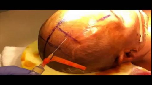
This video describes, step by step, how to place an external ventricular drain. This is a common neurosurgical procedure used to relieve intracranial pressure.

The superior sagittal sinus (also known as the superior longitudinal sinus), within the human head, is an unpaired area along the attached margin of falx cerebri. It allows blood to drain from the lateral aspects of anterior cerebral hemispheres to the confluence of sinuses.
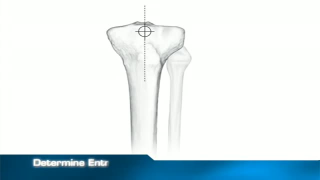
fixation of a tibial fracture utilizing the Titanium Cannulated Tibial Nail

Here's Why Your Skin Doesn't Rip Easily

Claudication is pain caused by too little blood flow, usually during exercise. Sometimes called intermittent claudication, this condition generally affects the blood vessels in the legs, but claudication can affect the arms, too. At first, you'll probably notice the pain only when you're exercising, but as claudication worsens, the pain may affect you even when you're at rest. Although it's sometimes considered a disease, claudication is technically a symptom of a disease. Most often, claudication is a symptom of peripheral artery disease, a potentially serious but treatable circulation problem in which the vessels that supply blood flow to your legs or arms are narrowed. Fortunately, with treatment, you may be able to maintain an active lifestyle without pain.

Make A Girl Squirt Fact
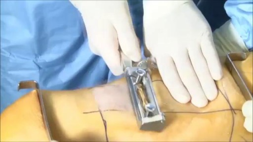

kin grafting is a type of graft surgery involving the transplantation of skin. The transplanted tissue is called a skin graft. Skin grafting is often used to treat: Extensive wounding or trauma Burns Areas of extensive skin loss due to infection such as necrotizing fasciitis or purpura fulminans[2] Specific surgeries that may require skin grafts for healing to occur - most commonly removal of skin cancers Skin grafts are often employed after serious injuries when some of the body's skin is damaged. Surgical removal (excision or debridement) of the damaged skin is followed by skin grafting. The grafting serves two purposes: reduce the course of treatment needed (and time in the hospital), and improve the function and appearance of the area of the body which receives the skin graft.

The term subclavian steal describes retrograde blood flow in the vertebral artery associated with proximal ipsilateral subclavian artery stenosis or occlusion, usually in the setting of subclavian artery occlusion or stenosis proximal to the origin of the vertebral artery. Alternatively, innominate artery disease has also been associated with retrograde flow in the ipsilateral vertebral artery, particularly where the subclavian artery origin is involved. Subclavian steal is frequently asymptomatic and may be discovered incidentally on ultrasound or angiographic examination for other indications, or it may be prompted by a clinical examination finding of reduced unilateral upper limb pulse or blood pressure. In some cases, patients may develop upper limb ischemic symptoms due to reduced arterial flow in the setting of subclavian artery occlusion, or they may develop neurologic symptoms due to posterior circulation ischemia associated with exercise of the ipsilateral arm.[1] Treatment has traditionally consisted of open subclavian artery revascularization, typically via carotid-subclavian bypass or subclavian artery transposition, which are generally durable procedures. Newer, less invasive options include endovascular intervention with recanalization as appropriate and angioplasty and stenting if required. The clinical relevance of subclavian steal was described in 1961 by Reivich, Holling and Roberts; however, the recognition of retrograde vertebral artery flow dates back another 100 years to Harrison and Smyth. Some papers, including a previous version of this article, advocate restricting the term subclavian steal to patients with neurologic symptoms only, but this is incorrect in view of the substantial literature using this term to describe the hemodynamic scenario of retrograde vertebral flow and proximal subclavian artery disease.
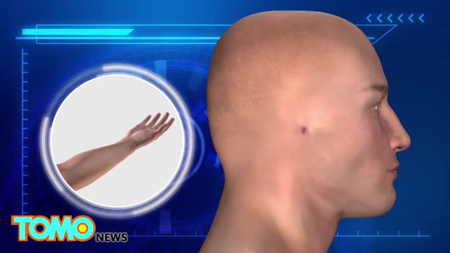
A Chinese hospital in the process of creating a human ear almost entirely through the human anatomy alone.

http://www.landging.com/car-crash-animations-front.html Car crash animations, accident reconstruction, front end collision.

ADH's job is to act on the kidneys to promote water reabsorption. In this lesson, we'll compare and contrast diabetes insipidus, or DI, in which there is too little ADH, and syndrome of inappropriate antidiuretic hormone secretion , or SIADH, in which there is too much ADH.
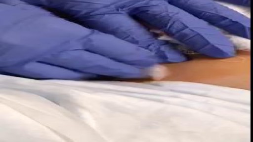

This type of gait is most often seen in peripheral nerve disease where the distal lower extremity is most affected. Because the foot dorsiflexors are weak, the patient has a high stepping gait in an attempt to avoid dragging the toe on the ground.
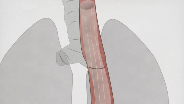
Esophageal atresia is an abnormality, or birth defect, of the esophagus that occurs early in pregnancy, as the baby is developing. The esophagus forms in the first few months of fetal life as a long, hollow, continuous tube joining the mouth to the stomach. In newborns with this birth defect, formation of this continuous esophageal tube is interrupted. esophageal-artresia-2In most cases, two separate tubes are formed, an upper (proximal) tube connected to the mouth and a lower (distal) tube connected to the stomach. This seperated tubes are sealed off creating a pouch on either side; the gap between these pounches can be short or very long. Saliva can accumulate in the upper pouch as it cannot drain into the stomach.
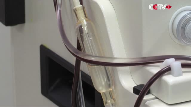
Australian Blood Donor Saves 2 Millions Babies with Special Right Arm
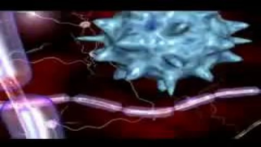
Multiple sclerosis (MS) involves an immune-mediated process in which an abnormal response of the body’s immune system is directed against the central nervous system (CNS). The CNS is made up of the brain, spinal cord and optic nerves. Within the CNS, the immune system causes inflammation that damages myelin — the fatty substance that surrounds and insulates the nerve fibers — as well as the nerve fibers themselves, and the specialized cells that make myelin. When myelin or nerve fibers are damaged or destroyed in MS, messages within the CNS are altered or stopped completely. Damage to areas of the CNS may produce a variety of neurological symptoms that will vary among people with MS in type and severity The damaged areas develop scar tissue which gives the disease its name – multiple areas of scarring or multiple sclerosis. The cause of MS is not known, but it is believed to involve genetic susceptibility, abnormalities in the immune system and environmental factors that combine to trigger the disease. People with MS typically experience one of four disease courses. There are over a dozen treatments to help modify the MS disease process.
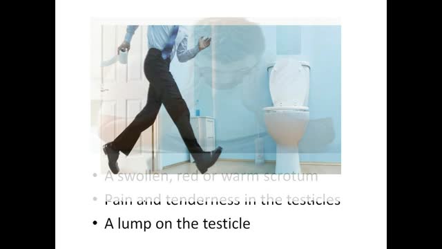
Epididymitis is infection or less frequently, inflammation of the epididymis (the coiled tube on the back of the testicle). The majority of men that develop epididymitis develop it because of a bacterial infection. Although males of any age can develop epididymitis, it occurs most frequently between ages of 20 to 39.

Dont worry sister!
