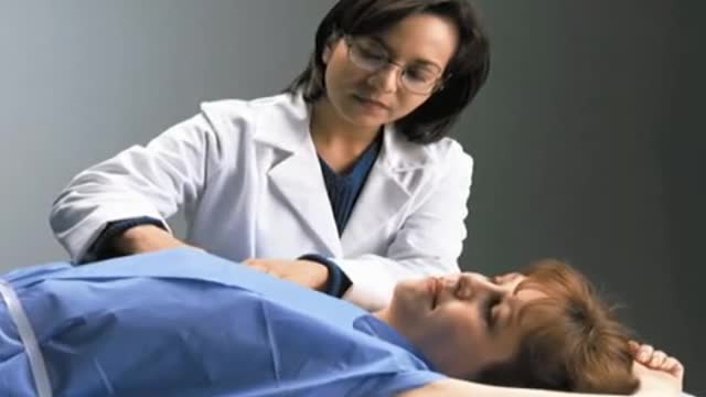
- Physical Examination
- Surgical Examination
- Ophthalmology
- Clinical Skills
- Orthopedics
- Surgery Videos
- Laparoscopy
- Pediatrics
- Funny Videos
- Cardiothoracic Surgery
- Nursing Videos
- Plastic Surgery
- Otorhinolaryngology
- Histology and Histopathology
- Neurosurgery
- Dermatology
- Pediatric Surgery
- Urology
- Dentistry
- Oncology and Cancers
- Anatomy Videos
- Health and Fitness
- Radiology
- Anaesthesia
- Physical Therapy
- Pharmacology
- Interventional Radiology
- Cardiology
- Endocrinology
- Gynecology
- Emergency Medicine
- Psychiatry and Psychology
- Childbirth Videos
- General Medical Videos
- Nephrology
- Physiology
- Diet and Food Health
- Diabetes Mellitus
- Neurology
- Women Health
- Osteoporosis
- Gastroenterology
- Pulmonology
- Hematology
- Rheumatology
- Toxicology
- Nuclear Medicine
- Infectious Diseases
- Vascular Disease
- Reproductive Health
- Burns and Wound Healing
- Other
Top videos
Tests. This test tracks electrical signals from the brain. There are a number of blood tests that may be recommended as part of your epilepsy diagnosis and treatment. A positron emission tomography (PET) scan may be used to locate the part of the brain that is causing seizures.

Triglycerides are a type of fat (lipid) found in your blood. When you eat, your body converts any calories it doesn't need to use right away into triglycerides. The triglycerides are stored in your fat cells. Later, hormones release triglycerides for energy between meals. If you regularly eat more calories than you burn, particularly "easy" calories like carbohydrates and fats, you may have high triglycerides (hypertriglyceridemia).

A ventricular septal defect (VSD) is an opening or hole in the wall that separates the two lower chambers of the heart. This wall is called the ventricular septum. The hole causes oxygen-rich blood to leak from the left side of the heart to the right side. This causes extra work for the right side of the heart, since more blood than necessary is flowing through the right ventricle to the lungs. The hole is usually closed with surgery. However, in certain situations, your child's cardiologist and surgeon may think it is best to close the hole with a special device. This procedure is done in the heart catheterization lab.
Try these tips from top fertility experts to increase the odds you'll be prego ASAP…that is, if you want to be. Take Prenatal Vitamins. ... Try to Time It. ... Skip the Lube. ... Cut Back on Caffeine. ... Don't Increase Your Exercise Routine. ... Go Easy on the Alcohol. ... Try to Chill Out.

Blackheads are small bumps that appear on your skin due to clogged hair follicles. These bumps are called “blackheads” because the surface looks dark or black. Blackheads are a mild type of acne that usually form on the face, but they can also appear on the back, chest, neck, arms, and shoulders
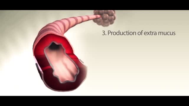
Asthma is a condition in which your airways narrow and swell and produce extra mucus. This can make breathing difficult and trigger coughing, wheezing and shortness of breath.

The cat's stomach is a sac-like structure designed to store large volumes of food and continue the digestive process. The esophagus carries food to the stomach, where it enters via a valve-like structure called the cardiac sphincter. On the interior surface of the stomach is a series of folds called gastric folds. These folds function to help grind and digest food. The inner stomach lining secretes acids and enzymes to break down food. Once the initial stomach digestive process is complete, the partially digested food exits the stomach through the pyloric sphincter area and then enters the duodenum (first segment of the small intestine). Once eaten, most food leaves the stomach within twelve hours after entering.
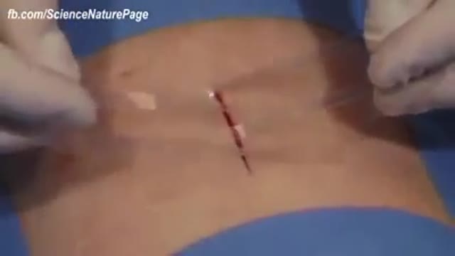
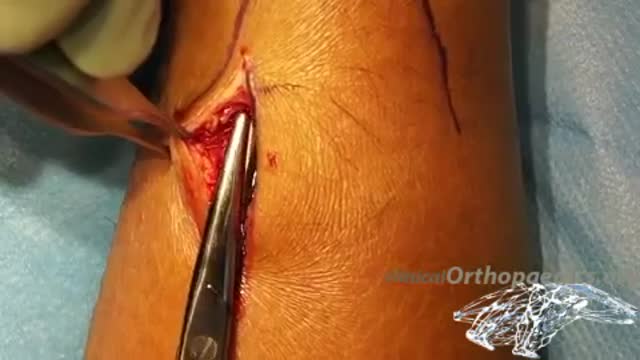
Wound-closure technologies are becoming less painful and more efficient at closing wounds.
A new report analyzing FDA-approved monoclonal antibodies (mAbs) produced by a select group of leading biotechnology companies shows that clinical development times – specifically the duration of Phase II and Phase III trials – are lengthening, while FDA review times have remained constant. The average time from investigational new drug (IND) filing to market was 6.7 years for 11 mABs approved between 1994 and 2003 but shot up to 8.3 years for 12 mAbs approved between 2004 and March 9, 2011, according to Deloitte Recap LLC’s analysis, Therapeutic Monoclonal Antibodies – Insights, Strategies and Data.
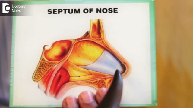
A deviated septum occurs when the thin wall (nasal septum) between your nasal passages is displaced to one side. In many people, the nasal septum is displaced — or deviated — making one nasal passage smaller. When a deviated septum is severe, it can block one side of your nose and reduce airflow, causing difficulty breathing. The additional exposure of a deviated septum to the drying effect of airflow through the nose may sometimes contribute to crusting or bleeding in certain individuals. Nasal obstruction can occur from a deviated nasal septum, from swelling of the tissues lining the nose, or from both. Treatment of nasal obstruction may include medications to reduce the swelling or nasal dilators that help open the nasal passages. To correct a deviated septum, surgery is necessary.
t’s the brain, after all, that devises experiments and interprets their results. How the brain perceives, how it makes decisions and judgments, and how those judgments can go awry are at least as important to science as knowing the intricacies of nonbiotic experimental machinery. And as any brain scientist will tell you, there’s still a long way to go before understanding the brain will get crossed off science’s to-do list. But there has been progress. A recent special issue of the journal Neuron offers a convenient set of “perspective” papers exploring the current state of understanding of the brain’s inner workings. Those papers show that a lot is known. But at the same time they emphasize that there’s a lot we don’t know.

Happy New Year 2017
Mini-invasive surgical repair of a ruptured Achilles tendon
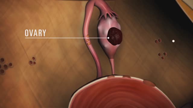
Endometriosis (en-doe-me-tree-O-sis) is an often painful disorder in which tissue that normally lines the inside of your uterus — the endometrium — grows outside your uterus. Endometriosis most commonly involves your ovaries, fallopian tubes and the tissue lining your pelvis. Rarely, endometrial tissue may spread beyond pelvic organs.
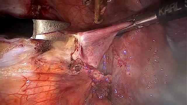
LAPAROSCOPIC END TO END URETERAL ANASTOMOSIS
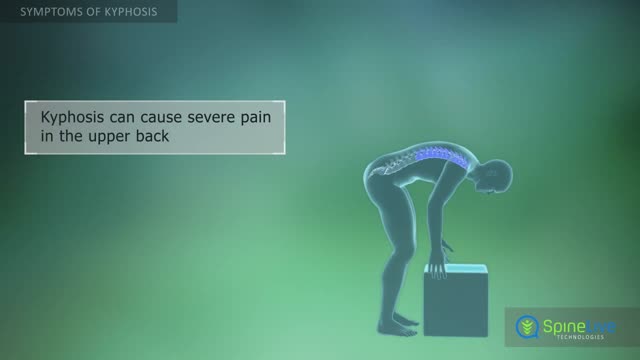
Kyphosis, also known as a round back or hunchback, is a condition in which the spine in the upper back has an excessive curvature. The upper back, or thoracic region of the spine, is supposed to have a slight natural curve.
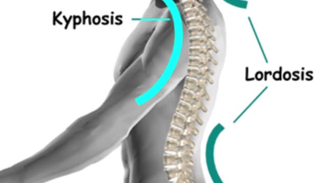
The gradual curves of the human spine allow the body to absorb many shocks and stresses in daily life. It’s a delicate balance, though, and if part of the spine curves too much, pain and limited mobility may result.
Dermatomyositis (dur-muh-toe-my-uh-SY-tis) is an uncommon inflammatory disease marked by muscle weakness and a distinctive skin rash. Dermatomyositis affects adults and children alike. In adults, dermatomyositis usually occurs from the late 40s to early 60s. In children, the disease most often appears between 5 and 15 years of age. Dermatomyositis affects more females than males. There's no cure for dermatomyositis, but periods of remission — when symptoms improve spontaneously — may occur. Treatment can clear the skin rash and help you regain muscle strength and function. Symptoms ShareTweet June 17, 2014 References Products and Services Newsletter: Mayo Clinic Health Letter See also Dysphagia Electromyography Fatigue MRI Muscle pain Peptic ulcer Prednisone risks, benefits Show more Advertisement Mayo Clinic does not endorse companies or products. Advertising revenue supports our not-for-profit mission. Advertising & Sponsorship PolicyOpportunitiesAd Choices Mayo Clinic Store Check out these best-sellers and special offers on books and newsletters from Mayo Clinic. NEW! – The Mayo Clinic Diet, Second Edition Treatment Strategies for Arthritis Mayo Clinic on Better Hearing and Balance Keeping your bones healthy and strong The Mayo Clinic Diet Online
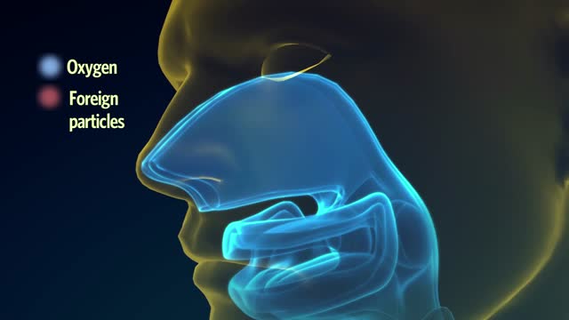
The lungs and respiratory system allow oxygen in the air to be taken into the body, while also enabling the body to get rid of carbon dioxide in the air breathed out. Respiration is the term for the exchange of oxygen from the environment for carbon dioxide from the body's cells.

Head to SimpleNursing’s OFFICIAL website here: https://bit.ly/3TzGwF0
SimpleNursing memberships have 1,200+ animated videos, 900+ colorful study guides, 3,000+ practice questions, and more! See why SimpleNursing is trusted by over 1,000,000 nursing students.
Today’s video is all about peritoneal dialysis vs hemodialysis for Nursing Students and NCLEX Review.
Two common treatments for kidney failure are hemodialysis and peritoneal dialysis. With the right nursing assessments and interventions, your kidney failure patient can have a better chance at recovery.
We’re going over the roles that potassium plays in each of these two types of dialysis, as well as how stenosis monitoring can be used to prevent complications.
00:00 Introduction
01:10 Hyperkalemia in Hemodialysis
02:27 Assessing Fluid Status
03:35 Medications to Hold Before Hemodialysis
04:50 Medications Removed During Hemodialysis
05:45 Dialysis Disequilibrium Syndrome
07:20 Caring for a Fistula
09:12 Avoiding Fistula Complications
10:35 Peritoneal Dialysis
11:23 Peritonitis Risk
12:31 Respiratory Distress With Peritoneal Dialysis
13:39 Repositioning With Outflow Problems
#KidneyFailure #Dialysis #Hemodialysis #Peritonealdialysis
