- Physical Examination
- Surgical Examination
- Ophthalmology
- Clinical Skills
- Orthopedics
- Surgery Videos
- Laparoscopy
- Pediatrics
- Funny Videos
- Cardiothoracic Surgery
- Nursing Videos
- Plastic Surgery
- Otorhinolaryngology
- Histology and Histopathology
- Neurosurgery
- Dermatology
- Pediatric Surgery
- Urology
- Dentistry
- Oncology and Cancers
- Anatomy Videos
- Health and Fitness
- Radiology
- Anaesthesia
- Physical Therapy
- Pharmacology
- Interventional Radiology
- Cardiology
- Endocrinology
- Gynecology
- Emergency Medicine
- Psychiatry and Psychology
- Childbirth Videos
- General Medical Videos
- Nephrology
- Physiology
- Diet and Food Health
- Diabetes Mellitus
- Neurology
- Women Health
- Osteoporosis
- Gastroenterology
- Pulmonology
- Hematology
- Rheumatology
- Toxicology
- Nuclear Medicine
- Infectious Diseases
- Vascular Disease
- Reproductive Health
- Burns and Wound Healing
- Other
Top videos

Quran describes the stages of human creation accurately

Blackheads are small bumps that appear on your skin due to clogged hair follicles. These bumps are called “blackheads” because the surface looks dark or black. Blackheads are a mild type of acne that usually form on the face, but they can also appear on the back, chest, neck, arms, and shoulders
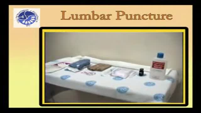
Pediatric Lumbar puncture

In this video, Professor Dan Reinstein performs a bilateral LASIK procedure filmed in real-time to demonstrate the full 8 and-a-half minute procedure from multiple angles. The superior design and experience of the Carl Zeiss Meditec Visumax femtosecond Laser for flap creation is seen, where the patient is only in contact with the device for about 30 seconds with extremely low contract force such that the patient feels effectively nothing, there are no red splodges (subconjunctival haemorages) left behind. From the surgeons' standpoint there is no device that is easier to use or faster for LASIK flap creation. The Carl Zeiss Meditec MEL80 excimer laser portion of the procedure is seamlessly integrated and incorporates all the features that make clinical outcomes so reproducible including the unique cone-for-controlled-atmosphere (CCA) and high efficiency, high sensitivity calibration test which can be performed for each individual patient to compensate for minor changes in energy that occur with excimer laser devices during the course of a day.
For reference to the clinical outcomes for LASIK with the MEL80 in presbyopia using PRESBYOND Laser Blended Vision see:
Reading glasses presbyopia (ageing eyes) only:
LASIK for presbyopia correction in emmetropic patients using aspheric ablation profiles and a micro-monovision protocol with the Carl Zeiss Meditec MEL 80 and VisuMax.
J Refract Surg. 2012 Aug;28(8):531-41. Reinstein DZ, Carp GI, Archer TJ, Gobbe M.
http://www.ncbi.nlm.nih.gov/pubmed/22869232
Short sighted, astigmatism and presbyopia (ageing eyes)
LASIK for Myopic Astigmatism and Presbyopia Using Non-Linear Aspheric Micro-Monovision with the Carl Zeiss Meditec MEL 80 Platform.
J Refract Surg. 2011 Jan;27(1):23-37. Epub 2010 Mar 1.
Reinstein DZ, Archer TJ, Gobbe M.
http://www.ncbi.nlm.nih.gov/pubmed/20205360
Long-sighted, astigmatism and presbyopia (ageing eyes)
LASIK for hyperopic astigmatism and presbyopia using micro-monovision with the Carl Zeiss Meditec MEL80 platform.
J Refract Surg. 2009 Jan;25(1):37-58. Reinstein DZ, Couch DG, Archer TJ.
http://www.ncbi.nlm.nih.gov/pubmed/19244952
For more information about laser eye surgery and PRESBYOND Laser Blended Vision, please contact the London Vision Clinic on 020 7224 1005.
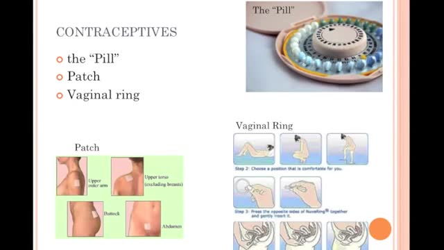
Choosing not to have sex provides 100 percent protection from HIV, STIs, and pregnancy. For some, this means avoiding vaginal, anal, and oral-genital intercourse altogether. Others may choose to avoid any type of sexual or intimate contact, including hugging and kissing. Choosing not to have sex is often referred to as “abstinence.” WHAT ARE THE ADVANTAGES OF CHOOSING NOT TO HAVE SEX (ABSTINENCE)? Choosing not to have sex is free and available to all. Not having sex is extremely effective at preventing both infection and pregnancy. It is the only 100% effective method of preventing sexually transmitted infections (STIs) and unintended pregnancy. Not having sex can be practiced at any time in one's life. Not having sex may encourage people to build relationships in other ways. Not having sex may be the course of action which you feel is right for you and makes you feel good about yourself.
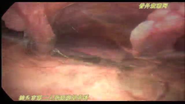
腹腔镜十二指肠球部溃疡穿孔修补术
soaking the wound in cool water for five minutes or longer. taking acetaminophen or ibuprofen for pain relief. applying lidocaine (an anesthetic) with aloe vera gel or cream to soothe the skin. using an antibiotic ointment and loose gauze to protect the affected area.
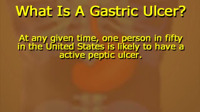
What Causes Ulcers? No single cause has been found for ulcers. However, it is now clear that an ulcer is the end result of an imbalance between digestive fluids in the stomach and duodenum. Most ulcers are caused by an infection with a type of bacteria called Helicobacter pylori (H. pylori). Factors that can increase your risk for ulcers include: Use of painkillers called nonsteroidal anti-inflammatory drugs (NSAIDs), such as aspirin, naproxen (Aleve, Anaprox, Naprosyn, and others), ibuprofen (Motrin, Advil, some types of Midol, and others), and many others available by prescription; even safety-coated aspirin and aspirin in powered form can frequently cause ulcers. Excess acid production from gastrinomas, tumors of the acid producing cells of the stomach that increases acid output (seen in Zollinger-Ellison syndrome) Excessive drinking of alcohol Smoking or chewing tobacco Serious illness Radiation treatment to the area What Are the Symptoms of an Ulcer? An ulcer may or may not have symptoms. When symptoms occur, they may include: A gnawing or burning pain in the middle or upper stomach between meals or at night Bloating Heartburn Nausea or vomiting In severe cases, symptoms can include: Dark or black stool (due to bleeding) Vomiting blood (that can look like "coffee-grounds") Weight loss Severe pain in the mid to upper abdomen

Vesicoureteral (ves-ih-koe-yoo-REE-tur-ul) reflux is the abnormal flow of urine from your bladder back up the tubes (ureters) that connect your kidneys to your bladder. Normally, urine flows only down from your kidneys to your bladder. Vesicoureteral reflux is usually diagnosed in infants and children. The disorder increases the risk of urinary tract infections, which, if left untreated, can lead to kidney damage. Vesicoureteral reflux can be primary or secondary. Children with primary vesicoureteral reflux are born with a defect in the valve that normally prevents urine from flowing backward from the bladder into the ureters. Secondary vesicoureteral reflux is due to a urinary tract malfunction, often caused by infection. Children may outgrow primary vesicoureteral reflux. Treatment, which includes medication or surgery, aims at preventing kidney damage.
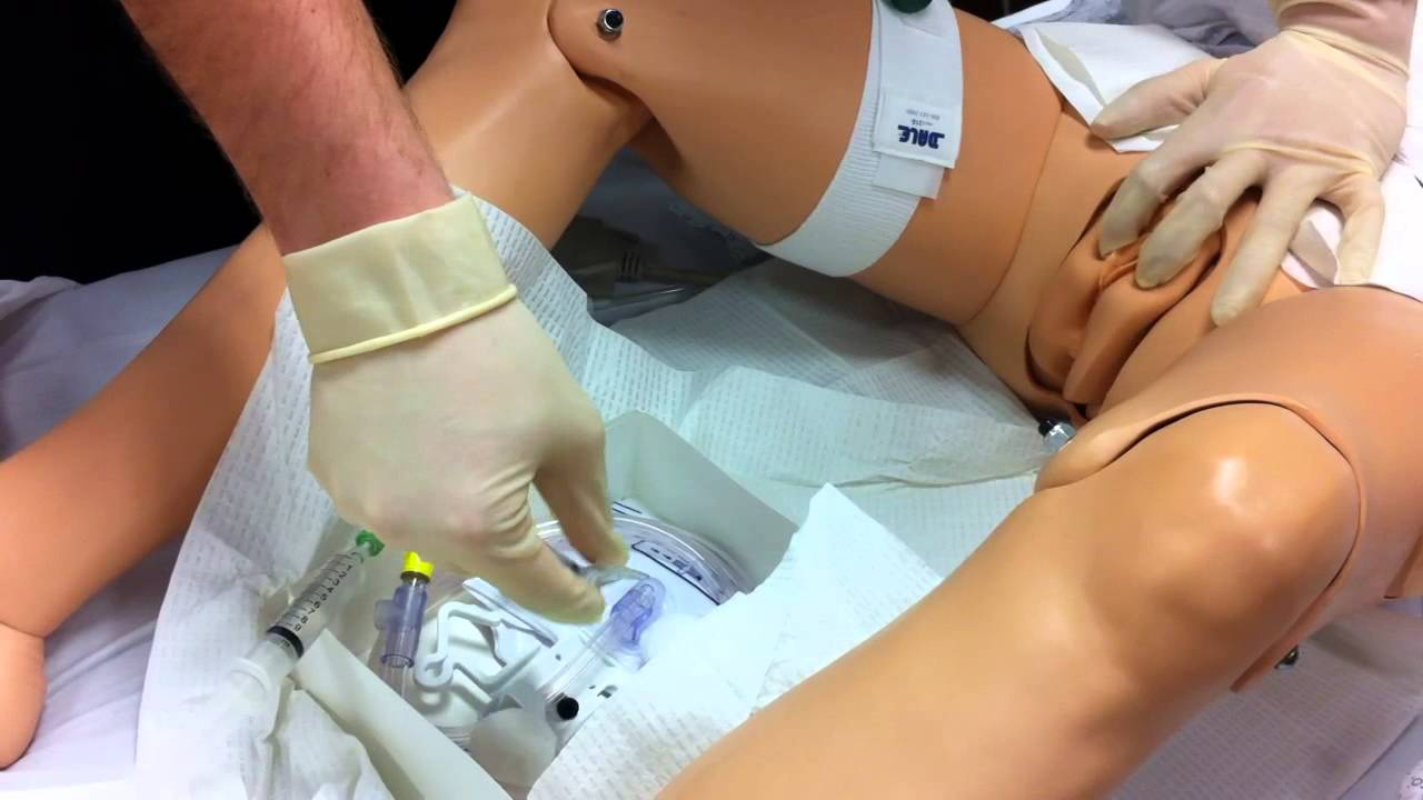
Watch that Female Foley Genital Catheter Insertion Procedure
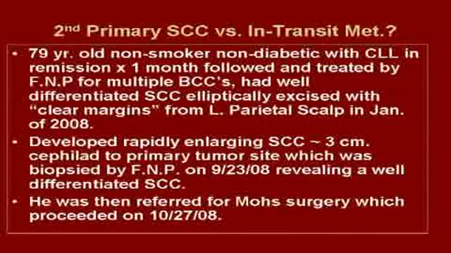
Graphic content of Mohs surgical removal of a large Squamous Cell Carcinoma on scalp followed by reconstruction with 10 week follow up. Visit us @ skincancercentre.com.

A coma is a prolonged state of unconsciousness. During a coma, a person is unresponsive to his or her environment. The person is alive and looks like he or she is sleeping. However, unlike in a deep sleep, the person cannot be awakened by any stimulation, including pain.

wide resection of giant cell tumor ,then strut grafting using free fibula graft,knowles pinning of the graft.
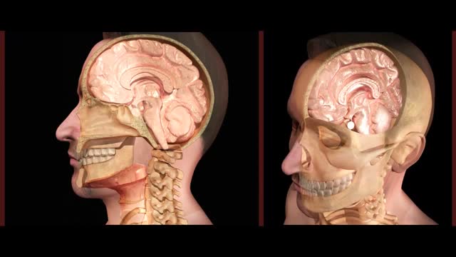
This was a Nasogastric Intubation that went very wrong. The tube went up into the brain, causing severe damage, instead of going down through the throat.

Demonstration of horizontal mattress suturing technique for laceration repair or wound closure in the operating room.
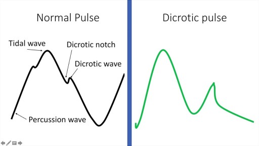
A detailed description of the Arterial Pulse including its waveform and pathological subtypes. Also discussed are the abnormal rates (tachycardia and bradycardia) and their causes, abnormal rhythm (including regularly regular and irregularly irregular pulses) and abnormal character (including pulses bisferiens, pulses parvus et tarsus, pulsus alternans, pulses paradoxus and others.) Description of pulse in various pathological states including Aortic stenosis and aortic regurgitation is also included. Finally there is also a description of the peripheral signs of aortic regurgitation.
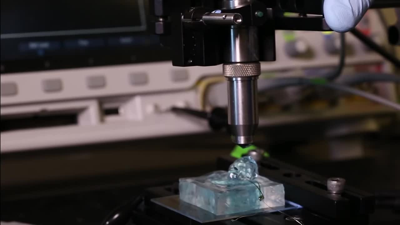
University of California, Berkeley engineers have built the first dust-sized, wireless sensors that can be implanted in the body, bringing closer the day when a Fitbit-like device could monitor internal nerves, muscles or organs in real time.
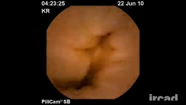
Meckels Diverticulum

There's only one group of people who really know what happens when you die: the dead. And since the dead won't be revealing their secrets anytime soon, it's up to scientists to explain what happens when a person dies. Death, just like life, is a process, scientists say. The first stage of this process is known as clinical death. It lasts from four to six minutes, beginning when a person stops breathing and the heart stops pumping blood. During this time, there may be enough oxygen in the brain that no permanent brain damage occurs. Other organs, such as the kidneys and eyes, also remain alive throughout clinical death.

Diastasis recti often occurs during pregnancy and can persist after pregnancy. It affects core strength and the appearance of the abdominal muscles.
Dr. Erick Sanchez repairs the abdominal muscles with every tummy tuck. This short video shows the muscle repair portion of the surgery with a bonus after photo at the end!
To request a consultation with Dr. Sanchez, visit sanchezplasticsurgery.com and click Request a Consultation. Fill out the form and someone will get in touch with you to answer all your questions.
Expected cost can be found at the bottom of each procedure page on our website.
