- Physical Examination
- Surgical Examination
- Ophthalmology
- Clinical Skills
- Orthopedics
- Surgery Videos
- Laparoscopy
- Pediatrics
- Funny Videos
- Cardiothoracic Surgery
- Nursing Videos
- Plastic Surgery
- Otorhinolaryngology
- Histology and Histopathology
- Neurosurgery
- Dermatology
- Pediatric Surgery
- Urology
- Dentistry
- Oncology and Cancers
- Anatomy Videos
- Health and Fitness
- Radiology
- Anaesthesia
- Physical Therapy
- Pharmacology
- Interventional Radiology
- Cardiology
- Endocrinology
- Gynecology
- Emergency Medicine
- Psychiatry and Psychology
- Childbirth Videos
- General Medical Videos
- Nephrology
- Physiology
- Diet and Food Health
- Diabetes Mellitus
- Neurology
- Women Health
- Osteoporosis
- Gastroenterology
- Pulmonology
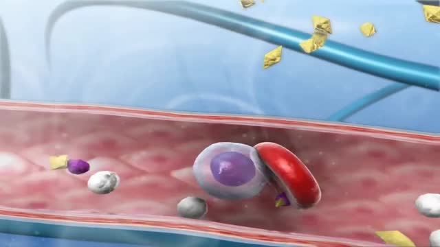
- Hematology
- Rheumatology
- Toxicology
- Nuclear Medicine
- Infectious Diseases
- Vascular Disease
- Reproductive Health
- Burns and Wound Healing
- Other
Top videos

The preferred route of access for temporary transvenous pacing is the internal jugular vein followed by subclavian and femoral veins. However, all the major venous access sites (internal and external jugular, subclavian, brachial, femoral) have been used and each is associated with particular problems.
The AutoPulse® Resuscitation System provides high-quality automated CPR to victims of sudden cardiac arrest. Easy to use and battery operated, the AutoPulse squeezes the patient’s entire chest to improve blood flow to the heart and brain.1,2,3 The only device of its kind, the AutoPulse automatically sizes to the patient, and has shown improved outcomes in numerous clinical trials.4,5 Designed for Patient Movement and Transport When the AutoPulse’s stabilizing board is placed on a soft stretcher, rescuers can continue providing high-quality CPR down steep stairwells, around sharp corners, or even in a cramped elevator. Compared with manual CPR, the AutoPulse has been shown to reduce interruptions in compressions during transport by more than 85%.6 The AutoPulse is made for resuscitation on the move.
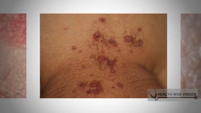
HPV is a different virus than HIV and HSV (herpes). HPV is so common that nearly all sexually active men and women get it at some point in their lives. There are many different types of HPV. Some types can cause health problems including genital warts and cancers.
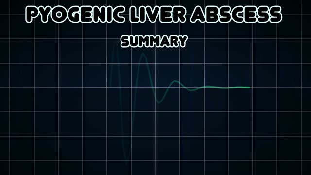
Pyogenic liver abscess Email this page to a friend Email this page to a friend Facebook Twitter Google+ Pyogenic liver abscess is a pus-filled area in the liver. Causes There are many potential causes of liver abscesses, including: Abdominal infection, such as appendicitis, diverticulitis, or a perforated bowel Infection in the blood Infection of the bile draining tubes Recent endoscopy of the bile draining tubes Trauma that damages the liver The most common bacteria that cause liver abscesses are: Escherichia coli Bacteroides Enterococcus Klebsiella pneumoniae Staphylococcus aureus Streptococcus In most cases, more than one type of bacteria is found.
How To Apply Contact Lenses

Today I will discuss about hemodialysis.
Start with a free 3-day trial at ReMarNurse.com/FREE
Follow & Subscribe for more weekly nursing and NCLEX content every Monday and Wednesday with Regina MSN, RN!
00:00 Introduction
02:53 Hemodialysis
06:06 Dialysis Apparatus
07:59 Dialysis Mechanism
13:27 Vascular Access
18:55 Nursing Considerations
25:07 Nursing Management for HD
27:57 NCLEX Practice Questions
Hemodialysis is a procedure where a dialysis machine and a special filter called an artificial kidney, or a dialyzer, are used to clean your blood.
I will also discuss about hemodialysis procedure, how hemodialysis machine works and its benefits for patients.
If you're interested in learning more about hemodialysis, or if this just seems like something you should know for nursing school or for the NCLEX exam, check out this video!
Join the #1 community of nursing students on the planet with 12,000+ students studying now inside of the NCLEX Virtual Trainer review on sale now at http://www.ReMarNurse.com
► Subscribe to JOIN the ReMar YouTube Channel: http://bit.ly/ReMar-Subscription
Your NCLEX RN & LPN Study Tools:
► Get NCLEX Virtual Trainer: http://www.ReMarNurse.com/NCLEXVT
► Get the Question Bank: http://www.ReMarNurse.com/NCLEXQBank
► Get Quick Facts for NCLEX: http://bit.ly/QuickFactsNCLEX
Get MORE from Regina MSN, RN:
► WATCH MORE: http://bit.ly/PassNCLEXPlayList/
► GET THE PODCAST: https://remarnurse.podbean.com/
► WATCH LESSONS: http://bit.ly/ReMarNCLEXLectures/
► FOLLOW ReMar on Instagram: https://www.instagram.com/ReMarNurse/
► LIKE ReMar on Facebook: https://www.facebook.com/ReMarReview/
#nursingstudent #hemodialysis #nursing #remarreview
ReMar Review features weekly NCLEX review questions and lectures from Regina M. Callion MSN, RN. ReMar is the #1 content-based NCLEX review and has helped thousands of repeat-testers pass NCLEX with a 99.2% student success rate!
ReMar focuses on 100% core nursing content and as a result, has the best review to help nursing students pass boards - fast!
A hormone is a chemical messenger that enables communication between cells. Hormones are secreted by the glands of the endocrine system and they serve to maintain homeostasis and to regulate numerous other systems and processes, including reproduction and development.

In most instances, STDs are passed from an infected person to another person during sexual activities, through contact with the mucous membranes of the penis, vagina, mouth and rectum. Such activity includes vaginal, oral and anal intercourse. Gonorrhea and chlamydia also can be transmitted by fingers to eyes. A sexually transmissible infection (STI) is any infection or disease that can be passed from one person to another during sexual activity. Sexually transmissible infections include chlamydia, herpes, gonorrhoea, syphilis, genital herpes, scabies, pubic lice (crabs), hepatitis and HIV (the virus that causes AIDS).
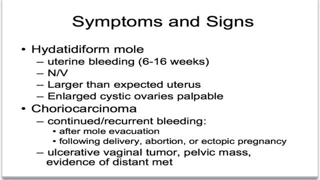
What is gestational trophoblastic disease? Cancer starts when cells in the body begin to grow out of control. Cells in nearly any part of the body can become cancer, and can spread to other areas of the body. To learn more about how cancers start and spread, see What Is Cancer? Gestational trophoblastic (jeh-STAY-shuh-nul troh-fuh-BLAS-tik) disease (GTD) is a group of rare tumors that involve abnormal growth of cells inside a woman's uterus. GTD does not develop from cells of the uterus like cervical cancer or endometrial (uterine lining) cancer do. Instead, these tumors start in the cells that would normally develop into the placenta during pregnancy. (The term gestational refers to pregnancy.) GTD begins in the layer of cells called the trophoblast (troh-fuh-BLAST) that normally surrounds an embryo. (Tropho- means nutrition, and -blast means bud or early developmental cell.) Early in normal development, the cells of the trophoblast form tiny, finger-like projections known as villi. The villi grow into the lining of the uterus. In time, the trophoblast layer develops into the placenta, the organ that protects and nourishes the growing fetus.

Cholesterol is a fat-like, waxy substance that can be found in all parts of your body. It helps your body make cell membranes, many hormones, and vitamin D. The cholesterol in your blood comes from two sources: the foods you eat and your liver. But your liver makes all the cholesterol your body needs.
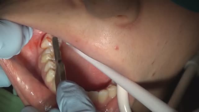
How to pull a wisdom tooth fully impacted

The timing of the nausea or vomiting can indicate the cause. When appearing shortly after a meal, nausea or vomiting may be caused by food poisoning, gastritis (inflammation of the stomach lining), an ulcer, or bulimia. Nausea or vomiting one to eight hours after a meal may also indicate food poisoning.
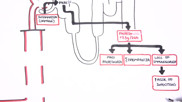
Nephrotic syndrome is a kidney disorder that causes your body to excrete too much protein in your urine. Nephrotic syndrome is usually caused by damage to the clusters of small blood vessels in your kidneys that filter waste and excess water from your blood. Nephrotic syndrome causes swelling (edema), particularly in your feet and ankles, and increases the risk of other health problems. Treatment for nephrotic syndrome includes treating the underlying condition that's causing it and taking medications. Nephrotic syndrome can increase your risk of infections and blood clots. Your doctor may recommend medications and dietary changes to prevent these and other complications of nephrotic syndrome.
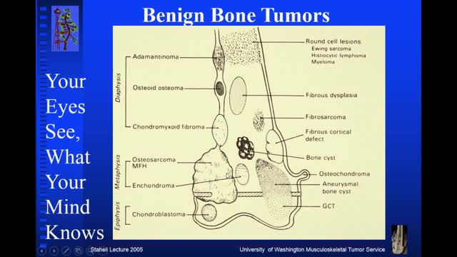
Osteochondroma. Osteochondromas (osteocartilaginous exostoses), the most common benign bone tumors, may arise from any bone but tend to occur near the ends of long bones. ... Enchondroma. ... Chondroblastoma. ... Chondromyxofibroma. ... Osteoid osteoma. ... Nonossifying fibroma (fibrous cortical defect) ... Benign giant cell tumor of bone.
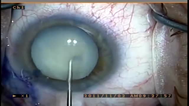
A cataract is a clouding of the lens in the eye that affects vision. Most cataracts are related to aging. Cataracts are very common in older people. By age 80, more than half of all Americans either have a cataract or have had cataract surgery. A cataract can occur in either or both eyes. It cannot spread from one eye to the other.

Alcohol not broken down by the liver goes to the rest of the body, including the brain. Alcohol can affect parts of the brain that control movement, speech, judgment, and memory. These effects lead to the familiar signs of drunkenness: difficulty walking, slurred speech, memory lapses, and impulsive behavior.
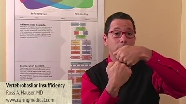
Vertebrobasilar insufficiency is typically secondary to emboli, thrombi, or arterial dissection. The labyrinth and brainstem are commonly affected, and symptoms may include vertigo, dizziness, dysarthria, diplopia, and numbness.
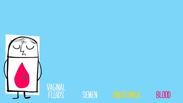
-Almost all the cases of occupational transmission of HIV have been due to transmission via exposure to blood and certain body fluids. The body fluids wherein standard precautions have been recommended include semen, vaginal secretions, and any other body fluid containing visible blood. Other standard precautions, according to the Center for Disease Control and Prevention (CDC), also apply to cerebrospinal, peritoneal, pleural, pericardia!, synovial fluid, or any other tissue, even though the epidemiologic data regarding the risk of HIV transmission from these fluids is insufficient. Standard precautions do not apply to urine, sweat, tears, sputum, vomitus, and nasal secretions or feces, as long as there is no gross visible blood. The occupational transmission of HIV has never been documented from the above sources.
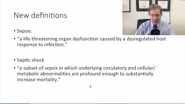
new sepsis definitions
