- Physical Examination
- Surgical Examination
- Ophthalmology
- Clinical Skills
- Orthopedics
- Surgery Videos
- Laparoscopy
- Pediatrics
- Funny Videos
- Cardiothoracic Surgery
- Nursing Videos
- Plastic Surgery
- Otorhinolaryngology
- Histology and Histopathology
- Neurosurgery
- Dermatology
- Pediatric Surgery
- Urology
- Dentistry
- Oncology and Cancers
- Anatomy Videos
- Health and Fitness
- Radiology
- Anaesthesia
- Physical Therapy
- Pharmacology
- Interventional Radiology
- Cardiology
- Endocrinology
- Gynecology
- Emergency Medicine
- Psychiatry and Psychology
- Childbirth Videos
- General Medical Videos
- Nephrology
- Physiology
- Diet and Food Health
- Diabetes Mellitus
- Neurology
- Women Health
- Osteoporosis
- Gastroenterology
- Pulmonology
- Hematology
- Rheumatology
- Toxicology
- Nuclear Medicine
- Infectious Diseases
- Vascular Disease
- Reproductive Health
- Burns and Wound Healing
- Other
Top videos

The pituitary is a small gland found inside the skull just below the brain and above the nasal passages, which are above the fleshy back part of the roof of the mouth (known as the soft palate). The pituitary sits in a tiny bony space called the sella turcica. The nerves that connect the eyes to the brain, called the optic nerves, pass close by it.

Lots of people wonder: does the pull out method work to prevent pregnancy? Pull out method effectiveness depends on whether or not you do it correctly. Learn more about pulling out in this video.
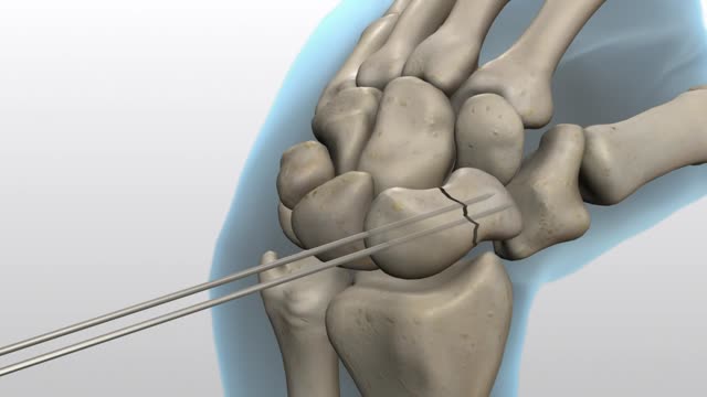
An animation for Acumed demonstrating their new line of screws for fixing fractures. This one is focused on a Scaphoid fracture in the hand.
Duct tape is one home remedy. Put a small strip over the wart and leave it on for six days. Then, remove the tape, soak the wart in water, and then gently debride it with a pumice stone or emory board. Repeat the process many times until the wart is gone.
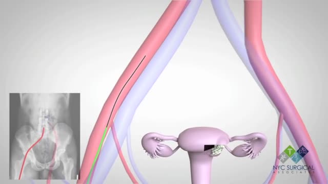
There's no single best approach to uterine fibroid treatment — many treatment options exist. If you have symptoms, talk with your doctor about options for symptom relief. Watchful waiting Many women with uterine fibroids experience no signs or symptoms, or only mildly annoying signs and symptoms that they can live with. If that's the case for you, watchful waiting could be the best option. Fibroids aren't cancerous. They rarely interfere with pregnancy. They usually grow slowly — or not at all — and tend to shrink after menopause, when levels of reproductive hormones drop. Medications Medications for uterine fibroids target hormones that regulate your menstrual cycle, treating symptoms such as heavy menstrual bleeding and pelvic pressure. They don't eliminate fibroids, but may shrink them. Medications include: Gonadotropin-releasing hormone (Gn-RH) agonists. Medications called Gn-RH agonists (Lupron, Synarel, others) treat fibroids by blocking the production of estrogen and progesterone, putting you into a temporary postmenopausal state. As a result, menstruation stops, fibroids shrink and anemia often improves. Your doctor may prescribe a Gn-RH agonist to shrink the size of your fibroids before a planned surgery. Many women have significant hot flashes while using Gn-RH agonists. Gn-RH agonists typically are used for no more than three to six months because symptoms return when the medication is stopped and long-term use can cause loss of bone. Progestin-releasing intrauterine device (IUD). A progestin-releasing IUD can relieve heavy bleeding caused by fibroids. A progestin-releasing IUD provides symptom relief only and doesn't shrink fibroids or make them disappear. It also prevents pregnancy. Tranexamic acid (Lysteda). This nonhormonal medication is taken to ease heavy menstrual periods. It's taken only on heavy bleeding days. Other medications. Your doctor might recommend other medications. For example, oral contraceptives or progestins can help control menstrual bleeding, but they don't reduce fibroid size. Nonsteroidal anti-inflammatory drugs (NSAIDs), which are not hormonal medications, may be effective in relieving pain related to fibroids, but they don't reduce bleeding caused by fibroids. Your doctor may also suggest that you take vitamins and iron if you have heavy menstrual bleeding and anemia
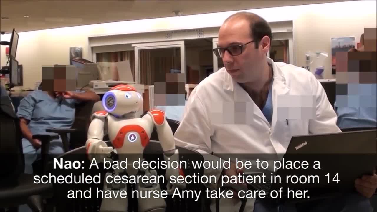
Medical Robot Assistants, new technology
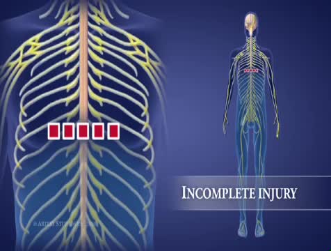
High-Cervical Nerves (C1 – C4) Most severe of the spinal cord injury levels Paralysis in arms, hands, trunk and legs Patient may not be able to breathe on his or her own, cough, or control bowel or bladder movements. Ability to speak is sometimes impaired or reduced. When all four limbs are affected, this is called tetraplegia or quadriplegia. Requires complete assistance with activities of daily living, such as eating, dressing, bathing, and getting in or out of bed May be able to use powered wheelchairs with special controls to move around on their own Will not be able to drive a car on their own Requires 24-hour-a-day personal care
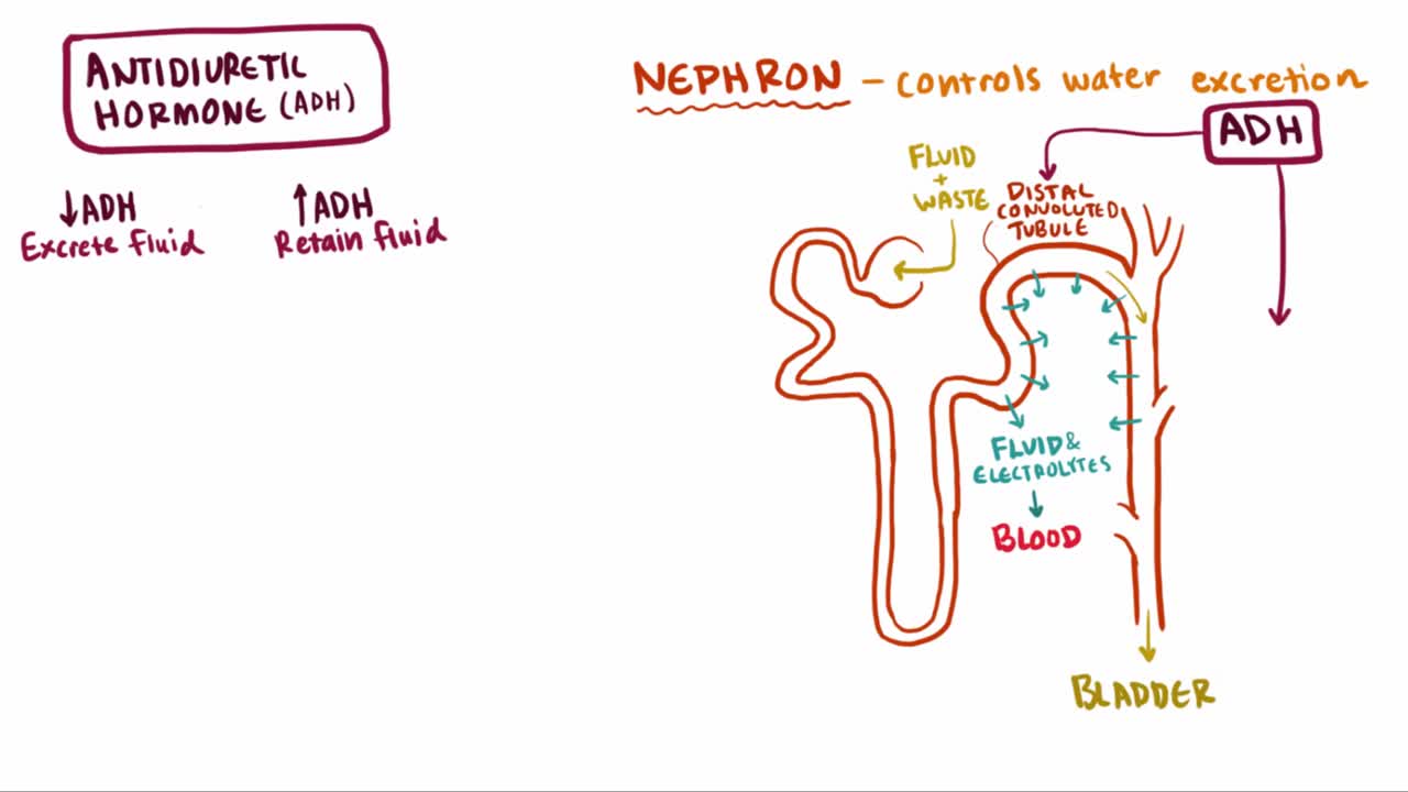
What is syndrome of inappropriate antidiuretic hormone (SIADH)? Well, SIADH is a condition where too much ADH hormone is released, which causes an increase in blood volume and ultimately leads to a series of complications related to the blood osmolality and osmolarity

This video describe the clinical managment of a patient with hyperprolactinemia, including the approach to diagnosis, important endocrine testing, and management options.
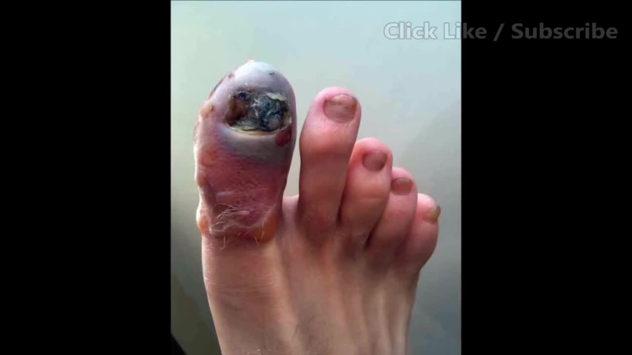
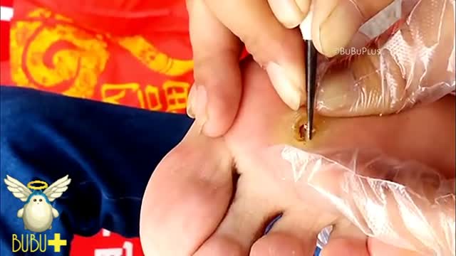
Worst Ingrown Toenail! What Caused It?
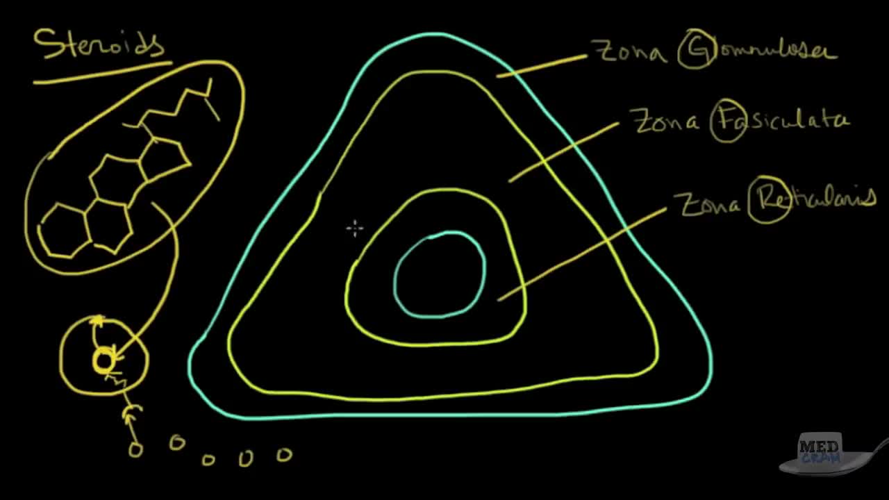
Understand the adrenal gland with a focus on the adrenal cortex with this clear explanation .
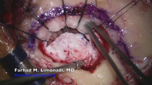
Brain Surgery: Microvascular Decompression of facial nerve for hemifacial spasm
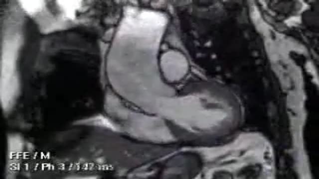
Video of aortic valve repair surgery

Cannula are often introduced into blood vessels in 80% of patients in the hospital for treatment. This can be a daunting experience to patients and stressful to doctors as multiple attempts are used. This may result in introducing spreading MRSA, E Coli & Chlostredium living on your skin into blood and results in Invasive MRSA infection.
Skin is often not adequatly cleaned during subsequent atempts as doctors/nurses do not wait for 1 min after applying cleaning solution on the skin before they puncture your skin.
Multiple punctured sites allow CA-MRSA to enter blood stream resulting in bacteremia and death.
Our mission is to reduce spreading invasive CA-MRSA in the hospitals by developing alternative technique to introduce cannulae.
Medifix was created by doctors with a mission to reduce the threat of spreading antibiotic resustant bacteria to mankind.


Cosmetic surgeryVideo

Histology of Eye
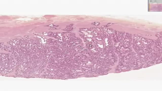
Histology of Prostate

Portal hypertension is an increase in the blood pressure within a system of veins called the portal venous system. ... If the vessels in the liver are blocked due to liver damage, blood cannot flow properly through the liver. As a result, high pressure in the portal system develops

new sepsis definitions
