
- Physical Examination
- Surgical Examination
- Ophthalmology
- Clinical Skills
- Orthopedics
- Surgery Videos
- Laparoscopy
- Pediatrics
- Funny Videos
- Cardiothoracic Surgery
- Nursing Videos
- Plastic Surgery
- Otorhinolaryngology
- Histology and Histopathology
- Neurosurgery
- Dermatology
- Pediatric Surgery
- Urology
- Dentistry
- Oncology and Cancers
- Anatomy Videos
- Health and Fitness
- Radiology
- Anaesthesia
- Physical Therapy
- Pharmacology
- Interventional Radiology
- Cardiology
- Endocrinology
- Gynecology
- Emergency Medicine
- Psychiatry and Psychology
- Childbirth Videos
- General Medical Videos
- Nephrology
- Physiology
- Diet and Food Health
- Diabetes Mellitus
- Neurology
- Women Health
- Osteoporosis
- Gastroenterology
- Pulmonology
- Hematology
- Rheumatology
- Toxicology
- Nuclear Medicine
- Infectious Diseases
- Vascular Disease
- Reproductive Health
- Burns and Wound Healing
- Other
Top videos


Sliced Fingertip Makeup Tutorial
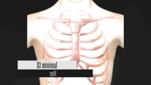
https://www.youtube.com/watch?v=0N7Yy1UYEWk
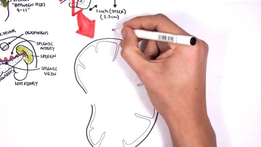
The spleen is an organ in the upper far left part of the abdomen, to the left of the stomach. The spleen varies in size and shape between people, but it’s commonly fist-shaped, purple, and about 4 inches long. Because the spleen is protected by the rib cage, you can’t easily feel it unless it’s abnormally enlarged. The spleen plays multiple supporting roles in the body. It acts as a filter for blood as part of the immune system. Old red blood cells are recycled in the spleen, and platelets and white blood cells are stored there. The spleen also helps fight certain kinds of bacteria that cause pneumonia and meningitis.
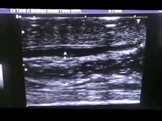
The epididymis is a long coiled tube that lies above and behind each testicle. The epididymis collects and transports sperm from the testis to the vas deferens (tubes that transport sperm to the urethra). An epididymal cyst is a cyst-like mass in the epididymis that contains clear fluid. Typically, epididymal cysts and spermatoceles do not cause symptoms. When discovered, the epididymal cyst is usually about the size of a pea and feels separate from the top of the testis. Spermatoceles typically arise from the head of the epididymis, and are felt on the top portion of the testicle. Epididymal cysts and spermatoceles are often incidental findings on testicular self-examination or routine physical examination. It is important that any mass noted in the scrotum be examined by a urologist in order to obtain an accurate diagnosis, especially a mass on the testicle itself. Our team in the Division of Urology will typically be able to confirm the diagnosis on physical exam. However, a scrotal ultrasound may also be used in order to rule out other conditions.

The preferred route of access for temporary transvenous pacing is the internal jugular vein followed by subclavian and femoral veins. However, all the major venous access sites (internal and external jugular, subclavian, brachial, femoral) have been used and each is associated with particular problems.
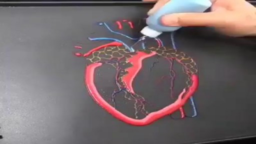
Pancake by a Cardiologist
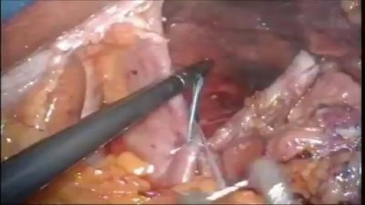
Hiatal hernias occur when contents of the abdominal cavity protrude through the esophageal hiatus of the diaphragm. Factors that contribute to the development of a hiatal hernia include an enlargement of the esophageal hiatus due to developmental defects, an increased abdominal thoracic pressure gradient, and the depletion of elastic fibers in the phrenoesophageal membrane with aging. There are four different types of hiatal hernias and management varies depending on the type. Type I, also known as a sliding hernia, is a simple displacement of the gastroesophageal junction into the thoracic cavity. The stomach remains in the abdominal cavity. This is the most common type of hiatal hernia, accounting for about 95% of all hiatal hernias. Types II-IV are classified as paraesophageal hernias. Type II occurs when the gastroesophageal junction maintains its position but the gastric fundus herniates through the diaphragmatic hiatus. Type III has both the gastroesophageal junction and the stomach herniate above the diaphragm. When more than 30% of the stomach is herniated into the thoracic cavity, it is termed a “giant” paraesophageal hernia. A patient has a type IV hernia when other organs, such as the colon, in addition to the stomach herniate above the diaphragm.

Dr. Arthur Handal explains both an open and closed approach to a Rhinoplasty and which is more effective, in his opinion.

Systemic Lupus Erythematosus Animation 3d
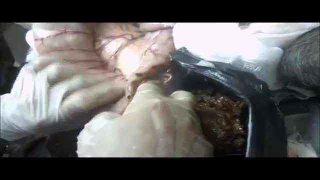
Megacolon, as well as megarectum, is a descriptive term. It denotes dilatation of the colon that is not caused by mechanical obstruction.[1, 2] Although the definition of megacolon has varied in the literature, most researchers use the measurement of greater than 12 cm for the cecum as the standard. Because the diameter of the large intestine varies, the following definitions would also be considered: greater than 6.5 cm in the rectosigmoid region and greater than 8 cm for the ascending colon. Megacolon can be divided into the following 3 categories: Acute megacolon ( pseudo-obstruction) Chronic megacolon, which includes congenital, acquired, and idiopathic causes Toxic megacolon
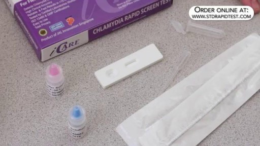
How to use a Chlamydia rapid test kit for self-diagnosis of Chlamydia (swab test). Convenient, Easy to Use, and over 95% Accurate. Certified GMP and ISO13485. Test yourself at home with Complete Privacy. Buy online today at: http://www.stdrapidtest.com
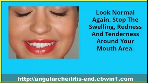
Cracked Corners Of Mouth, Cheilitis, Angular Cheilitis Remedy, Angular Cheilitis Medicine, Cheilitis--- http://angularcheilitis-end.cbwin1.com --- Foods Which Can Limit the Occurrence of Angular Cheilitis. People suffering from Angular Cheilitis know that this is one of the most troubling and annoying skin condition one can experience. It prevents you from eating, drinking and speaking normally. Many people even refuse to go out of the house when suffering from this condition, thus becoming isolated from the rest of the world. This is why it is better to prevent it then having to treat it. If you have had it long time ago and are afraid that will come back, if you have it and want to treat it faster or if you do not want to have this terrible experience ever, you should start by eating the foods listed below. They will provide your body with all the vitamins and nutrients necessary to effectively fight this disease and prevent it from appearing ever again. Most of the times, Angular Cheilitis appears as a result of a weak immune system. Thus, you will need to have a balanced diet, filled with fruits and vegetables that will supply you with all the things you need to remain healthy and have a strong immune system. The first thing that you will need to have in your body to fight Angular Cheilitis is iron. If you no longer want to have those anesthetic and painful cracks around your mouth, if you want to eat, drink and speak normally without experiencing any pain when opening your mouth, then check out this new and revolutionary treatment! It will get you rid of Angular Cheilitis in just a few days and you will be able to enjoy life to its fullest again, without worrying about those otiose cracks! Click Here. http://angularcheilitis-end.cbwin1.com

Stay active and push your body to its limit – tips on how you can mend strained muscles and prevent injury.

Most people with TS are not significantly impaired by their symptoms and therefore do not require treatment with medication. However, several medications are available to control TS symptoms that interfere with functioning. Except in more severe cases when tics are sometimes painful, the main reasons for medication are to improve appearance and lessen embarrassing social interactions. As with all medications, there are possible side effects that should be monitored carefully by the physician. Patients should always be included in the decision to take medication, as they are the best judge of how disruptive the symptoms are to them. Early diagnosis and treatment are crucial to help the person with TS cope with his or her condition. Often, people make fun of a person with TS. Parents of children with TS also may find it difficult to cope with their child's behavior. When people with TS are diagnosed correctly and treated for the condition early, they can learn to cope with their disorder and accept that they are ok and normal.

The procedure is very simple, safe & relatively pain free. The technique takes 5 to 8 hours depending on grafts quantity. Most of the transplanted hair goes through a healing phase of couple of months after that these transplanted hair starts growing same like non-transplanted hair. The best benefit of the procedure is that they have different genetic make-up than the lost hair and are genetically permanent. Patient can cut, dye and colour these hair and grow continuously like non-transplanted hair.
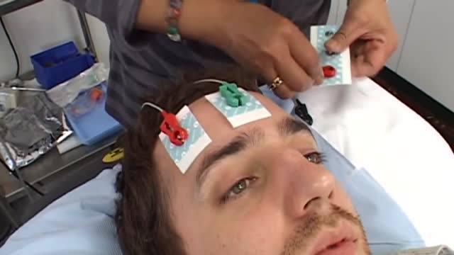
Electroconvulsive therapy (ECT) is a procedure, done under general anesthesia, in which small electric currents are passed through the brain, intentionally triggering a brief seizure. ECT seems to cause changes in brain chemistry that can quickly reverse symptoms of certain mental illnesses. It often works when other treatments are unsuccessful. Much of the stigma attached to ECT is based on early treatments in which high doses of electricity were administered without anesthesia, leading to memory loss, fractured bones and other serious side effects. ECT is much safer today. Although ECT still causes some side effects, it now uses electric currents given in a controlled setting to achieve the most benefit with the fewest possible risks.
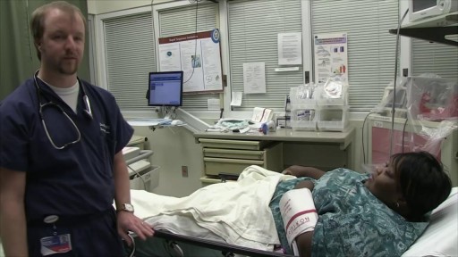
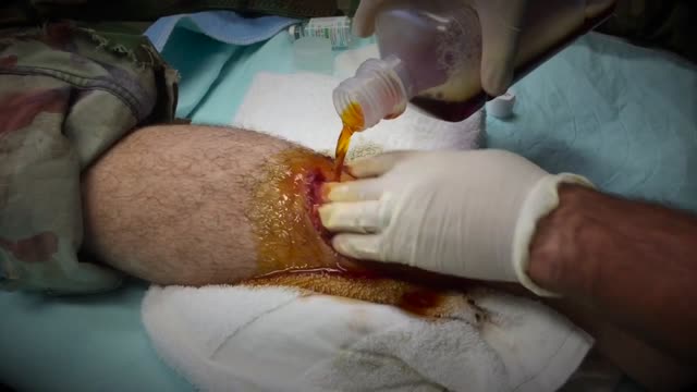
Chainsaw Accident! Lacerations, Cysts, Blackheads & Surgerys
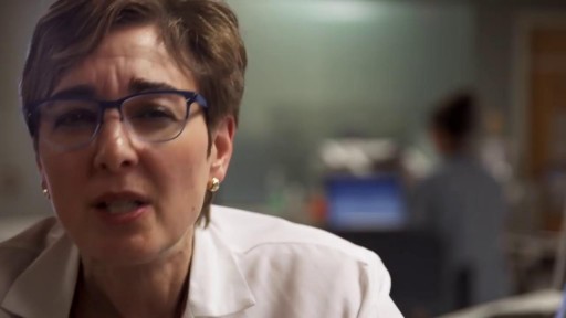

Peritoneal dialysis (per-ih-toe-NEE-ul die-AL-uh-sis) is a way to remove waste products from your blood when your kidneys can no longer do the job adequately. A cleansing fluid flows through a tube (catheter) into part of your abdomen and filters waste products from your blood. After a prescribed period of time, the fluid with filtered waste products flows out of your abdomen and is discarded. Peritoneal dialysis differs from hemodialysis, a more commonly used blood-filtering procedure. With peritoneal dialysis, you can give yourself treatments at home, at work or while traveling. Peritoneal dialysis isn't an option for everyone wit
