- Physical Examination
- Surgical Examination
- Ophthalmology
- Clinical Skills
- Orthopedics
- Surgery Videos
- Laparoscopy
- Pediatrics
- Funny Videos
- Cardiothoracic Surgery
- Nursing Videos
- Plastic Surgery
- Otorhinolaryngology
- Histology and Histopathology
- Neurosurgery
- Dermatology
- Pediatric Surgery
- Urology
- Dentistry
- Oncology and Cancers
- Anatomy Videos
- Health and Fitness
- Radiology
- Anaesthesia
- Physical Therapy
- Pharmacology
- Interventional Radiology
- Cardiology
- Endocrinology
- Gynecology
- Emergency Medicine
- Psychiatry and Psychology
- Childbirth Videos
- General Medical Videos
- Nephrology
- Physiology
- Diet and Food Health
- Diabetes Mellitus
- Neurology
- Women Health
- Osteoporosis
- Gastroenterology
- Pulmonology
- Hematology
- Rheumatology
- Toxicology
- Nuclear Medicine
- Infectious Diseases
- Vascular Disease
- Reproductive Health
- Burns and Wound Healing
- Other
Top videos

Future Technologies and Medical Advances That Will Change The World
Shoulder impingement syndrome, also called subacromial impingement, painful arc syndrome, supraspinatus syndrome, swimmer's shoulder, and thrower's shoulder, is a clinical syndrome which occurs when the tendons of the rotator cuff muscles become irritated and inflamed as they pass through the subacromial space ...
During surgery to repair the hernia, the bulging tissue is pushed back in. Your abdominal wall is strengthened and supported with sutures (stitches), and sometimes mesh. This repair can be done with open or laparoscopic surgery. You and your surgeon can discuss which type of surgery is right for you.
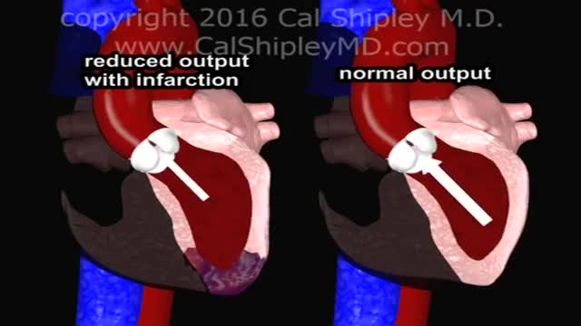
Cardiogenic shock is a condition in which your heart suddenly can't pump enough blood to meet your body's needs. The condition is most often caused by a severe heart attack. Cardiogenic shock is rare, but it's often fatal if not treated immediately. If treated immediately, about half the people who develop the condition survive.
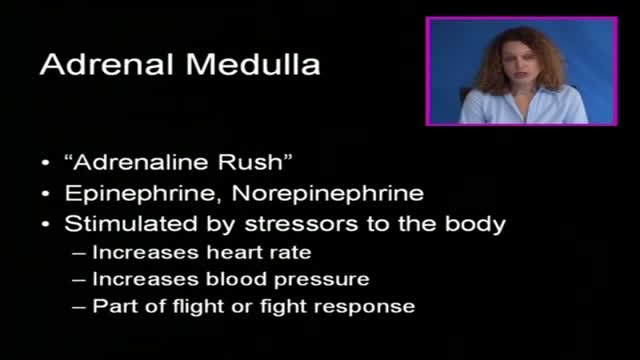
A pheochromocytoma (fee-o-kroe-moe-sy-TOE-muh) is a rare, usually noncancerous (benign) tumor that develops in cells in the center of an adrenal gland. You have two adrenal glands, one above each kidney. Your adrenal glands produce hormones that give instructions to virtually every organ and tissue in your body. If you have a pheochromocytoma, an adrenal gland releases hormones that cause persistent or episodic high blood pressure. If left untreated, a pheochromocytoma can result in severe or life-threatening damage to other body systems, especially the cardiovascular system. Most people with a pheochromocytoma are between the ages of 20 and 50, but the tumor can develop at any age. Surgical treatment to remove a pheochromocytoma usually returns blood pressure to normal.
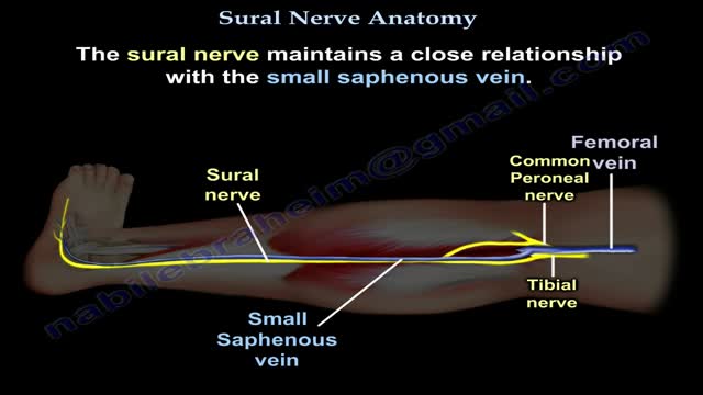
Nerves are the organs that make up the peripheral nervous system (PNS). They serve as information pipelines that allow the brain and spinal cord to communicate with other tissues and organs. Inside the nerves are the axon processes of sensory and motor neurons (nerve cells).
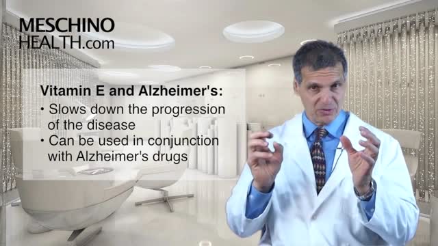
Vitamin E is an antioxidant. It may help protect your cells from damage. This essential nutrient occurs naturally in many foods. It’s also available as a dietary supplement. Sometimes, it’s in processed foods. Vitamin E is fat-soluble. This means your body stores and uses it as needed. The term “vitamin E” describes eight different compounds. Alpha-tocopherol is the most active one in humans
Endometrial cancer is a type of cancer that begins in the uterus. The uterus is the hollow, pear-shaped pelvic organ in women where fetal development occurs. Endometrial cancer begins in the layer of cells that form the lining (endometrium) of the uterus. Endometrial cancer is sometimes called uterine cancer. Other types of cancer can form in the uterus, including uterine sarcoma, but they are much less common than endometrial cancer. Endometrial cancer is often detected at an early stage because it frequently produces abnormal vaginal bleeding, which prompts women to see their doctors. If endometrial cancer is discovered early, removing the uterus surgically often cures endometrial cancer.
Shoulder dystocia is a rare emergency that can happen during the end of the second stage of labour. It's all to do with how your baby moves down through your vagina and out into the world. Shoulder dystocia happens when your baby's head has been born, but one of her shoulders becomes stuck.
If they are damaged, waste and fluids build up in your blood instead of leaving your body. Kidney damage from diabetes is called diabetic nephropathy. It begins long before you have symptoms. An early sign of it is small amounts of protein in your urine.

The eyes A close up of a young person's eyes. The eyes are responsible for four-fifths of all the information our brain receives. Here you can find out a bit more about how they work, common problems that affect vision and the work Sightsavers does to treat and prevent avoidable blindness. You can also find out more about the people whose lives have been changed thanks to donations from people like you. How do eyes work? (click image to see enlarged version or click here for text alternative) Graphic of an eye with information about its different parts The images we see are made up of light reflected from the objects we look at. This light enters the eye through the cornea. Because this part of the eye is curved, it bends the light, creating an upside down image on the retina (this is eventually put the right way up by the brain). The retina is a complex part of the eye, but only the very back of it is light sensitive. This part of the retina has roughly the area of a 10p coin, and is packed with photosensitive cells called rods and cones. Cones are the cells responsible for daylight vision. There are three kinds – each responding to a different wavelength of light: red, green and blue. The cones allow us to see images in colour and detail. Rods are responsible for night vision. They are sensitive to light but not to colour. In darkness, the cones do not function at all. How do we see an image? The lens focuses the image. It can do this because it is adjustable – using muscles to change shape and help us focus on objects at different distances. The automatic focusing of the lens is a reflex response and is not controlled by the brain. Once the image is clearly focused on the sensitive part of the retina, energy in the light that makes up that image creates an electrical signal. Nerve impulses can then carry information about that image to the brain through the optic nerve.
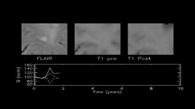
If you have multiple sclerosis (MS), you probably had several tests done before you received your diagnosis. There isn’t one test to diagnosis MS, so testing can vary. Doctors can use neurological exams, information about previous symptoms, blood tests, and spinal fluid tests. A magnetic resonance imaging (MRI) scan isn’t used to diagnose MS but rather to rule out other diseases. A diagnosis of MS requires more information than what a scan alone can give. By looking at more than one test or exam result, doctors can get a clearer picture of what’s going on in your body.
A heart attack is a medical emergency. A heart attack usually occurs when a blood clot blocks blood flow to the heart. Without blood, tissue loses oxygen and dies. Symptoms include tightness or pain in the chest, neck, back, or arms, as well as fatigue, lightheadedness, abnormal heartbeat, and anxiety. Women are more likely to have atypical symptoms than men. Treatment ranges from lifestyle changes and cardiac rehabilitation to medications, stents, and bypass surgery.
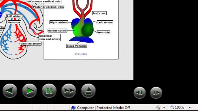
Embryonic cardiovascular system. ... The human arterial and venous systems develop from different embryonic areas. Aortic Arches. The aortic arches—or pharyngeal arch arteries—are a series of six, paired, embryological vascular structures that give rise to several major arteries .
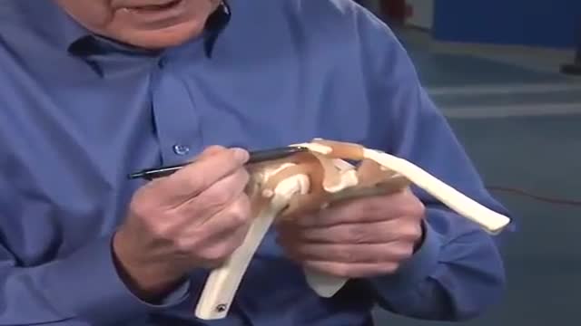
The rotator cuff is a group of muscles and tendons that surround the shoulder joint, keeping the head of your upper arm bone firmly within the shallow socket of the shoulder. A rotator cuff injury can cause a dull ache in the shoulder, which often worsens when you try to sleep on the involved side. Rotator cuff injuries occur most often in people who repeatedly perform overhead motions in their jobs or sports. Examples include painters, carpenters, and people who play baseball or tennis. The risk of rotator cuff injury also increases with age. Many people recover from rotator cuff disease with physical therapy exercises that improve flexibility and strength of the muscles surrounding the shoulder joint. Sometimes, rotator cuff tears may occur as a result of a single injury. In those circumstances, medical care should be provided as soon as possible. Extensive rotator cuff tears may require surgical repair, transfer of alternative tendons or joint replacement.
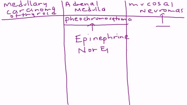
Multiple endocrine neoplasia type 2 (MEN2) is a hereditary condition associated with three primary types of tumors: medullary thyroid cancer, parathyroid tumors, and pheochromocytoma. MEN2 is classified into three subtypes based on clinical features. MEN2A, which affects 60% to 90% of MEN2 families Medullary thyroid cancer: 98% to 100% with MEN2A are affected Pheochromocytoma, a typically benign (noncancerous) tumor of the adrenal glands: 50% with MEN2A affected Parathyroid adenoma (benign tumor) or hyperplasia, meaning increased size, of the parathyroid gland: 5% to 10% with MEN2A affected MEN2B, which affects 5% of MEN2 families Medullary thyroid cancer: 98% to 100% with MEN2B affected Pheochromocytoma: 50% with MEN2B affected Mucosal neuromas, which is a benign tumor of nerve tissue on the tongue, lips and throughout the gastrointestinal tract: 95% to 98% affected Digestive problems caused by disordered nerves in the gastrointestinal tract: 75% to 90% affected Muscle, joint, and spinal problems: 95% affected Typical facial features, including swollen lips and thick eyelids: 75% to 90% affected Familial medullary thyroid cancer (FMTC), which affects 5% to 35% of MEN2 families Medullary thyroid carcinoma only Sources: Gagel RF, Marx SJ. “Multiple endocrine neoplasia.” Williams Textbook of Endocrinology, Chapter 40, 11th ed., Philadelphia, 2008, and Eng C, Clayton D, et al. Grubbs EG, Gagel RF. My, How Things Have Changed in Multiple Endocrine Neoplasia Type 2A! J Clin Endocrinol Metab 100(7):2532-5, 7/2015. PMID: 26151398. What causes MEN2? MEN2 is a genetic condition. This means that the cancer risk and other features of MEN2 can be passed from generation to generation in a family. The gene associated with MEN2 is called RET. A mutation (alteration) in the RET gene gives a person an increased risk of developing medullary thyroid cancer and other tumors associated with MEN2.

Watch that Massive Skin Jiggers Removals
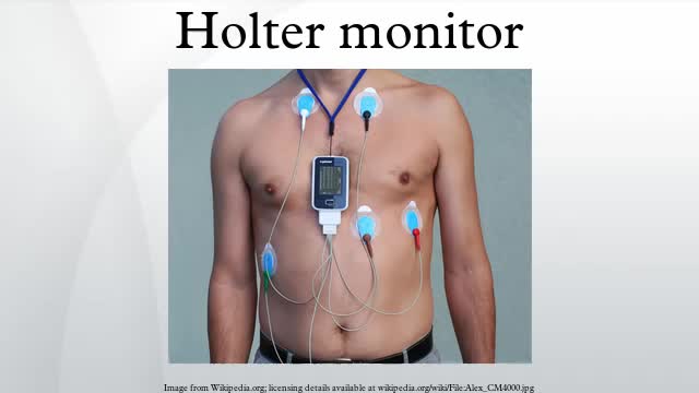
Holter monitoring, electrocardiogram or echocardiogram are only recommended if a cardiac cause (e.g., arrhythmias, possible cardiac syncope, myocardial ischemia) is suspected.
Bizarre Body Modifications
