Top videos
Loyola oral Presentation for Rounds video
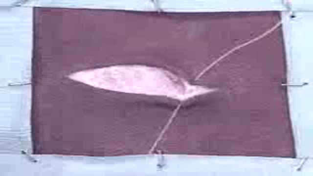
Subcutaneous Pattern Suture

How to read ECG:
Part 1 Shows:
1-All
2-Introduction
3-Rate and Axis
4-Chamber Hypertrophy
5-Bundle Branch Block
6-Myocardial Infarction

How to read ECG Part 2:
1-All
2-Myocardial Ischaemia
3-Ectopics, Sinus Pause
4-Atrial Arrhythmias
5-Ventricular Arrhythmia
6-A-V Block
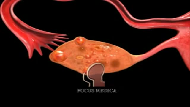
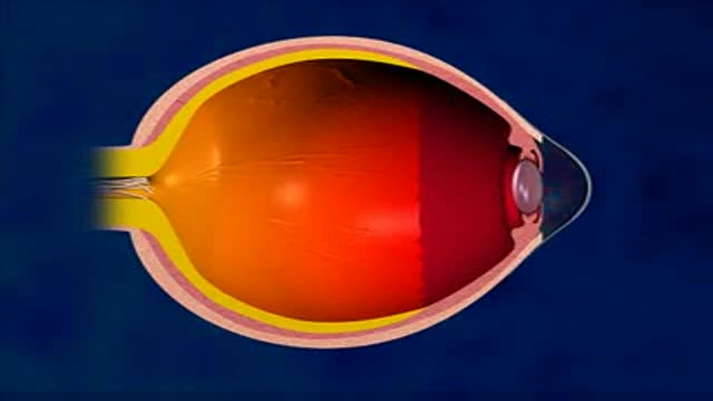
An animation showing what PCO is
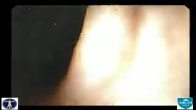
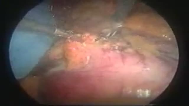
The colonoscope is slowly withdrawn during this screening colonoscopy down from the transverse colon, back around the splenic flexure, and down the descending colon, and reveals this finding a colonic diverticula. Diverticulosis is a common, acquired, age-related occurrence affecting over 50% of the... western adult population over the age of 50. It is seen rarely in Africa and Asia where the dietary fiber content is traditionally higher. Thus most investigators feel that low fiber diets are related to the development of this condition. Ironically, colonic diverticula are not true diverticula but rather pseudodiverticula in that the sac includes layers of the mucosa and submucosa that push through rather than include the outer muscular layer. As with the small bowel the colon has an inner circular muscular layer, but the outer longitudinal layer is composed of three bands of muscle that run the length of the colon known as teniae. Diverticula occur in rows between the mesenteric and two antimesenteric teniae where the colonic wall is further weakened by the defect caused by the perforating vasa recti artery which supplies the colonic mucosa. Occasionally, the anatomic propensity of diverticula to form in rows is quite apparent as seen when this clip is replayed in slow motion. Most often, however, the arrangement of the diverticula appears random due to the angulation of the bowel and thickening of the semi lunar folds. The conditions that cause these pulsion diverticula are not know with certainty but may include high intrahaustral pressures, muscular hypertrophy, and age related alterations in collagen cross linking. Diverticula can bleed or can abscess and perforate. The incidence of diverticulitis or diverticular bleeding is in the range of 1:1,000 patients with diverticulosis.
Laparoscopic Sleeve Gastrectomy Operation
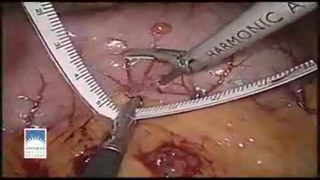
Cholecystectomy
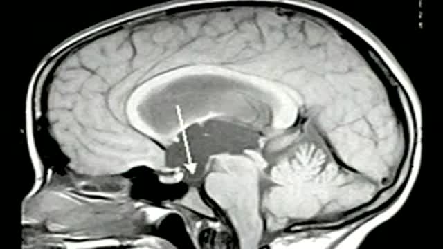
Endoscopic third ventriculostomy in a patient with obstructive hydrocephalus

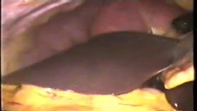
Removal of the superficial lobe is performed on a child presenting with a mass
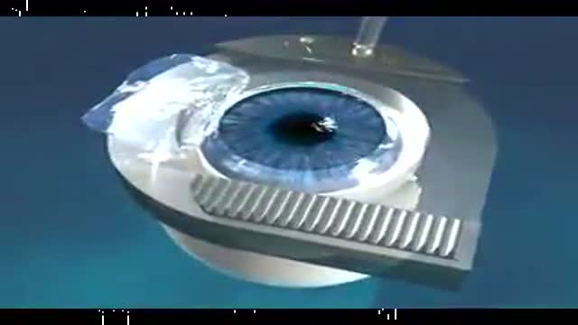
Microkeratome in Lasik
LASIK or Laser-Assisted In Situ Keratomileusis is a surgical procedure intended to reduce a person's dependency on glasses or contact lenses.
LASIK surgery is most commonly performed as a cure for myopia (nearsightedness), but can also be used to cure hyperopia (farsightedness) or astigmatism (corneal irregularities).
LASIK is a procedure that permanently changes the shape of the cornea using a special laser and thus focusing the light rays exactly on the retina.
The steps of the procedure are as follows:
A suction ring is placed on the eye to stabilize and check the eye pressure.
The microkeratome, a cutting instrument, is attached to the suction ring.
The blade of the microkeratome is used to cut a flap in the cornea.
The exposed inner layer of the cornea is then reshaped with an excimer laser.
The corneal flap is returned to its original position.
LASIK is an ambulatory procedure; the patient can walk into the surgery center, have the procedure and walk out again and is awake the whole time. Occasionally, the doctor may administer a mild oral sedative.

Oral Surgery and Dental Implants
Mini Gastric Bypass surgery Operation

1year follow up Video of Scott Kopperud who underwent Hip resurfacing Surgeon:- Dr.Vijay C Bose, ARCH Asian Regional ...
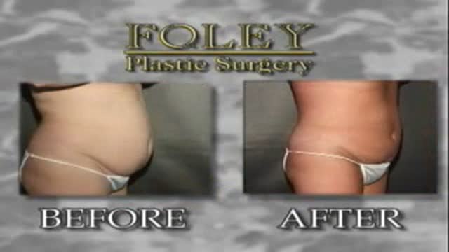
This is an Abdominal Liposuction surgery performed by Dr. Art Foley. Liposuction is a procedure that can help sculpt the body by removing unwanted fat from specific areas including the abdomen, hips, buttocks, thighs, knees, upper arms and neck. Although no type of liposuction is a substitute for dieting and exercise, liposuction can remove stubborn areas of fat that don't respond to traditional weight loss methods.
Laser Liposuction for Weight Loss
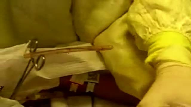
nurses removing chest tube from surgery after spontaneous pneumothorax
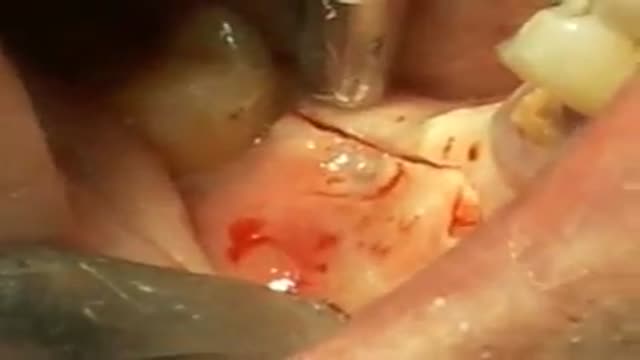
Fractured implant extraction
Less pain and no incisions are just two benefits of robotically assisted surgery thanks to the da Vinci Surgical System. ~ Detroit Medical Center
