- Physical Examination
- Surgical Examination
- Ophthalmology
- Clinical Skills
- Orthopedics
- Surgery Videos
- Laparoscopy
- Pediatrics
- Funny Videos
- Cardiothoracic Surgery
- Nursing Videos
- Plastic Surgery
- Otorhinolaryngology
- Histology and Histopathology
- Neurosurgery
- Dermatology
- Pediatric Surgery
- Urology
- Dentistry
- Oncology and Cancers
- Anatomy Videos
- Health and Fitness
- Radiology
- Anaesthesia
- Physical Therapy
- Pharmacology
- Interventional Radiology
- Cardiology
- Endocrinology
- Gynecology
- Emergency Medicine
- Psychiatry and Psychology
- Childbirth Videos
- General Medical Videos
- Nephrology
- Physiology
- Diet and Food Health
- Diabetes Mellitus
- Neurology
- Women Health
- Osteoporosis
- Gastroenterology
- Pulmonology
- Hematology
- Rheumatology
- Toxicology
- Nuclear Medicine
- Infectious Diseases
- Vascular Disease
- Reproductive Health
- Burns and Wound Healing
- Other
Top videos

How to Get Rid of Mucus in Lungs
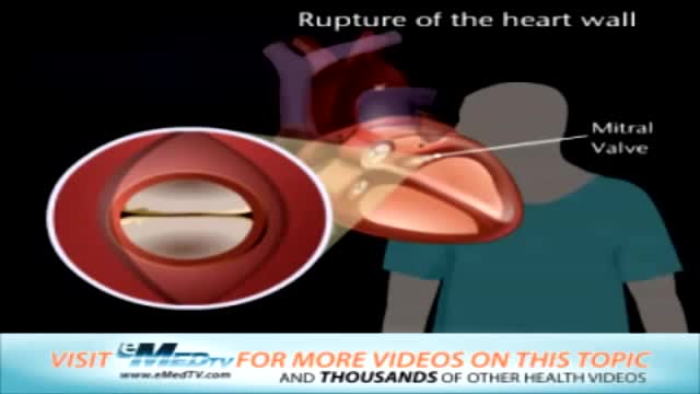
Rupture of the Heart Wall and Mitral Valve Replacement
Heart Attack vs Cardiac Arrest vs Stroke
Carbon monoxide poisoning occurs after too much inhalation of carbon monoxide (CO). Carbon monoxide is a toxic gas, but, being colorless, odorless, tasteless, and initially non-irritating, it is very difficult for people to detect.
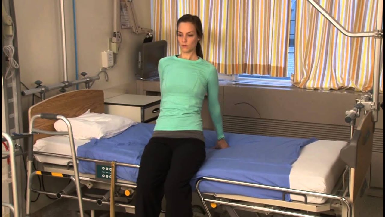
Patient information from Sunnybrook's Holland Musculoskeletal Program. For more, visit: http://sunnybrook.ca/holland
If a patient comes to you with a painful, throbbing, swollen, red face (a ''fat face'), perhaps with fever, trismus and lymphadenitis, he is probably suffering from an acute dental or oral infection, most probably an alveolar abscess. He may have: (1) An alveolar abscess begins as an infection in the bone around a non-vital infected tooth. He has severe pain, which becomes less as pus is released into more superficial tissues and his face starts to swell. After 36 hours of cellulitis he usually has a fluctuant abscess which needs draining. If drainage is delayed, the pus in his abscess discharges spontaneously through a sinus (26-8) in his gum or face, which may become chronic. First, control infection with antibiotics, and then drain the abscess, either by incising it where it is pointing, or by removing the infected tooth, which acts as a cork to prevent the pus escaping, or by doing both these things. If you remove a tooth before you have controlled the infection with antibiotics, and while his face is still severely swollen, you may spread the infection; your task will also be more difficult. (2) A periodontal abscess at the side of a tooth, caused by spread from an infected gum. (3) A pericoronal abscess caused by infection of the gum over the crown of an unerupted and impacted tooth, usually a lower third molar (''an infected wisdom tooth'). Often, an abscess does not form, and the gum round the tooth is merely inflamed.

Many women with hair loss suffer in silence, altering their hairstyle to hide thinning or patches. But the sooner you seek care, the better the chances of successfully treating it, says Mary Gail Mercurio, MD, associate professor of dermatology at the University of Rochester in Rochester, N.Y. It's not as uncommon as you may think: As many as 5% of women under 30 and 60% of those older than 70 are affected, she says. At the recent meeting of the American Academy of Dermatology in Miami Beach, Fla., Mercurio discussed common forms of hair loss in women and treatment options.
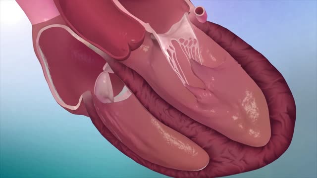
Hypertrophic cardiomyopathy (HCM) is very common and can affect people of any age. It affects men and women equally. It is a common cause of sudden cardiac arrest in young people, including young athletes. Hypertrophic cardiomyopathy occurs if heart muscle cells enlarge and cause the walls of the ventricles (usually the left ventricle) to thicken. The ventricle size often remains normal, but the thickening may block blood flow out of the ventricle. If this happens, the condition is called obstructive hypertrophic cardiomyopathy. Sometimes the septum, the wall that divides the left and right sides of the heart, thickens and bulges into the left ventricle. This can block blood flow out of the left ventricle. Then the ventricle must work hard to pump blood. Symptoms can include chest pain, dizziness, shortness of breath, or fainting. Hypertrophic cardiomyopathy also can affect the heart's mitral valve, causing blood to leak backward through the valve. Sometimes, the thickened heart muscle doesn't block blood flow out of the left ventricle. This is referred to as non-obstructive hypertrophic cardiomyopathy. The entire ventricle may thicken, or the thickening may happen only at the bottom of the heart. The right ventricle also may be affected. In both obstructive and non-obstructive HCM, the thickened muscle makes the inside of the left ventricle smaller, so it holds less blood. The walls of the ventricle may stiffen, and as a result, the ventricle is less able to relax and fill with blood.

A stroke occurs when the blood supply to your brain is interrupted or reduced. This deprives your brain of oxygen and nutrients, which can cause your brain cells to die. A stroke may be caused by a blocked artery (ischemic stroke) or the leaking or bursting of a blood vessel (hemorrhagic stroke)
Anaphylaxis is a severe, potentially life-threatening allergic reaction. It can occur within seconds or minutes of exposure to something you're allergic to, such as a peanut or the venom from a bee sting. The flood of chemicals released by your immune system during anaphylaxis can cause you to go into shock; your blood pressure drops suddenly and your airways narrow, blocking normal breathing. Signs and symptoms of anaphylaxis include a rapid, weak pulse, a skin rash, and nausea and vomiting. Common triggers of anaphylaxis include certain foods, some medications, insect venom and latex. Anaphylaxis requires an immediate trip to the emergency department and an injection of epinephrine. If anaphylaxis isn't treated right away, it can lead to unconsciousness or even death.
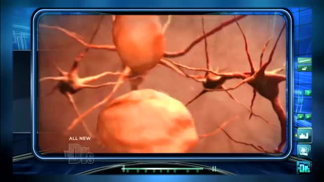
Memory Loss & the Brain. It's not just a movement disorder. Besides causing tremors and other motion-related symptoms, Parkinson's disease affects memory, learning, and behavior. Parkinson's disease is notorious for so-called motor symptoms like muscle rigidity, tremor, slowed movement, and unsteady posture and gait.
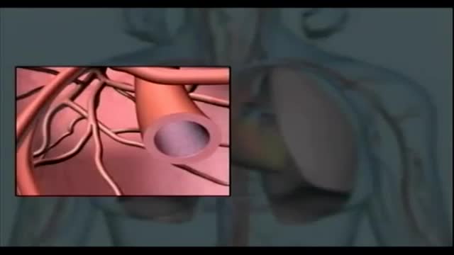
Cardiac cath is performed to find out if you have disease of the heart muscle, valves or coronary (heart) arteries. During the procedure, the pressure and blood flow in your heart can be measured. Coronary angiography is done during cardiac catheterization.
Technically, there's no formal definition for a "Code", but doctors often use the term as slang for a cardiopulmonary arrest happening to a patient in a hospital or clinic, requiring a team of providers (sometimes called a "code team") to rush to the specific location and begin immediate resuscitative efforts.

Breast Implants Bottoming Out? Steps to Reduce The Risks
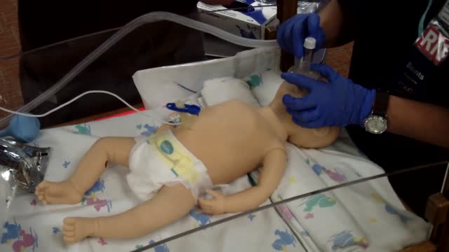
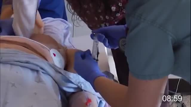
Neonatal resuscitation skills are essential for all health care providers who are involved in the delivery of newborns. The transition from fetus to newborn requires intervention by a skilled individual or team in approximately 10% of all deliveries. This figure is concerning because 81% of all babies in the United States are born in nonteaching, nonaffiliated level I or II hospitals. In such hospitals, the volume of delivery service may not be perceived as sufficient economic justification for the continuous in-hospital presence of personnel with high-risk delivery room experience, as recommended by the American Academy of Pediatrics (AAP) and the American College of Obstetricians and Gynecologists (ACOG). [1] Perinatal asphyxia and extreme prematurity are the 2 complications of pregnancy that most frequently necessitate complex resuscitation by skilled personnel. However, only 60% of asphyxiated newborns can be predicted ante partum. The remaining newborns are not identified until the time of birth. Additionally, approximately 80% of low-birth-weight infants require resuscitation and stabilization at delivery. Nearly one half of newborn deaths (many of which involve extremely premature infants) occur during the first 24 hours after birth. Many of these early deaths also have a component of asphyxia or respiratory depression as an etiology. For the surviving infants, effective management of asphyxia in the first few minutes of life may influence long-term outcome. Even though prenatal care can identify many potential fetal difficulties ante partum, allowing maternal transfer to the referral center for care, many women who experience preterm labor are not identified prospectively and therefore are not appropriately transferred to a tertiary perinatal center. Consequently, many deliveries of extremely premature infants occur in smaller hospitals. For this reason, all personnel involved in delivery room care of the newborn should be trained adequately in all aspects of neonatal resuscitation. Additionally, equipment that is appropriately sized to resuscitate infants of all gestational ages should be available in all delivering institutions, even if the institution does not care for preterm or intensive care infants. Along with the necessary skills, the practitioner should approach any resuscitation with a good comprehension of transitional physiology and adaptation, as well as an understanding of the infant's response to resuscitation. Resuscitation involves much more than possessing an ordered list of technical skills and having a resuscitation team; it requires excellent assessment skills and a grounded understanding of physiology.

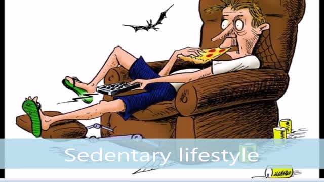
Weight loss is the most effective nonpharmacologic measure to decrease blood pressure in overweight individuals. Weight loss with other nonpharmacologic measures can prevent or delay the onset of hypertension and reduce the overall risk of cardiovascular events in such patients. In some patients with established hypertension, lifestyle changes alone may control their blood pressure.

Pain in the affected bone is the most common complaint of patients with bone cancer. At first, the pain is not constant. It may be worse at night or when the bone is used (for example, leg pain when walking). As the cancer grows, the pain will be there all the time. The pain increases with activity and the person might limp if a leg is involved.
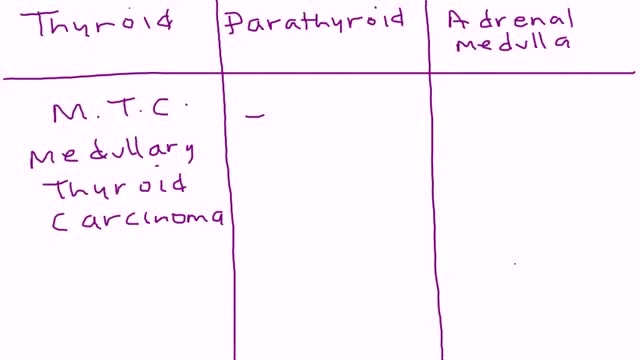
Multiple endocrine neoplasia type 2 (MEN2) (also known as "Pheochromocytoma and amyloid producing medullary thyroid carcinoma", "PTC syndrome," and "Sipple syndrome") is a group of medical disorders associated with tumors of the endocrine system. The tumors may be benign or malignant (cancer).
A febrile seizure is a convulsion in a child that may be caused by a spike in body temperature, often from an infection. Your child's having a febrile seizure can be alarming, and the few minutes it lasts can seem like an eternity. Febrile seizures represent a unique response of a child's brain to fever, usually the first day of a fever. Fortunately, they're usually harmless and typically don't indicate an ongoing problem. You can help by keeping your child safe during a febrile seizure and by comforting him or her afterward.

Migraine headaches are recurrent throbbing or pulsatile headaches often associated with a prodrome, nausea, vomiting, photophobia, and phonophobia. When they occur, the prodromes are characterized by visual scintillations, scotomas, dizziness, or tinnitus
