- Physical Examination
- Surgical Examination
- Ophthalmology
- Clinical Skills
- Orthopedics
- Surgery Videos
- Laparoscopy
- Pediatrics
- Funny Videos
- Cardiothoracic Surgery
- Nursing Videos
- Plastic Surgery
- Otorhinolaryngology
- Histology and Histopathology
- Neurosurgery
- Dermatology
- Pediatric Surgery
- Urology
- Dentistry
- Oncology and Cancers
- Anatomy Videos
- Health and Fitness
- Radiology
- Anaesthesia
- Physical Therapy
- Pharmacology
- Interventional Radiology
- Cardiology
- Endocrinology
- Gynecology
- Emergency Medicine
- Psychiatry and Psychology
- Childbirth Videos
- General Medical Videos
- Nephrology
- Physiology
- Diet and Food Health
- Diabetes Mellitus
- Neurology
- Women Health
- Osteoporosis
- Gastroenterology
- Pulmonology
- Hematology
- Rheumatology
- Toxicology
- Nuclear Medicine
- Infectious Diseases
- Vascular Disease
- Reproductive Health
- Burns and Wound Healing
- Other
Top videos

Doctor Ricky Brown reacts to this surgery simulation of an inguinal hernia repair where they repair the hernia sack and create a mesh for the organ to comfortably rest on.
3D Animation powered by:
3DMedWorld - 3dmedworld.com
#shorts #doctor #education #surgery #medical

A stress ulcer is a single or multiple mucosal defect which can become complicated by upper gastrointestinal bleeding during the physiologic stress of serious illness.

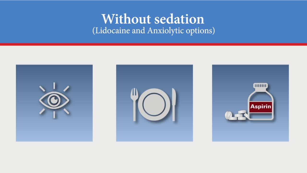
Aspirin is used to reduce fever and relieve mild to moderate pain from conditions such as muscle aches, toothaches, common cold, and headaches. It may also be used to reduce pain and swelling in conditions such as arthritis. Aspirin is known as a salicylate and a nonsteroidal anti-inflammatory drug (NSAID).

Vatche, Minassian, MD, MPH, Chief of Urogynecology, and Sarah Cohen, MD, MPH, Director of the Minimally Invasive Gynecologic Surgery Fellowship Program at Brigham and Women’s Hospital, perform a laparoscopic burch colposuspension, a procedure used to correct stress urinary incontinence.
Stress urinary incontinence is one of the most common types of incontinence and is characterized by urinary leakage during physical activities including coughing, sneezing, exercising, lifting, and laughing. As the condition progresses, it can become severe enough to happen with simple acts such as bending and walking. This condition is due to an anatomic weakness of the bladder neck which typically maintains the seal of urine during activity. Stress incontinence can result from a variety of conditions including vaginal childbirth, aging, menopause and obesity. As this is an anatomic condition, primary treatment may involve pelvic floor exercises and/or minimally invasive surgery.
Learn more about treatment for stress urinary incontinence:
Division of Urogynecology: http://www.brighamandwomens.or....g/Departments_and_Se
Division of Minimally Invasive Gynecologic Surgery: http://www.brighamandwomens.or....g/Departments_and_Se
A bone marrow biopsy removes a small amount of bone and a small amount of fluid and cells from inside the bone (bone marrow). A bone marrow aspiration removes only the marrow. These tests are often done to find the reason for many blood disorders and may be used to find out if cancer or infection has spread to the bone marrow. Bone marrow aspiration removes a small amount of bone marrow fluid and cells through a needle put into a bone. The bone marrow fluid and cells are checked for problems with any of the blood cells made in the bone marrow. Cells can be checked for chromosome problems. Cultures can also be done to look for infection. A bone marrow biopsy removes bone with the marrow inside to look at under a microscope. The aspiration (taking fluid) is usually done first, and then the biopsy.
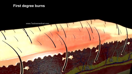
What are the classifications of burns? Burns are classified as first-, second-, or third-degree, depending on how deep and severe they penetrate the skin's surface. First-degree (superficial) burns. First-degree burns affect only the epidermis, or outer layer of skin. The burn site is red, painful, dry, and with no blisters. Mild sunburn is an example. Long-term tissue damage is rare and usually consists of an increase or decrease in the skin color. Second-degree (partial thickness) burns. Second-degree burns involve the epidermis and part of the dermis layer of skin. The burn site appears red, blistered, and may be swollen and painful. Third-degree (full thickness) burns. Third-degree burns destroy the epidermis and dermis and may go into the subcutaneous tissue. The burn site may appear white or charred Fourth degree burns. Fourth degree burns also damage the underlying bones, muscles, and tendons. There is no sensation in the area since the nerve endings are destroyed.
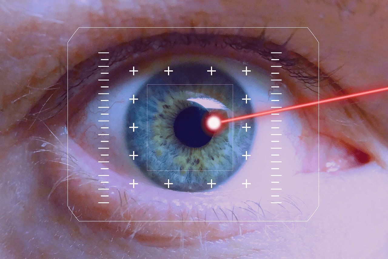
LASIK eye procedure for correcting vision

What is hemodialysis, and why would someone need it? How does hemodialysis work? Can people perform hemodialysis at home? John Kevin Tucker, M.D., Nephrologist at Brigham and Women's Hospital and Vice President for Education at Mass General Brigham, discusses hemodialysis and how it helps people who have lost their kidney function to maintain normal lives.
Subscribe Link: https://www.youtube.com/channe....l/UCYrLjATd88gPwIKnt
0:00 - Intro
0:26 - The Condition
2:06 - Hemodialysis: How It Works
4:37 - In-Center Hemodialysis Care Team
About Mass General Brigham:
Mass General Brigham combines the strength of two world-class academic medical centers, five nationally ranked specialty hospitals, 11 community hospitals, and dozens of health centers. Our doctors and researchers accelerate medical breakthroughs and drive innovations in patient care. They are leaders in medical education, serving as Harvard Medical School faculty and training the next generation of physicians. Mass General Brigham’s mission is to deliver the best, affordable health care to patients everywhere. Together, we transform the health of our communities and beyond.
#MassGeneralBrigham #MGB #Hemodialysis
Visit Mass General Brigham: https://www.massgeneralbrigham.org/
Find us on social:
Twitter: https://twitter.com/MassGenBrigham
Instagram: https://www.instagram.com/massgeneralbrigham/
Facebook: https://www.facebook.com/MassGeneralBrigham/
LinkedIn: https://www.linkedin.com/compa....ny/mass-general-brig
Mass General Brigham:
https://www.youtube.com/massgeneralbrigham
Kidney Failure: Signs, Dialysis Options, and Hemodialysis Explained | Mass General Brigham
https://youtu.be/azy7yc19QYQ
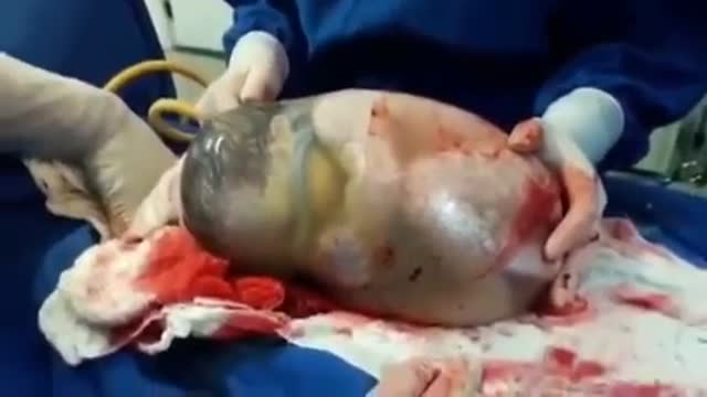
This is the incredible moment a new-born baby arrived still inside its amniotic sac, completely intact. The tiny infant can be seen moving and stretching still inside the sac, as medics prepare to snip the new born free. The amniotic sac is a thin but durable membrane filled with fluid which helps keep a baby warm and safe from bumps during pregnancy. When it breaks, this is typically referred to as a woman's 'waters breaking' shortly before she gives birth. But in rare cases, less than 1-in-80,000 births, the baby is delivered with the membranes still intact and this is known as a 'caul birth'. Some babies are born with part of the membrane still attached to them, but to be born completely encased in the intact membrane is incredibly rare. Many people still believe the phenomenon to be a good omen for the child's infancy and it is has even been suggested, but not proven, that caul babies will always have a natural affinity for water. The video was taken in Spain on Saturday and captures the rare moment the baby was born with the membrane covering its entire body, just minutes after its twin was delivered normally.
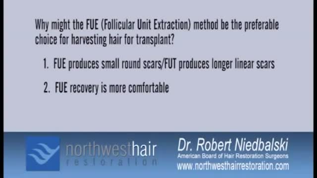
Today, hair transplant physicians are able to make use of different techniques to extract and transplant large numbers of hair follicles (follicular units). There are two primary techniques for hair transplantation currently in use. The FUE (Follicular Unit Extraction) and the FUT (Follicular Unit Transplantation) methods. They differ primarily in the way hair follicles are extracted from the donor area. Follicular Unit Transplantation (FUT) The FUT process involves removing a small strip of tissue from the back of the head, from which the donor hair follicles will be extracted. The hair follicles are harvested from the strip by a skilled clinical team before being individually transplanted to the recipient areas. In most cases, and especially cases of advanced hair loss, FUT is the preferred method because it allows the physician to fully utilize the scalp area to deliver results consistent with patient expectations. FUT typically allows for the greatest number of grafts to be transplanted in a single session. Pain Management Some patients report higher levels of discomfort with FUT procedures compared to FUE due the potential swelling in the area where the strip of tissue was removed, but both methods have a very manageable recovery period and pain medication can be prescribed by your physician if needed. Both techniques of hair transplantation are relatively simple. Hair transplantation procedures are outpatient surgeries with some patients going back to work as soon as the very next day. Scarring The FUT strip extraction method typically results in a very narrow linear scar in the back of the head (typically 1mm in diameter or less in size). Since the scar is very thin, it’s easily concealed by all but the shortest of haircut styles. A short to moderate crop setting on most clippers is sufficient to conceal the scar for the majority of patients, and over time the scar will become less noticeable as it fades. Costs The industry norm for pricing is on a per-graft basis. This allows each individual to pay for only what they need and receive in number of grafts, and not a flat rate that in the end may cost you more. The per-graft cost of a FUT procedure is generally lower than that of a FUE procedure. Lately however, in response to the rising popularity of the FUE technique, many hair transplantation clinics have started lowering the per graft cost on FUE procedures, so that the cost difference between the two types of procedure are not as much as most people think. The costs of medical procedures always vary by patients’ conditions, needs and objectives. For the most accurate assessment of your hair loss and the associated cost of your hair restoration, you will need to speak to a physician. Follicular Unit Extraction (FUE) In an FUE hair transplantation, each follicular unit is individually taken directly from the scalp with no strip of tissue being removed. Hair follicles are removed in a random fashion and the result is less density in the donor area that many say is not even noticeable. This is the main difference between FUE & FUT. Since follicles are removed one at a time, fewer follicles can be harvested during a typical session, making FUE a better option to restore hair in smaller cases (number of grafts) compared to the traditional FUT method. FUE is constantly evolving and what was once utilized for only smaller cases is now being utilized for larger and larger cases. Some people that prefer the FUE method may have the option of splitting their procedure into two days in order to complete their recommended transplantation goals. Pain Management With no stitches required and no linear scar left to heal, FUE procedures do have a faster healing time and less post-procedure discomfort compared to the traditional FUT procedure. Scarring Since FUE procedures involve removing hairs individually from the scalp, there is no linear scar left behind. However, there will be tiny 1mm in diameter or less puncture marks that tend to heal by themselves after scabbing-over in the days following the procedure. These tiny wounds typically heal within three to seven days. Costs Since the physician must remove each hair follicle individually, the time-sensitive nature of an FUE procedure typically makes it more expensive than an FUT procedure. As stated earlier, FUE technology is improving as well as gaining popularity and many hair restoration practices (including Bosley) have started to lower the cost per graft price for FUE procedures. Nowadays, the cost difference between a FUE and a FUT procedures is less disparate.
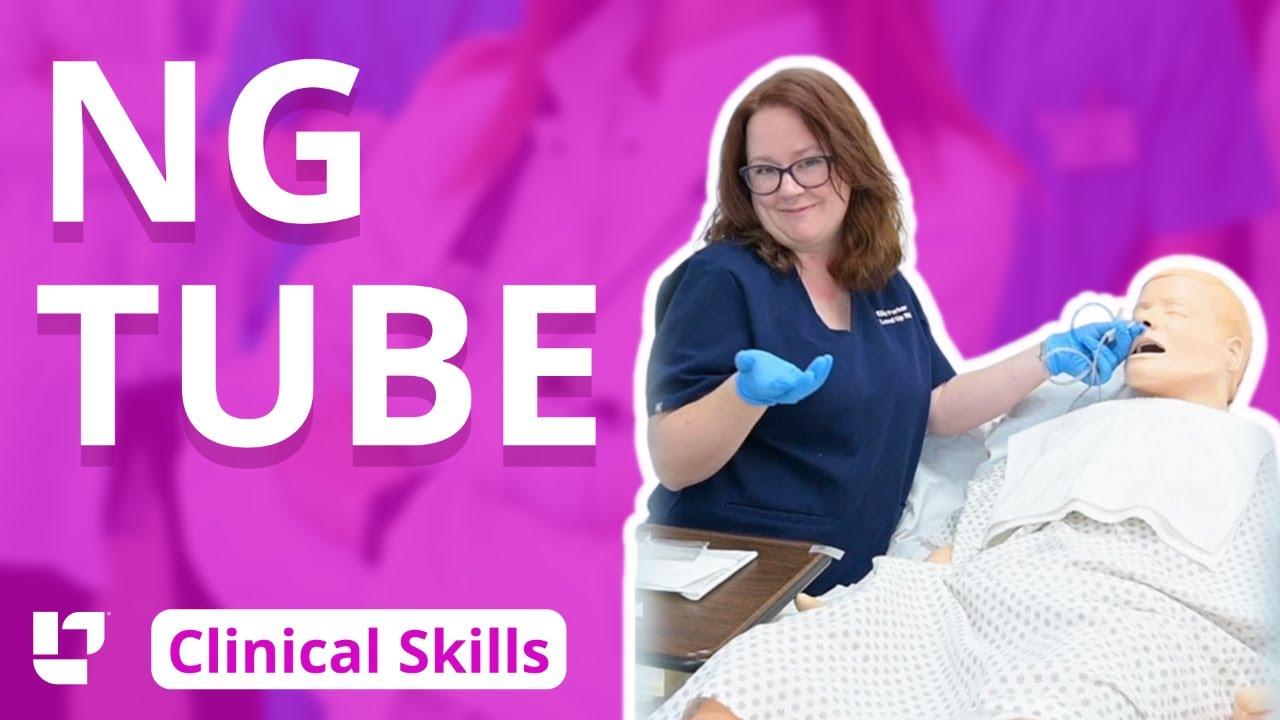
Ellis demonstrates how to insert and then remove an NG tube. This includes drawing gastric residual and checking the pH. After the demonstration, Ellis provides additional tips about clamping the NG tube and using the blue pigtail.
Our Critical Nursing Skills video tutorial series is taught by Ellis Parker MSN, RN-BC, CNE, CHS and intended to help RN and PN nursing students study for your nursing school exams, including the ATI, HESI and NCLEX.
#NCLEX #HESI #Kaplan #ATI #NursingSchool #NursingStudent #Nurse #RN #PN #Education #LVN #LPN #ClinicalSkills #NGTube #nurseeducator
00:00 What to expect
00:30 Preparing NG tube patient
00:56 Preparing NG tube equipment
1:29 Measuring the NG tube
2:02 Preparing for NG tube insertion
2:28 Inserting the NG tube
3:17 Checking placement with pH
4:23 Anchoring with split-tape
5:32 Connecting to suction
6:05 Disconnecting from suction
6:17 What to do before removal?
7:03 Removing NG tube
7:40 Additional tips on clamping
8:31 The blue pigtail
🚨 Reminder: shipping deadlines are looming 👀
🎁 Regular Shipping: Order by Friday, December 15
🚀 Expedited Shipping: Order by Monday, December 18
🔍 Still searching for last-minute gifts? Consider a Level Up RN Gift Card! 💌 It’s not only a thoughtful present but also the perfect way to share treasures like Pharmacology Flashcards OR digital treasures like Flashables Digital Nursing Flashcards & the Level Up RN membership. Give the gift of knowledge this holiday season! 🧠⚡️💖 bit.ly/LevelUpRNGC
🚪 Access our Cram Courses, Quizzes and Videos all in one ad free space with Level Up RN Membership https://bit.ly/LevelUpRNMembership
Want more ways to MASTER Clinical Skills? Check out our flashcards & videos!
👇👇👇👇👇👇👇👇👇👇
👉 https://bit.ly/clinicalnursingskills 👈
☝️👆☝️👆☝️👆☝️👆☝️👆
This is your one-stop-shop for materials to help you LEARN & REVIEW so you can PASS Nursing School.
🤔🤔🤔 DO YOU WANT TO PASS your classes, proctored exams and the NCLEX? 🤔🤔🤔 Our resources are the best you can buy. They are built with a single goal: help you pass with no fluff. Everything you need, and nothing you don’t. Don’t take our word for it, though! Check out our hundreds of ⭐️⭐️⭐️⭐️⭐️ reviews from nurses who passed their exams and the NCLEX with Level Up RN.
🗂️ Our Ultimate Nursing School Survival kit is your number 1 resource to get through nursing school and to pass the NCLEX. Whether you're just starting school or you’re already prepping for the NCLEX, this bundle of flashcards is the best you can buy. It covers all the information you need to know to pass all your exams and it has FREE shipping!
➡️ https://bit.ly/TUNSSK ⬅️
L👀king for EVEN MORE resources to survive Nursing School? Make your Nursing School experience your own! Life’s difficult enough—learning shouldn’t be.
🪅 Games https://nursesquad.com
💻 Digital resources https://bit.ly/NursingStudyCourses
📅 Organizational tools https://bit.ly/OrganizingSchool
✨Want perks? Join our channel!
https://youtube.com/leveluprn/join
🏷 Head to https://leveluprn.com/specials for all our latest deals!🥳️
📧 LOOKING FOR FREE RESOURCES TO HELP WITH YOUR EXAMS? Get exclusive tips, latest video releases and more delivered to your email!
➡️ https://leveluprn.com/signup ⬅️
⚕ 👩 LEVEL UP NURSE SQUAD 👩⚕️
All of the nurses at Level Up RN are here to help! Cathy Parkes started helping her fellow classmates back when she was in nursing school, tutoring so they could pass their exams and graduate. After she got her BSN and started working as an RN at Scripps Encinitas Hospital, she started this YouTube channel to help nursing students around the world. Since then she has built a team of top-notch dedicated nurses and nurse educators who are focused on improving nursing education and supporting career advancement for nurses everywhere. With flashcards, videos, courses, organizational tools and more, we are singularly focused on helping students and nurses Level Up on their exams and nursing careers.
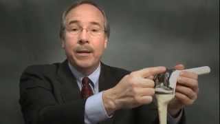
Timothy Lovell, MD, an orthopedic surgeon, talks to Spokane, WA knee replacement surgery patients about the procedure, possible risks and complications of surgery, and about your recovery time.
Dr. Lovell addresses anesthesia, the size and location of the incision, and shows you what the knee replacement ball and socket joint looks like. He'll talk about the recovery process; using a crutches, a walker or a cane to get around; movements to avoid; and how long it takes to feel better and return to your normal, active life.
To learn more about Dr. Lovell, visit http://washington.providence.o....rg/find-a-provider/l
And, to learn more about having orthopedic surgery in Spokane, WA, visit http://washington.providence.o....rg/clinics/providenc
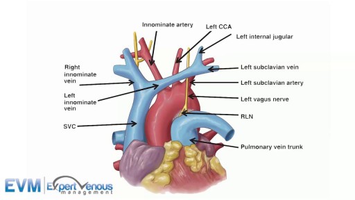
The superior vena cava (SVC, also known as the cava or cva) is a short, but large diameter vein located in the anterior right superior mediastinum.
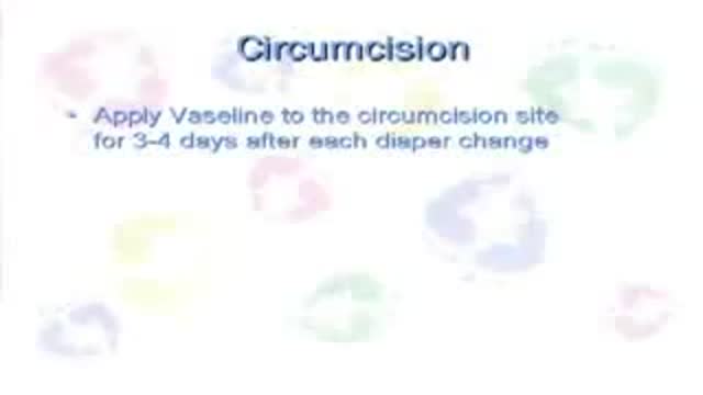
An OB/GYN nurse from Erlanger Hospital discusses caring for a newborn baby after a circumcision.
49-years old patient complaining of cough, fever and pleuritic pain for 2 weeks. At admission he was febrile and tachypnic. Chest X-Ray showed left pleural effusion. Thoracocentesis revealed purulent fluid. Chest CT-scan showed large and loculated left pleural effusion and pleural thickening. VATS decortication was performed through three incisions.

This video demonstrates why ears become clogged and why ear popping helps. The video also explains why ear popping may become difficult resulting in a persistent clogged or muffled ear especially after an ear infection.
Excision of Pilonidal Cyst. Open method.
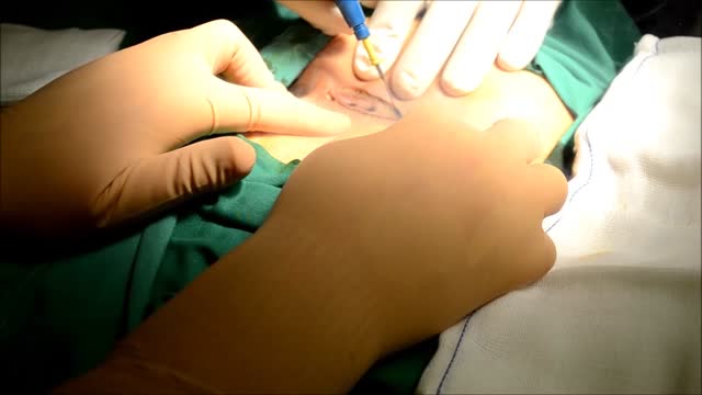
Vasectomy is a minor surgical procedure wherein the vasa deferentia of a man are severed, and then tied or sealed in a manner such to prevent sperm from entering the seminal stream (ejaculate). Typically done in an outpatient setting, a traditional vasectomy involves numbing (local anesthetic) of the scrotum after which 1 (or 2) small incisions are made, allowing a surgeon to gain access to the vas deferens.
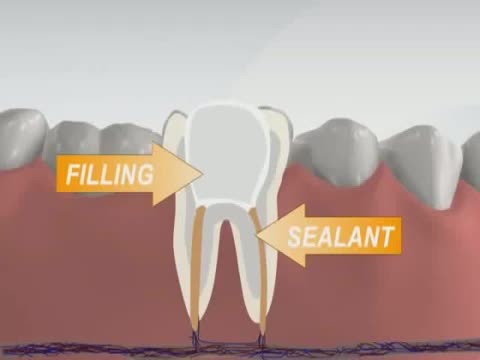
Has your dentist or endodontist told you that you need root canal treatment? If so, you're not alone. Millions of teeth are treated and saved each year with root canal, or endodontic, treatment. Remember, root canal treatment doesn't cause pain, it relieves it. Watch our videos below to learn more! Inside the tooth, under the white enamel and a hard layer called the dentin, is a soft tissue called the pulp. The pulp contains blood vessels, nerves and connective tissue, and helps to grow the root of your tooth during development. In a fully developed tooth, the tooth can survive without the pulp because the tooth continues to be nourished by the tissues surrounding it.
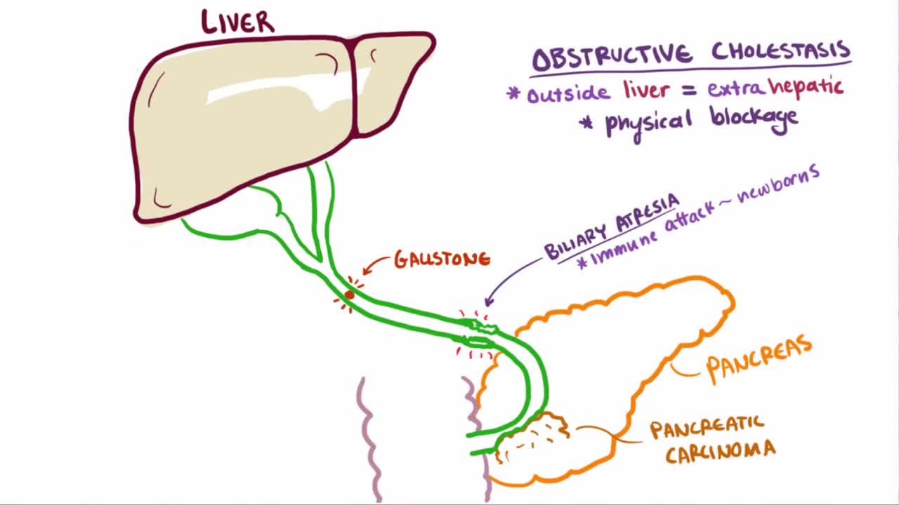
Cholestatic liver disease is a condition that results from an impairment of bile formation or bile flow to the gallbladder and duodenum (first section of the small intestine). ... The effects of cholestasis are profound and widespread, leading to worsening liver disease and systemic illness.
