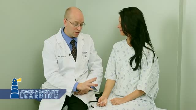
- Physical Examination
- Surgical Examination
- Ophthalmology
- Clinical Skills
- Orthopedics
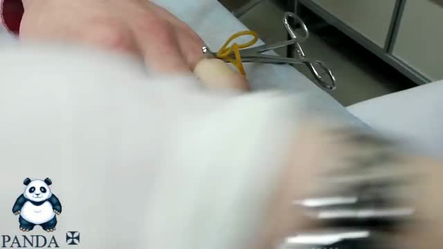
- Surgery Videos
- Laparoscopy
- Pediatrics
- Funny Videos
- Cardiothoracic Surgery
- Nursing Videos
- Plastic Surgery
- Otorhinolaryngology
- Histology and Histopathology
- Neurosurgery
- Dermatology
- Pediatric Surgery
- Urology
- Dentistry
- Oncology and Cancers
- Anatomy Videos
- Health and Fitness
- Radiology
- Anaesthesia
- Physical Therapy
- Pharmacology
- Interventional Radiology
- Cardiology
- Endocrinology
- Gynecology
- Emergency Medicine
- Psychiatry and Psychology
- Childbirth Videos
- General Medical Videos
- Nephrology
- Physiology
- Diet and Food Health
- Diabetes Mellitus
- Neurology
- Women Health
- Osteoporosis
- Gastroenterology
- Pulmonology
- Hematology
- Rheumatology
- Toxicology
- Nuclear Medicine
- Infectious Diseases
- Vascular Disease
- Reproductive Health
- Burns and Wound Healing
- Other
Top videos
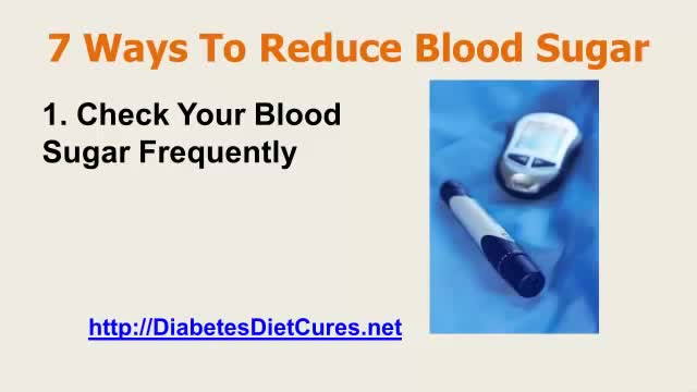
Before deciding how to treat one episode of high blood glucose, it is important to figure out why the number is high. Some possible causes include eating a heavy meal, not getting enough physical activity, forgetting to take diabetes medication, and dealing with illness and stress. Insulin is the medication that will bring blood glucose down the fastest. Someone who uses mealtime insulin can take correction doses to lower blood glucose. This requires a thorough understanding of when to inject, how often to give correction doses, and how much insulin to use. You will need to work with your doctor or diabetes educator to learn how to do this. Apart from administering insulin, the fastest way to lower your blood glucose is to engage in physical activity. Exercise results in an increased sensitivity to insulin. It causes your muscle cells to take up more glucose, leaving less of it to circulate in your bloodstream during and after the physical activity (which means a lower blood glucose when you test). Frequent, regular exercise is very important to good blood glucose control no matter what type of diabetes you have. Research has shown that it is vital in warding off long-term complications like neuropathy, retinopathy, and heart and kidney diseases. Don't forget to check with a doctor, though, before making any major changes to your exercise routine. And, if you have type 1 diabetes and your glucose is 250 mg/dl or higher, check for urine ketones. You should not exercise if ketones are present.
http://sciatica-rimedi.good-info.co Nervo Sciatico, Accavallamento Nervi, Lombosciatalgia Sintomi E Cure, Dolore Coscia, Sciatica. Come curare la sciatica a casa Se hai avuto abbastanza sciatalgia a dirigere la tua vita, non disperare! ti mostrerò tre dei più comuni trattamenti casalinghi per la sciatica, e come usarli per ridurre il dolore in modo rapido. La parte migliore di questi trattamenti è che possono curare la sciatica, e non solo coprire il dolore. Quindi, cominciamo... 1. Programma di esercizi a casa I programmi di esercizio sono una componente importante di qualsiasi piano di trattamento della sciatica. Con l'allungamento e il rafforzamento di parti del corpo che possono causare l'irritazione del nervo sciatico, è possibile ridurre il dolore e accelerare il recupero. Gli esercizi più efficaci dipendono dalla ragione di fondo per cui soffri di sciatica. La sciatica causata da un'ernia del disco, per esempio, non viene trattata con gli stessi esercizi della sciatica causata da stenosi spinale. È anche importante mantenere il corpo rilassato, per consentirgli di guarire. Un modo grandioso per farlo, senza aggravare la tua condizione, è camminare a ritmo sostenuto. Altre attività leggere possono avere un effetto simile, ma se qualcosa fa male è necessario fermarsi immediatamente. Suggerimento gratuito: è essenziale che non ci si riduca a letto a causa del dolore. Stare sdraiati a letto per più di due giorni ha dimostrato peggiorare la sciatica, perché i muscoli si irrigidiscono e si indeboliscono. 2. Bilancia la tua dieta Curare la sciatica in modo permanente, spesso significa trattare più che la semplice causa fisica. Per impedire che il dolore si ripresenti, dovrai anche migliorare la tua dieta. Uno dei modi più semplici per ridurre il dolore associato con sciatica è quello di bere più acqua. Quando si è disidratati, parti della colonna vertebrale si sgonfiano. Questo può causare ulteriore pressione sul nervo sciatico. Se possibile, si dovrebbe anche cercare di evitare alimenti infiammatori. Gli alimenti infiammatori sono troppi, per elencarli in questo articolo, ma qualsiasi alimento dotato di elevato contenuto di zucchero può, potenzialmente, portare a infiammazione e ad aumento del dolore. 3. Rimedi casalinghi I rimedi casalinghi possono fare una grande differenza per tua sciatalgia, spesso in tempi relativamente brevi. La cosa grandiosa dei rimedi casalinghi è che non richiedono prescrizione o ingredienti costosi. Uno dei più semplici rimedi casalinghi sono le noccioline. Questo perché le arachidi contengono un sacco di magnesio, che è cruciale per consentire muscoli di rilassarsi.
Here are seven ways to start reining in your risks today, before a stroke has the chance to strike. Lower blood pressure. ... Lose weight. ... Exercise more. ... Drink — in moderation. ... Treat atrial fibrillation. ... Treat diabetes. ... Quit smoking.
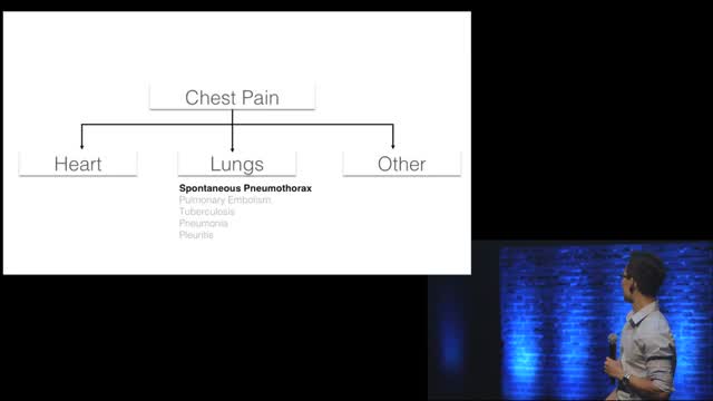
Chest pain is a frequent complaint of patients seeking urgent medical assistance, and accounts for an estimated 2-4 per cent of all A&E visits in the UK (Becker, 2000). Generally, acute chest pain should be considered cardiovascular in origin until proven otherwise and it is common in clinical practice to err on the conservative or ‘safe’ side when evaluating people with chest pain. Individuals with suspected ischaemic chest pain must be evaluated rapidly for several reasons: - Myocardial ischaemia, if prolonged and severe, can cause myocardial infarction (necrosis); - Treatment strategies that achieve myocardial salvage (thrombolytic therapy or primary coronary angioplasty) are available for patients with acute coronary syndromes and these treatments reduce morbidity and mortality;
The examination room should be quiet, warm and well lit. After you have finished interviewing the patient, provide them with a gown (a.k.a. "Johnny") and leave the room (or draw a separating curtain) while they change. Instruct them to remove all of their clothing (except for briefs) and put on the gown so that the opening is in the rear. Occasionally, patient's will end up using them as ponchos, capes or in other creative ways. While this may make for a more attractive ensemble it will also, unfortunately, interfere with your ability to perform an examination! Prior to measuring vital signs, the patient should have had the opportunity to sit for approximately five minutes so that the values are not affected by the exertion required to walk to the exam room. All measurements are made while the patient is seated. Observation: Before diving in, take a minute or so to look at the patient in their entirety, making your observations, if possible, from an out-of-the way perch. Does the patient seem anxious, in pain, upset? What about their dress and hygiene? Remember, the exam begins as soon as you lay eyes on the patient. Temperature: This is generally obtained using an oral thermometer that provides a digital reading when the sensor is placed under the patient's tongue. As most exam rooms do not have thermometers, it is not necessary to repeat this measurement unless, of course, the recorded value seems discordant with the patient's clinical condition (e.g. they feel hot but reportedly have no fever or vice versa). Depending on the bias of a particular institution, temperature is measured in either Celcius or Farenheit, with a fever defined as greater than 38-38.5 C or 101-101.5 F. Rectal temperatures, which most closely reflect internal or core values, are approximately 1 degree F higher than those obtained orally. Respiratory Rate: Respirations are recorded as breaths per minute. They should be counted for at least 30 seconds as the total number of breaths in a 15 second period is rather small and any miscounting can result in rather large errors when multiplied by 4. Try to do this as surreptitiously as possible so that the patient does not consciously alter their rate of breathing. This can be done by observing the rise and fall of the patient's hospital gown while you appear to be taking their pulse. Normal is between 12 and 20. In general, this measurement offers no relevant information for the routine examination. However, particularly in the setting of cardio-pulmonary illness, it can be a very reliable marker of disease activity. Pulse: This can be measured at any place where there is a large artery (e.g. carotid, femoral, or simply by listening over the heart), though for the sake of convenience it is generally done by palpating the radial impulse. You may find it helpful to feel both radial arteries simultaneously, doubling the sensory input and helping to insure the accuracy of your measurements. Place the tips of your index and middle fingers just proximal to the patients wrist on the thumb side, orienting them so that they are both over the length of the vessel.
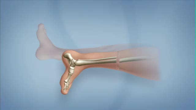
Rotationplasty is a type of autograft wherein a portion of a limb is removed, while the remaining limb below the involved portion is rotated and reattached. This procedure is used when a portion of an extremity is injured or involved with a disease, such as cancer. Typically, the ankle joint becomes the knee joint.
Rotator cuff repair is surgery to repair a torn tendon in the shoulder. The procedure can be done with a large (open) incision or with shoulder arthroscopy, which uses small buttonhole-sized incisions.

Sickle cell anemia causes pain, fatigue and delayed growth, all because of a lack of enough healthy red blood cells. And yet genetic mutations that cause it — recessive genes for the oxygen-carrying hemoglobin protein — have survived natural selection because they also seem to provide a natural defense against malaria.
A lumbar puncture (also called a spinal tap) is a procedure to collect and look at the fluid (cerebrospinal fluid, or CSF) surrounding the brain and spinal cord. During a lumbar puncture, a needle is carefully inserted into the spinal canal low in the back (lumbar area). Samples of CSF are collected.
Use warm water and sea salt. Soak the wart for 10 to 15 minutes in warm salt water to moisten the skin. Scrape the dead skin layers off the wart using a nail file, pumice stone or mild sandpaper. You could also use your fingers, but wash them thoroughly before and after, as warts can easily spread.
Varicose veins are caused by weakened valves and veins in your legs. Normally, one-way valves in your veins keep blood flowing from your legs up toward your heart. When these valves do not work as they should, blood collects in your legs, and pressure builds up. The veins become weak, large, and twisted.
Interventional Nephrology is a new and emerging subspecialty of Nephrology that mainly deals with ultrasonography of kidneys and ultrasound-guided renal biopsy, insertion of peritoneal dialysis catheters, tunneled dialysis catheters as a vascular access for patients undergoing hemodialysis as well as percutaneous ...
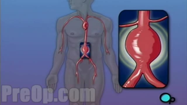
For this surgery, your doctor makes a large incision in the abdomen to expose the aorta. Once he or she has opened the abdomen, a graft can be used to repair the aneurysm. Open repair remains the standard procedure for an abdominal aortic aneurysm repair. Endovascular aneurysm repair (EVAR).

Inner Workings tells the story of the ceaseless pull of the human heart — even as it works against the very stoic realism of the brain.
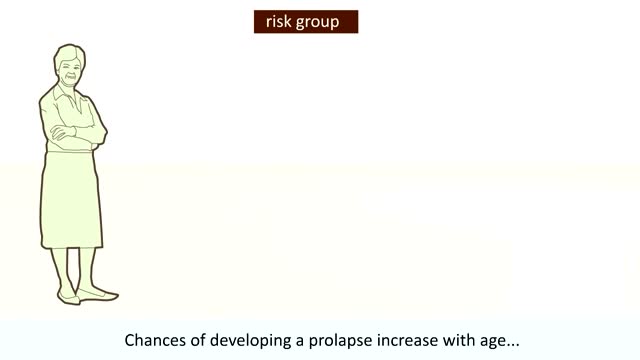
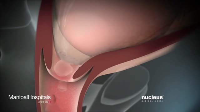
our uterus (or womb) is normally held in place inside your pelvis with various muscles, tissue, and ligaments. Because of pregnancy, childbirth or difficult labor and delivery, in some women these muscles weaken. Also, as a woman ages and with a natural loss of the hormone estrogen, her uterus can drop into the vaginal canal, causing the condition known as a prolapsed uterus.
Labor And Delivery During Vaginal Child Birth
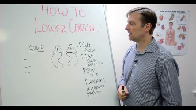
Assuming you haven't been diagnosed with Cushing's disease by your doctor, here are steps you can take to help lower high cortisol levels naturally: Switch to a Whole Foods, Anti-inflammatory Diet. Reduce and Manage Stress. Exercise Regularly. Use Adaptogen Herbs and Superfoods. Try Essential Oils to Promote Relaxation.
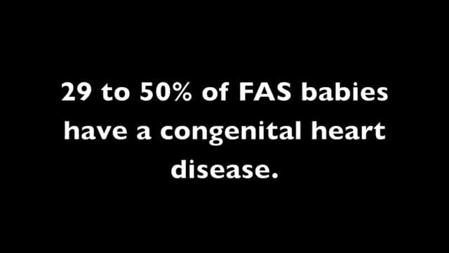
Like any syndrome, fetal alcohol syndrome (FAS) is a group of signs and symptoms that appear together and indicate a certain condition. In the case of FAS, the signs and symptoms are birth defects that result from a woman's use of alcohol during her pregnancy.
Diabetic neuropathy is a type of nerve damage that can occur if you have diabetes. High blood sugar (glucose) can injure nerve fibers throughout your body, but diabetic neuropathy most often damages nerves in your legs and feet. Depending on the affected nerves, symptoms of diabetic neuropathy can range from pain and numbness in your extremities to problems with your digestive system, urinary tract, blood vessels and heart. For some people, these symptoms are mild; for others, diabetic neuropathy can be painful, disabling and even fatal. Diabetic neuropathy is a common serious complication of diabetes. Yet you can often prevent diabetic neuropathy or slow its progress with tight blood sugar control and a healthy lifestyle.

The Hypertensive urgency must be distinguished from hypertensive emergency. Urgency is defined as severely elevated blood pressure (ie, systolic >220 mm Hg or diastolic >120 mm Hg) with no evidence of target organ damage.
