- Physical Examination
- Surgical Examination
- Ophthalmology
- Clinical Skills
- Orthopedics
- Surgery Videos
- Laparoscopy
- Pediatrics
- Funny Videos
- Cardiothoracic Surgery
- Nursing Videos
- Plastic Surgery
- Otorhinolaryngology
- Histology and Histopathology
- Neurosurgery
- Dermatology
- Pediatric Surgery
- Urology
- Dentistry
- Oncology and Cancers
- Anatomy Videos
- Health and Fitness
- Radiology
- Anaesthesia
- Physical Therapy
- Pharmacology
- Interventional Radiology
- Cardiology
- Endocrinology
- Gynecology
- Emergency Medicine
- Psychiatry and Psychology
- Childbirth Videos
- General Medical Videos
- Nephrology
- Physiology
- Diet and Food Health
- Diabetes Mellitus
- Neurology
- Women Health
- Osteoporosis
- Gastroenterology
- Pulmonology
- Hematology
- Rheumatology
- Toxicology
- Nuclear Medicine
- Infectious Diseases
- Vascular Disease
- Reproductive Health
- Burns and Wound Healing
- Other
Top videos
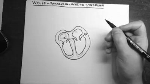
Wolff-Parkinson-White (WPW) syndrome, an extra electrical pathway between your heart's upper and lower chambers causes a rapid heartbeat. The extra pathway is present at birth and fairly rare. The episodes of fast heartbeats usually aren't life-threatening, but serious heart problems can occur. Treatment can stop or prevent episodes of fast heartbeats. A catheter-based procedure (ablation) can permanently correct the heart rhythm problems. Most people with an extra electrical pathway experience no fast heartbeat. This condition, called Wolff-Parkinson-White pattern, is discovered only by chance during a heart exam. Although WPW pattern is often harmless, doctors might recommend further evaluation before children with WPW pattern participate in high-intensity sports.
Patient information from Sunnybrook's Holland Musculoskeletal Program. For more, visit: http://sunnybrook.ca/holland
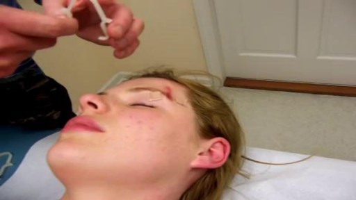
Demonstration of Burke-Baier wound closure forceps on simulated wound near eyebrow.
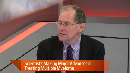
Multiple myeloma is a cancer formed by malignant plasma cells. Normal plasma cells are found in the bone marrow and are an important part of the immune system. The immune system is made up of several types of cells that work together to fight infections and other diseases. Lymphocytes (lymph cells) are the main cell type of the immune system. The major types of lymphocytes are T cells and B cells.

Breast Implants Bottoming Out? Steps to Reduce The Risks
Surgical Repair of Pectus Excavatum. Pectus excavatum is a condition in which a person's breastbone is sunken into his or her chest.

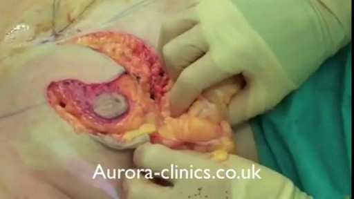
Skin Graft? Skin grafting is a surgical procedure that involves removing the skin from one area of the body and moving it, or transplanting it, to a different area of the body. This surgery may be done if a part of your body has lost its protective covering of skin due to burns, injury, or illness

new Anti-Choking Device.

This coating prevents blood from sticking on medical devices

Male babies leave their DNA in the mother
Occlusal Stamp Technique.Make Occlusal Anatomy Easily

Acanthamoeba keratitis is a rare disease in which amoebae invade the cornea of the eye. It may result in permanent visual impairment or blindness.
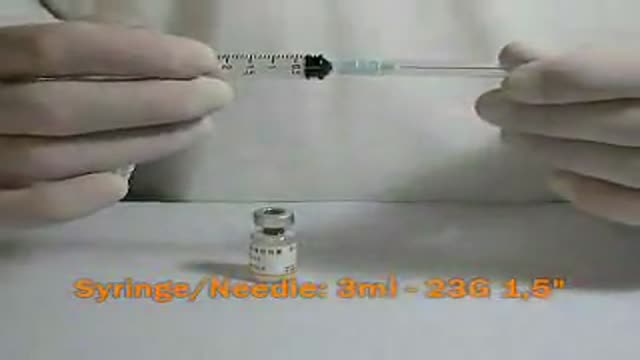
HCG Injection Procedure
In this video, the 1st Breast Reduction Surgery had been performed 10 years ago which had resulted in asymmetry. In this breast reduction surgery the breast asymmetry was corrected and the large size of both areolas was addressed.

Cracked Corners Of Mouth, Cheilitis, Angular Cheilitis Remedy, Angular Cheilitis Medicine, Cheilitis--- http://angularcheilitis-end.cbwin1.com --- Foods Which Can Limit the Occurrence of Angular Cheilitis. People suffering from Angular Cheilitis know that this is one of the most troubling and annoying skin condition one can experience. It prevents you from eating, drinking and speaking normally. Many people even refuse to go out of the house when suffering from this condition, thus becoming isolated from the rest of the world. This is why it is better to prevent it then having to treat it. If you have had it long time ago and are afraid that will come back, if you have it and want to treat it faster or if you do not want to have this terrible experience ever, you should start by eating the foods listed below. They will provide your body with all the vitamins and nutrients necessary to effectively fight this disease and prevent it from appearing ever again. Most of the times, Angular Cheilitis appears as a result of a weak immune system. Thus, you will need to have a balanced diet, filled with fruits and vegetables that will supply you with all the things you need to remain healthy and have a strong immune system. The first thing that you will need to have in your body to fight Angular Cheilitis is iron. If you no longer want to have those anesthetic and painful cracks around your mouth, if you want to eat, drink and speak normally without experiencing any pain when opening your mouth, then check out this new and revolutionary treatment! It will get you rid of Angular Cheilitis in just a few days and you will be able to enjoy life to its fullest again, without worrying about those otiose cracks! Click Here. http://angularcheilitis-end.cbwin1.com
Histology of Lymph Node

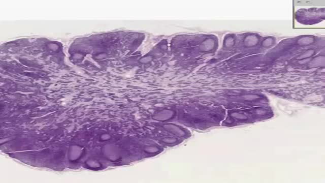
Histology of Liver

Menorrhagia is the medical term for menstrual periods with abnormally heavy or prolonged bleeding. Although heavy menstrual bleeding is a common concern, most women don't experience blood loss severe enough to be defined as menorrhagia. With menorrhagia, you can't maintain your usual activities when you have your period because you have so much blood loss and cramping. If you dread your period because you have such heavy menstrual bleeding, talk with your doctor. There are many effective treatments for menorrhagia.

Both selegiline and rasagiline can improve the symptoms of Parkinson's disease, although their effects are small compared with levodopa. They can be used alongside levodopa or dopamine agonists. MAO-B inhibitors are generally very well tolerated, but can occasionally cause side effects, including: nausea.

ADC was first identified early in the AIDS epidemic as a common and novel CNS syndrome.(4,5) The three components of the term, AIDS dementia complex embody central features of the condition. AIDS emphasizes its morbidity and poor prognosis, particularly when its severity is at stage 2 or greater (see Table 1), a severity comparable to other clinical AIDS-defining complications of HIV-1 infection. Dementia designates the acquired and persistent cognitive decline with preserved alertness that usually dominates the clinical presentation and determines its principal disability. Complex emphasizes that this disease not only impairs the intellect, but also concomitantly alters motor performance and, at times, behavior. This involvement of the nervous system beyond cognition is evidence of a wider involvement of the CNS than occurs in some other types of dementia such as Alzheimer's disease. Additionally, myelopathy may be an important, indeed predominating, aspect of ADC, and organic psychosis may also be a feature in a subset of patients (see Rheumatologic and Musculoskeletal Manifestations of HIV). These manifestations are therefore also encompassed within this term. By contrast, neither neuropathy nor functional psychiatric disturbance are included in ADC.
