- Physical Examination
- Surgical Examination
- Ophthalmology
- Clinical Skills
- Orthopedics
- Surgery Videos
- Laparoscopy
- Pediatrics
- Funny Videos
- Cardiothoracic Surgery
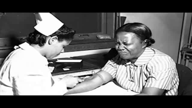
- Nursing Videos
- Plastic Surgery
- Otorhinolaryngology
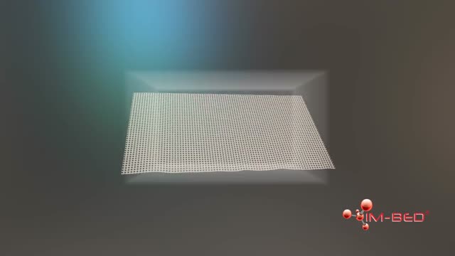
- Histology and Histopathology
- Neurosurgery
- Dermatology
- Pediatric Surgery
- Urology
- Dentistry
- Oncology and Cancers
- Anatomy Videos
- Health and Fitness
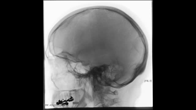
- Radiology
- Anaesthesia
- Physical Therapy
- Pharmacology
- Interventional Radiology
- Cardiology
- Endocrinology
- Gynecology
- Emergency Medicine
- Psychiatry and Psychology
- Childbirth Videos
- General Medical Videos
- Nephrology
- Physiology
- Diet and Food Health
- Diabetes Mellitus
- Neurology
- Women Health
- Osteoporosis
- Gastroenterology
- Pulmonology
- Hematology
- Rheumatology
- Toxicology
- Nuclear Medicine
- Infectious Diseases
- Vascular Disease
- Reproductive Health
- Burns and Wound Healing
- Other
Top videos

If levels of both vitamins are extremely high, there is more than a 17-fold greater risk that a child will develop autism, the researchers said. Most of the women in the study said they took multivitamins — which would include folic acid and vitamin B12 — throughout their pregnancy.
The management of acute ischemic stroke has advanced greatly over the past 2 decades. New interventions, including intravenous and endovascular treatment strategies, have evolved to recanalize arteries and salvage the ischemic brain. The evolution of interventional approaches to the treatment of acute stroke has been prompted by the limitations of intravenous therapy and intended to extend the treatment window, improve recanalization rates, and subsequently long-term clinical outcomes. The major techniques that have defined the current field of interventional acute stroke management and the relevant past and current data, and ongoing clinical trials on interventional stroke therapy will be reviewed. New issues, such as futile recanalization, and time to microcatheter, will also be discussed.

The best way to prevent hepatitis A is through vaccination with the hepatitis A vaccine. Vaccination is recommended for all children age 12 months or older, for travelers to certain countries, and for people at high risk for infection with the virus. The hepatitis A vaccine is given as two shots, six months apart.

This short course reviews the main features of EKG tracings. A method for analyzing EKGs is also presented. This method includes assessment of rhythm, calculating heart rate, observing P-wave forms, measurement of EKG intervals and segments and the evaluation of other relevant waves.
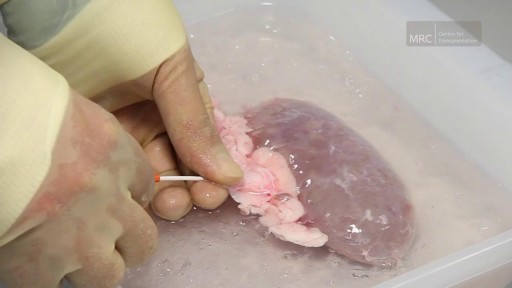
Preparing The Kidney for Transplant

Overactive bladder syndrome is common. Symptoms include an urgent feeling to go to the toilet, going to the toilet frequently and sometimes leaking urine before you can get to the toilet (urge incontinence). Treatment with bladder training often cures the problem. Sometimes medication may be advised in addition to bladder training to relax the bladder.

The pain of ovulation can range from a mild twinge to severe discomfort and usually lasts from minutes to hours. It is generally felt on one side of the abdomen and may vary each month, depending on which ovary is releasing the egg during that cycle.

A new Harvard study claims a breakthrough that could lead to a new treatment of Type 1 diabetes. WSJ's Ron Winslow explains what it means on Lunch Break. Photo: Getty
Wound healing is the process by which skin or other body tissue repairs itself after trauma. ... This process is divided into predictable phases: blood clotting (hemostasis), inflammation, tissue growth (proliferation) and tissue remodeling (maturation).
The cornea occupies the front center part of the outer wall of the eye. It is made of collagen fibers in a very special arrangement so that the cornea is clear. One looks through the cornea to see the iris and pupil. The cornea bends light coming into the eye so that it is focused on the retina.
INVIVO
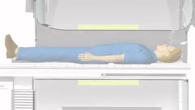
Magnetic resonance imaging (MRI) can be an important tool in the diagnosis of multiple sclerosis (MS). MRI can also be used to monitor the progression of the disease in people living with MS. How does it work? MRI uses very strong magnets, radio signals, and computer software to take 3-dimensional pictures of the inside of the body. Will I need contrast material? Maybe. Contrast material is a substance that temporarily changes the way imaging tools interact with the body. They are often used to visualize certain types of MS disease activity on the MRI. If your doctor thinks your scan requires this contrast material, you may get an injection before you get in the MRI machine. How long will it take? The time may vary based on the type of MRI. Be sure to discuss with your doctor in advance so he or she can provide you with exact timing. But don’t worry, you won’t have to stay still the whole time. The technician will let you know when they’re starting a new image.
The accumulation of ascitic fluid represents a state of total-body sodium and water excess, but the event that initiates the unbalance is unclear. Although many pathogenic processes have been implicated in the development of abdominal ascites, about 75% likely occur as a result of portal hypertension in the setting of liver cirrhosis, with the remainder due to infective, inflammatory, and infiltrative conditions. Three theories of ascites formation have been proposed: underfilling, overflow, and peripheral arterial vasodilation. The underfilling theory suggests that the primary abnormality is inappropriate sequestration of fluid within the splanchnic vascular bed due to portal hypertension and a consequent decrease in effective circulating blood volume. This activates the plasma renin, aldosterone, and sympathetic nervous system, resulting in renal sodium and water retention. The overflow theory suggests that the primary abnormality is inappropriate renal retention of sodium and water in the absence of volume depletion. This theory was developed in accordance with the observation that patients with cirrhosis have intravascular hypervolemia rather than hypovolemia. The most recent theory, the peripheral arterial vasodilation hypothesis, includes components of both of the other theories. It suggests that portal hypertension leads to vasodilation, which causes decreased effective arterial blood volume. As the natural history of the disease progresses, neurohumoral excitation increases, more renal sodium is retained, and plasma volume expands. This leads to overflow of fluid into the peritoneal cavity. The vasodilation theory proposes that underfilling is operative early and overflow is operative late in the natural history of cirrhosis. Although the sequence of events that occurs between the development of portal hypertension and renal sodium retention is not entirely clear, portal hypertension apparently leads to an increase in nitric oxide levels. Nitric oxide mediates splanchnic and peripheral vasodilation. Hepatic artery nitric oxide synthase activity is greater in patients with ascites than in those without ascites. Regardless of the initiating event, a number of factors contribute to the accumulation of fluid in the abdominal cavity. Elevated levels of epinephrine and norepinephrine are well-documented factors. Hypoalbuminemia and reduced plasma oncotic pressure favor the extravasation of fluid from the plasma to the peritoneal fluid, and, thus, ascites is infrequent in patients with cirrhosis unless both portal hypertension and hypoalbuminemia are present.

Obstetrics is the field of medicine which encompasses the care of a woman during pregnancy and childbirth. In that way it is very unique, as when assessing these patients, your actually also assessing another the child.
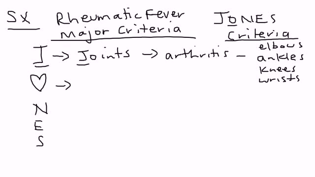
Rheumatic fever is an inflammatory disease that can develop as a complication of inadequately treated strep throat or scarlet fever. Strep throat and scarlet fever are caused by an infection with streptococcus bacteria. Rheumatic fever is most common in 5- to 15-year-old children, though it can develop in younger children and adults. Although strep throat is common, rheumatic fever is rare in the United States and other developed countries. However, rheumatic fever remains common in many developing nations. Rheumatic fever can cause permanent damage to the heart, including damaged heart valves and heart failure. Treatments can reduce damage from inflammation, lessen pain and other symptoms, and prevent the recurrence of rheumatic fever.
If it gets more severe and causes symptoms, your low hemoglobin count may indicate you have anemia. A low hemoglobin count is generally defined as less than 13.5 grams of hemoglobin per deciliter (135 grams per liter) of blood for men and less than 12 grams per deciliter (120 grams per liter) for women.
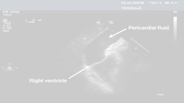
Pericardiocentesis is the aspiration of fluid from the pericardial space that surrounds the heart. This procedure can be life saving in patients with cardiac tamponade, even when it complicates acute type A aortic dissection and when cardiothoracic surgery is not available. [1] Cardiac tamponade is a time sensitive, life-threatening condition that requires prompt diagnosis and management. Historically, the diagnosis of cardiac tamponade has been based on clinical findings. Claude Beck, a cardiovascular surgeon, described 2 triads of clinical findings that he found associated with acute and chronic cardiac tamponade. The first of these triads consisted of hypotension, an increased venous pressure, and a quiet heart. It has come to be recognized as Beck's triad, a collection of findings most commonly produced by acute intrapericardial hemorrhage. Subsequent studies have shown that these classic findings are observed in only a minority of patients with cardiac tamponade. [2] The detection of pericardial fluid has been facilitated by the development and continued improvement of echocardiography. [3] Cardiac ultrasound is now accepted as the criterion standard imaging modality for the assessment of pericardial effusions and the dynamic findings consistent with cardiac tamponade. With echocardiography, the location of the effusion can be identified, the size can be estimated (small, medium, or large), and the hemodynamic effects can be examined by assessing for abnormal septal motion, right atrial or right ventricular inversion, and decreased respiratory variation of the diameter of the inferior vena cava.

Cancer starts when cells in a part of the body begins to grow out of control and can spread to other areas of the body. There are many kinds of cancer. Cells in nearly any part of the body can become cancer. To learn more about how cancers start and spread, see What Is Cancer? Leukemias are cancers that start in cells that would normally develop into different types of blood cells. Here we will talk about acute myeloid leukemia (AML). Acute myeloid leukemia (AML) has many other names, including acute myelocytic leukemia, acute myelogenous leukemia, acute granulocytic leukemia, and acute non-lymphocytic leukemia. “Acute” means that this leukemia can progress quickly if not treated, and would probably be fatal in a few months. “Myeloid” refers to the type of cell this leukemia starts from. Most cases of AML develop from cells that would turn into white blood cells (other than lymphocytes), but some cases of AML develop in other types of blood-forming cells. The different types of AML are listed in “ How is acute myeloid leukemia classified?” AML starts in the bone marrow (the soft inner part of certain bones, where new blood cells are made), but in most cases it quickly moves into the blood. It can sometimes spread to other parts of the body including the lymph nodes, liver, spleen, central nervous system (brain and spinal cord), and testicles. Other types of cancer can start in these organs and then spread to the bone marrow. But these cancers that start elsewhere and then spread to the bone marrow are not leukemias. Normal bone marrow, blood, and lymphoid tissue To understand the different types of leukemia, it helps to know about the blood and lymph systems.

Both selegiline and rasagiline can improve the symptoms of Parkinson's disease, although their effects are small compared with levodopa. They can be used alongside levodopa or dopamine agonists. MAO-B inhibitors are generally very well tolerated, but can occasionally cause side effects, including: nausea.

ADC was first identified early in the AIDS epidemic as a common and novel CNS syndrome.(4,5) The three components of the term, AIDS dementia complex embody central features of the condition. AIDS emphasizes its morbidity and poor prognosis, particularly when its severity is at stage 2 or greater (see Table 1), a severity comparable to other clinical AIDS-defining complications of HIV-1 infection. Dementia designates the acquired and persistent cognitive decline with preserved alertness that usually dominates the clinical presentation and determines its principal disability. Complex emphasizes that this disease not only impairs the intellect, but also concomitantly alters motor performance and, at times, behavior. This involvement of the nervous system beyond cognition is evidence of a wider involvement of the CNS than occurs in some other types of dementia such as Alzheimer's disease. Additionally, myelopathy may be an important, indeed predominating, aspect of ADC, and organic psychosis may also be a feature in a subset of patients (see Rheumatologic and Musculoskeletal Manifestations of HIV). These manifestations are therefore also encompassed within this term. By contrast, neither neuropathy nor functional psychiatric disturbance are included in ADC.
