- Physical Examination
- Surgical Examination
- Ophthalmology
- Clinical Skills
- Orthopedics
- Surgery Videos
- Laparoscopy
- Pediatrics
- Funny Videos
- Cardiothoracic Surgery
- Nursing Videos
- Plastic Surgery
- Otorhinolaryngology
- Histology and Histopathology
- Neurosurgery
- Dermatology
- Pediatric Surgery
- Urology
- Dentistry
- Oncology and Cancers
- Anatomy Videos
- Health and Fitness
- Radiology
- Anaesthesia
- Physical Therapy
- Pharmacology
- Interventional Radiology
- Cardiology
- Endocrinology
- Gynecology
- Emergency Medicine
- Psychiatry and Psychology
- Childbirth Videos
- General Medical Videos
- Nephrology
- Physiology
- Diet and Food Health
- Diabetes Mellitus
- Neurology
- Women Health
- Osteoporosis
- Gastroenterology
- Pulmonology
- Hematology
- Rheumatology
- Toxicology
- Nuclear Medicine
- Infectious Diseases
- Vascular Disease
- Reproductive Health
- Burns and Wound Healing
- Other
Top videos

More videos on my youtube channel
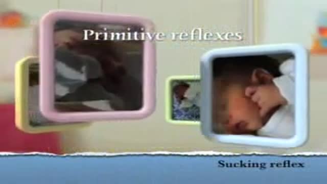
Sucking Reflex
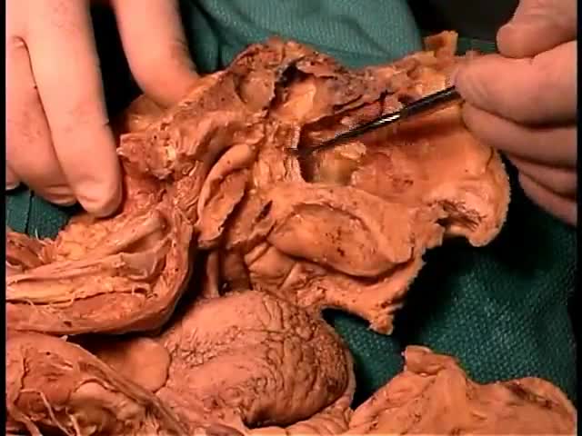
Anatomy of The Nasal Cavity and Sinuses
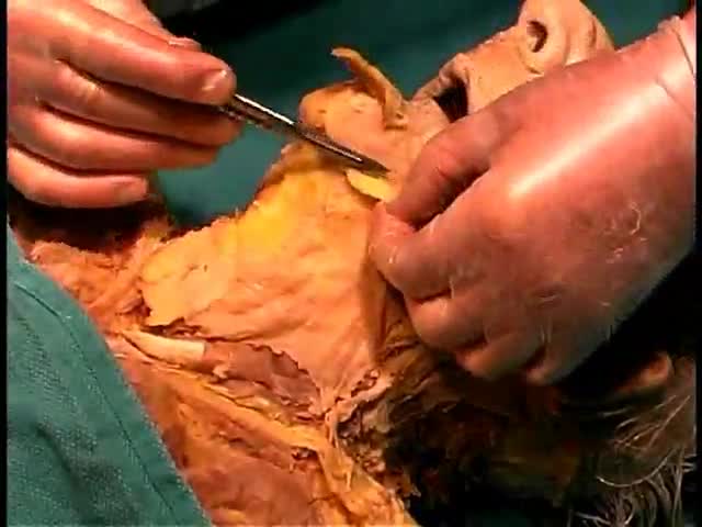
Anatomy of The Superficial Face

Hey everyone! When I started posting more squat and lower-body workouts last week, I got many requests for this video. I actually had already had a knee pain video, but I felt like I could make it a lot better, so I remade it.
For more information, check out my friend @ChrisRaynorMD aka @stablekneez on Instagram. Dr. Chris is a great surgeon, creative content creator, and all around good guy. He knows a LOT more than I do about this stuff. If you're interested, check him out!
Knee pain can be caused by MANY things, and this video is not intended to diagnose or treat any medical conditions. Some issues cannot be solved with exercise or physical therapy alone.
However, I've found that building up do doing squats with a full range of motion can help with knee pain. It's interesting, because there are some fitness figures that are adamantly against deep squatting because they claim it is BAD for the knees. I like it, and I've found it to be a very natural position. However, I do think that many people may not be prepared for it. Like any exercise, it can potentially hurt you if you're not acclimated to it. You can use your arm to assist you through the motion until you're able to do it without your arms with no pain. I have a full YouTube video on this: www.hybridcalisthenics.com/deepsquat.
Beyond this, sometimes strengthening our calves and hamstrings can "mysteriously" fix our knee pain. Both of these muscle groups support the knees.
On the other hand, sometimes these muscles are simply too TIGHT. You muscle fibers may be getting too tight and constantly trying to hyperextend your knees. Savor some calf and hamstring stretches.
I should point out that my chosen hamstring stretch in this video, standing toe touches, are controversial to some. Again, I like them, so they're shown here. If you are against them or they hurt you, you're very welcome to choose a different hamstring stretch.
Finally, I talk about isometric exercises like the horse stance and wall sit that seem to help with knee pain.
I touch upon synovial fluid, which is largely responsible for healing and nourishing our joints. Synovial fluid mostly circulates with movement, so I've included some knee "circles."
Hope this helps!
Legal Notice: Consult your doctor before beginning any kind of exercise program. This video does not replace a physical therapy program or consultation with a medical professional.
#shorts #hybridcalisthenics #kneepain
---
Free Fitness Routine: https://www.hybridcalisthenics.com/routine
Join our Discord community! https://www.hybridcalisthenics.com/discord
Shirts: https://www.bonfire.com/store/hybridcalisthenics
---
Instagram: https://www.hybridcalisthenics.com/instagram
YouTube: https://www.hybridcalisthenics.com/youtube
FaceBook: https://www.hybridcalisthenics.com/facebook
Twitter: https://www.hybridcalisthenics.com/twitter
Twitch: https://www.hybridcalisthenics.com/twitch
TikTok: https://www.hybridcalisthenics.com/tiktok
Tumblr: https://www.hybridcalisthenics.com/tumblr
Patreon: https://www.hybridcalisthenics.com/patreon
Subreddit: https://www.hybridcalisthenics.com/subreddit
Linkedin: https://www.linkedin.com/compa....ny/hybridcalisthenic
All Other Links:
https://www.linktr.ee/HybridCalisthenics
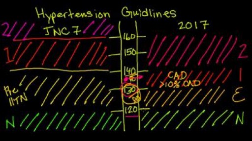
New 2017 Hypertension guidelines
Your body's immune system protects you from disease and infection. But if you have an autoimmune disease, your immune system attacks healthy cells in your body by mistake. Autoimmune diseases can affect many parts of the body. No one is sure what causes autoimmune diseases. They do tend to run in families. Women - particularly African-American, Hispanic-American, and Native-American women - have a higher risk for some autoimmune diseases. There are more than 80 types of autoimmune diseases, and some have similar symptoms. This makes it hard for your health care provider to know if you really have one of these diseases, and if so, which one. Getting a diagnosis can be frustrating and stressful. Often, the first symptoms are fatigue, muscle aches and a low fever. The classic sign of an autoimmune disease is inflammation, which can cause redness, heat, pain and swelling. The diseases may also have flare-ups, when they get worse, and remissions, when symptoms get better or disappear. Treatment depends on the disease, but in most cases one important goal is to reduce inflammation. Sometimes doctors prescribe corticosteroids or other drugs that reduce your immune response.
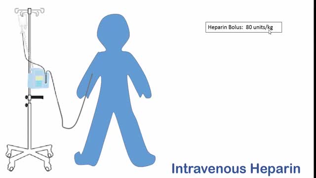
What is heparin injection? Heparin is an anticoagulant (blood thinner) that prevents the formation of blood clots. Heparin is used to treat and prevent blood clots in the veins, arteries, or lung. It is also used before surgery to reduce the risk of blood clots. Heparin works by inactivating thrombin in the clotting process. This stops the formation of fibrin and so stops blood clots forming. Heparin is used to treat blood clots that have formed abnormally inside the blood vessels. It can also be used to prevent these types of dangerous blood clots.
Depending on the underlying cause, some types of kidney disease can be treated. Often, though, chronic kidney disease has no cure. Treatment usually consists of measures to help control signs and symptoms, reduce complications, and slow progression of the disease. If your kidneys become severely damaged, you may need treatment for end-stage kidney disease. Treating the cause Your doctor will work to slow or control the cause of your kidney disease. Treatment options vary, depending on the cause. But kidney damage can continue to worsen even when an underlying condition, such as high blood pressure, has been controlled. Treating complications Kidney disease complications can be controlled to make you more comfortable. Treatments may include: High blood pressure medications. People with kidney disease may experience worsening high blood pressure. Your doctor may recommend medications to lower your blood pressure — commonly angiotensin-converting enzyme (ACE) inhibitors or angiotensin II receptor blockers — and to preserve kidney function. High blood pressure medications can initially decrease kidney function and change electrolyte levels, so you may need frequent blood tests to monitor your condition. Your doctor will likely also recommend a water pill (diuretic) and a low-salt diet. Medications to lower cholesterol levels. Your doctor may recommend medications called statins to lower your cholesterol. People with chronic kidney disease often experience high levels of bad cholesterol, which can increase the risk of heart disease. Medications to treat anemia. In certain situations, your doctor may recommend supplements of the hormone erythropoietin (uh-rith-roe-POI-uh-tin), sometimes with added iron. Erythropoietin supplements aid in production of more red blood cells, which may relieve fatigue and weakness associated with anemia. Medications to relieve swelling. People with chronic kidney disease may retain fluids. This can lead to swelling in the legs, as well as high blood pressure. Medications called diuretics can help maintain the balance of fluids in your body. Medications to protect your bones. Your doctor may prescribe calcium and vitamin D supplements to prevent weak bones and lower your risk of fracture. You may also take medication known as a phosphate binder to lower the amount of phosphate in your blood, and protect your blood vessels from damage by calcium deposits (calcification). A lower protein diet to minimize waste products in your blood. As your body processes protein from foods, it creates waste products that your kidneys must filter from your blood. To reduce the amount of work your kidneys must do, your doctor may recommend eating less protein. Your doctor may also ask you to meet with a dietitian who can suggest ways to lower your protein intake while still eating a healthy diet.
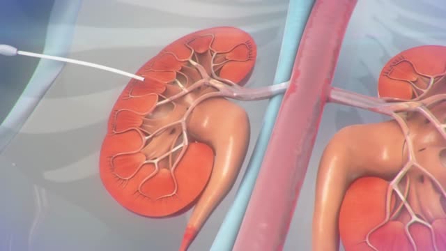
A ureteral stent is a thin, hollow tube that is placed in the ureter to help urine pass from the kidney into the bladder. Ureters are the tubes that connect the kidneys to the bladder. You may have a small amount of blood in your urine for 1 to 3 days after the procedure.
The pain of ovulation can range from a mild twinge to severe discomfort and usually lasts from minutes to hours. It is generally felt on one side of the abdomen and may vary each month, depending on which ovary is releasing the egg during that cycle.
Traumatic brain injury (TBI) is a nondegenerative, noncongenital insult to the brain from an external mechanical force, possibly leading to permanent or temporary impairment of cognitive, physical, and psychosocial functions, with an associated diminished or altered state of consciousness.
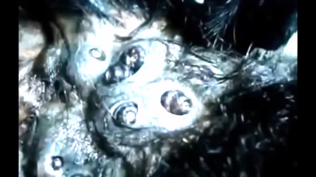
Blackheads, Cysts & Pimples

Comas are caused by an injury to the brain. Brain injury can be due to increased pressure, bleeding, loss of oxygen, or buildup of toxins. The injury can be temporary and reversible. It also can be permanent.
Post-streptococcal GN is a form of glomerulonephritis. It is caused by an infection with a type of streptococcus bacteria. The infection does not occur in the kidneys, but in a different part of the body, such as the skin or throat. The strep bacterial infection causes the tiny blood vessels in the filtering units of the kidneys (glomeruli) to become inflamed. This makes the kidneys less able to filter the urine. Post-streptococcal GN is uncommon today because infections that can lead to the disorder are commonly treated with antibiotics. The disorder may develop 1 to 2 weeks after an untreated throat infection, or 3 to 4 weeks after a skin infection. It may occur in people of any age, but it most often occurs in children ages 6 through 10. Although skin and throat infections are common in children, post-streptococcal GN is a rare complication of these infections. Risk factors include: Strep throat Streptococcal skin infections (such as impetigo)
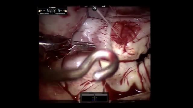
Mitral valve repair of anterior leaflet perforation and ruptured chordae
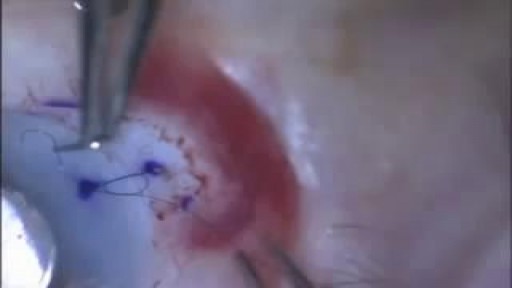
Video demonstrates the fundamental components of placing your first suture.
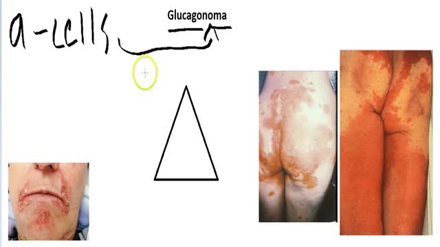
A glucagonoma is a rare tumor of the alpha cells of the pancreas that results in the overproduction of the hormone glucagon. Alpha cell tumors are commonly associated with glucagonoma syndrome, though similar symptoms are present in cases of pseudoglucagonoma syndrome in the absence of a glucagon-secreting tumor.

How To Improve Memory Power, How To Improve Concentration And Memory, Foods That Help The Brain---- http://brain-revitalizer.info-pro.co --- Brain Entrainment, For centuries humans have wondered at the connection between specific sound frequencies and the effect it can have on the brain and corresponding moods and emotions. From tribal drums to periodic stimulus tones the human brain taps into dominant external frequencies and when it does the mind can be altered to induce a host of different states including relaxation, sleep, creativity and excitement. The practice of causing brainwave frequencies to match a periodic stimulus to produce an intended state is called brainwave entrainment or brainwave synchronization and it is becoming more popular as life becomes more stressful. The study of sound and light and how it affects the human brain is nothing new. In the 1930's William Grey Walter used EEG equipment and strobe lights to detect the existence of high speed alpha waves and low speed delta waves and how each played a factor in human sleep patterns. In 1973 Gerald Oster published his discovery of binaural beats in Scientific American, a breakthrough article that defined binaural beats as apparent sounds which arise in the brain for specific physical stimuli. Though first discovered in 1839 by Heinrich Wilhelm Dove it wasn't until Oster's research that scientists began to speculate that binaural beats could be used to help induce relaxation, creativity and other desirable mental states. Today brainwave entrainment is gaining rapid popularity with people who feel over-stressed, depressed and unmotivated. With technology growing by leaps and bounds it's not hard to understand how a person can feel overwhelmed by information and sensory overload and instead of taking pills a growing segment of the population is turning to brainwave entrainment to produce a more natural and lasting feeling of relaxation. But YOU can be different! You can use Genius Brain Power to empower your brain so that you come alive with more energy, learn quicker, think more creatively, focus on your work like never before and drastically reduce stress with amazingly deep states of relaxation and meditation. click here: http://brain-revitalizer.info-pro.co
