- Physical Examination
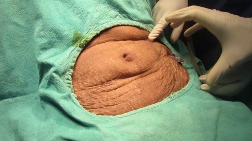
- Surgical Examination
- Ophthalmology
- Clinical Skills
- Orthopedics
- Surgery Videos
- Laparoscopy
- Pediatrics
- Funny Videos
- Cardiothoracic Surgery
- Nursing Videos
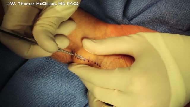
- Plastic Surgery
- Otorhinolaryngology
- Histology and Histopathology
- Neurosurgery
- Dermatology
- Pediatric Surgery
- Urology
- Dentistry
- Oncology and Cancers
- Anatomy Videos
- Health and Fitness
- Radiology
- Anaesthesia
- Physical Therapy
- Pharmacology
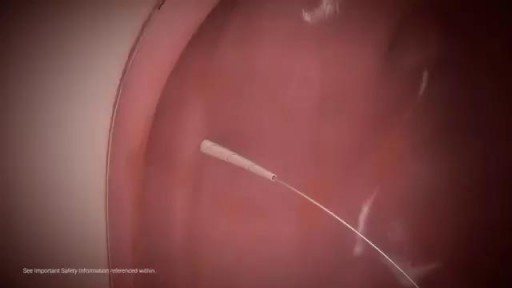
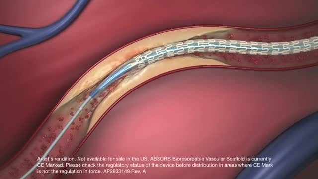
- Interventional Radiology
- Cardiology
- Endocrinology
- Gynecology
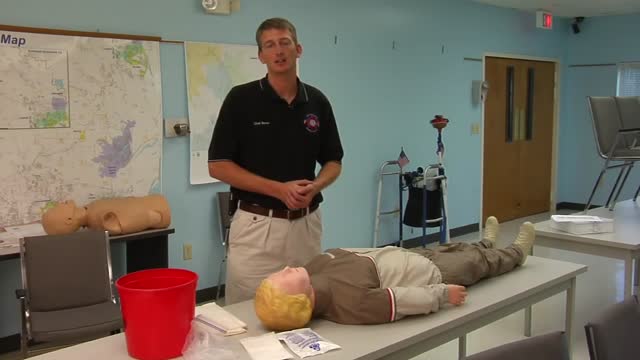
- Emergency Medicine
- Psychiatry and Psychology
- Childbirth Videos
- General Medical Videos
- Nephrology
- Physiology
- Diet and Food Health
- Diabetes Mellitus
- Neurology
- Women Health
- Osteoporosis
- Gastroenterology
- Pulmonology
- Hematology
- Rheumatology
- Toxicology
- Nuclear Medicine
- Infectious Diseases
- Vascular Disease
- Reproductive Health
- Burns and Wound Healing
- Other
Top videos
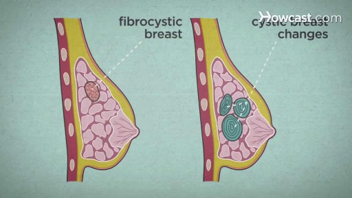
Over the course of a woman's lifetime, she may experience breast changes. While many end up being nothing to worry about, it's important to have any changes that you notice checked by a doctor -- just to be on the safe side. Here are the potential breast cancer symptoms to watch out for.
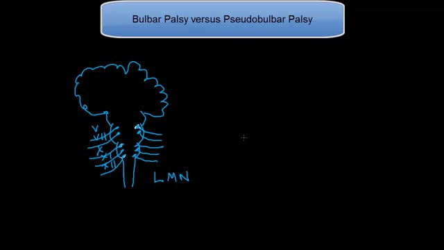
This tutorial explains the difference in mechanisms between the 2 palsies. Bulbar palsy is a lower motor neuron condition and pseudobulbar palsy is an upper motor neuron condidtion.
Pregnancy Stretch Marks Removal Treatment
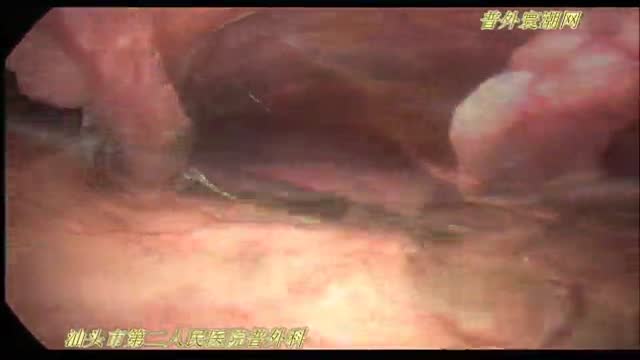
腹腔镜十二指肠球部溃疡穿孔修补术

This video: Polycystic ovary syndrome (PCOS) is a common endocrine disorder among women of reproductive age. Women with PCOS may have enlarged ovaries that contain small collections of fluid — called follicles — located in each ovary as seen during an ultrasound exam. Infrequent or prolonged menstrual periods, excess hair growth, acne, and obesity can all occur in women with polycystic ovary syndrome. In adolescents, infrequent or absent menstruation may raise suspicion for the condition. The exact cause of polycystic ovary syndrome is unknown. Early diagnosis and treatment along with weight loss may reduce the risk of long-term complications, such as type 2 diabetes and heart disease.
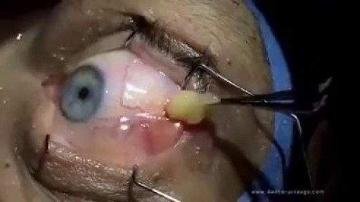
Eyeball cyst Removal
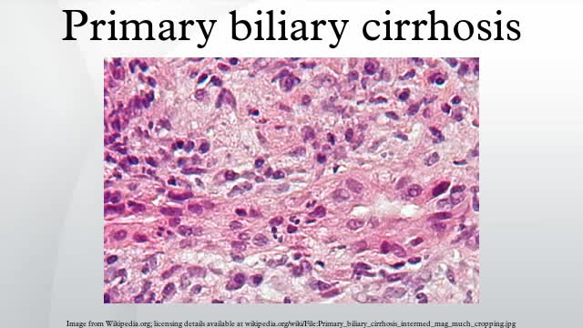
Primary biliary cirrhosis, sometimes called PBC, is a disease in which the bile ducts in your liver are slowly destroyed. Bile, a fluid produced in your liver, plays a role in digesting food and helps rid your body of worn-out red blood cells, cholesterol and toxins. When bile ducts are damaged, as in primary biliary cirrhosis, harmful substances can build up in your liver and sometimes lead to irreversible scarring of liver tissue (cirrhosis). Primary biliary cirrhosis is considered an autoimmune disease, in which the body turns against its own cells. Researchers think it is triggered by a combination of genetic and environmental factors. Primary biliary cirrhosis usually develops slowly and medication can slow its progression, especially if treatment begins early.

Finding a donor heart can be difficult. The heart must be donated by someone who is brain-dead but is still on life support. The donor heart must be matched as closely as possible to your tissue type to reduce the chance that your body will reject it. You are put into a deep sleep with general anesthesia, and a cut is made through the breastbone. Your blood flows through a heart-lung bypass machine while the surgeon works on your heart. This machine does the work of your heart and lungs while they are stopped, and supplies your body with blood and oxygen. Your diseased heart is removed and the donor heart is stitched in place. The heart-lung machine is then disconnected. Blood flows through the transplanted heart, which takes over supplying your body with blood and oxygen. Tubes are inserted to drain air, fluid, and blood out of the chest for several days, and to allow the lungs to fully re-expand.

Major signs and symptoms include enlargement of the liver and spleen (hepatosplenomegaly), a low number of red blood cells (anemia), easy bruising caused by a decrease in blood platelets (thrombocytopenia), lung disease, and bone abnormalities such as bone pain, fractures, and arthritis.

Are Glass Sex Toys Safe? | How to Use a Glass Dildo
Heat stroke is the most serious form of heat injury and is considered a medical emergency. If you suspect that someone has heat stroke -- also known as sunstroke -- call 911 immediately and give first aid until paramedics arrive. Heat stroke can kill or cause damage to the brain and other internal organs. Although heat stroke mainly affects people over age 50, it also takes a toll on healthy young athletes. Heat stroke often occurs as a progression from milder heat-related illnesses such as heat cramps, heat syncope (fainting), and heat exhaustion. But it can strike even if you have no previous signs of heat injury. Heat stroke results from prolonged exposure to high temperatures -- usually in combination with dehydration -- which leads to failure of the body's temperature control system. The medical definition of heat stroke is a core body temperature greater than 105 degrees Fahrenheit, with complications involving the central nervous system that occur after exposure to high temperatures. Other common symptoms include nausea, seizures, confusion, disorientation, and sometimes loss of consciousness or coma
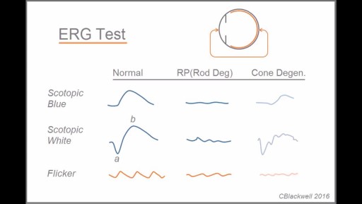
Retinitis pigmentosa is a rare, inherited degenerative eye disease that causes severe vision impairment. Symptoms often begin in childhood. They include decreased vision at night or in low light and loss of side vision (tunnel vision). There's no effective treatment for this condition. Wearing sunglasses may help protect remaining vision.
Mitral valve regurgitation, known as leaky heart valve, can be treated with the MitraClip procedure, especially if you're not a candidate for surgery. As premier heart specialists in the Rocky Mountains, Aurora Denver Cardiology Associates physicians perform this procedure and believe it can be an essential treatment for heart health.
Ganglion cysts are noncancerous lumps that most commonly develop along the tendons or joints of your wrists or hands. They also may occur in the ankles and feet. Ganglion cysts are typically round or oval and are filled with a jellylike fluid. Small ganglion cysts can be pea-sized, while larger ones can be around an inch (2.5 centimeters) in diameter. Ganglion cysts can be painful if they press on a nearby nerve. Their location can sometimes interfere with joint movement. If your ganglion cyst is causing you problems, your doctor may suggest trying to drain the cyst with a needle. Removing the cyst surgically also is an option. But if you have no symptoms, no treatment is necessary. In many cases, the cysts go away on their own.
When the arteries in your heart become blocked, the condition is called coronary artery disease. It can be a serious condition if not treated. Coronary artery disease puts you at risk for a heart attack. Be sure you pay attention to your symptoms and manage your heart health risks.
Slicosis is caused by inhalation of unbound (free) crystalline silica dust and is characterized by nodular pulmonary fibrosis. Chronic silicosis initially causes no symptoms or only mild dyspnea but over years can advance to involve most of the lung and cause dyspnea, hypoxemia, pulmonary hypertension, and respiratory impairment. Diagnosis is based on history and chest x-ray findings. No effective treatment exists except supportive care and, for severe cases, lung transplantation.
» A kidney stone, also known as a renal calculus (from the Latin rēnēs, “kidneys,” and calculus, “pebble”), is a solid concretion or crystal aggregation formed in the kidneys from dietary minerals in the urine. » Urolithiasis (UL) is one of the most common diseases, with approximately 7.6% incidence in Western India. Although most patients have only one stone episode, 25% of patients experience recurrent stone formation. UL therefore has a significant impact on quality of life and socioeconomic factors.
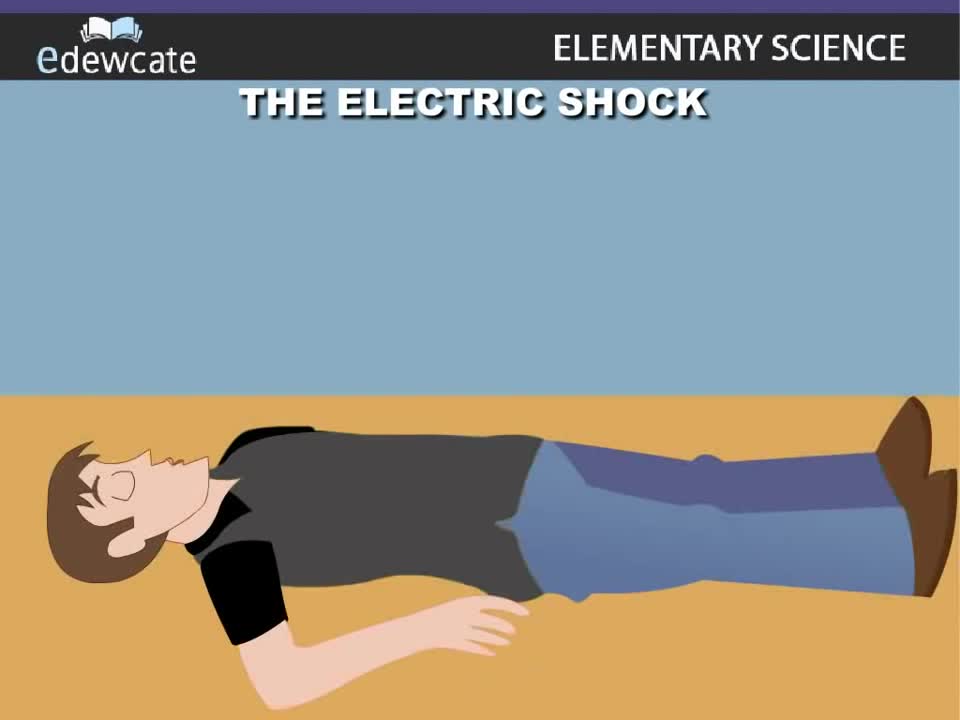
Very small currents can be imperceptible. Larger current passing through the body may make it impossible for a shock victim to let go of an energized object. Still larger currents can cause fibrillation of the heart and damage to tissues. Death caused by an electric shock is called electrocution.
