- Physical Examination
- Surgical Examination
- Ophthalmology
- Clinical Skills
- Orthopedics
- Surgery Videos
- Laparoscopy
- Pediatrics
- Funny Videos
- Cardiothoracic Surgery
- Nursing Videos
- Plastic Surgery
- Otorhinolaryngology
- Histology and Histopathology
- Neurosurgery
- Dermatology
- Pediatric Surgery
- Urology
- Dentistry
- Oncology and Cancers
- Anatomy Videos
- Health and Fitness
- Radiology
- Anaesthesia
- Physical Therapy
- Pharmacology
- Interventional Radiology
- Cardiology
- Endocrinology
- Gynecology
- Emergency Medicine
- Psychiatry and Psychology
- Childbirth Videos
- General Medical Videos
- Nephrology
- Physiology
- Diet and Food Health
- Diabetes Mellitus
- Neurology
- Women Health
- Osteoporosis
- Gastroenterology
- Pulmonology
- Hematology
- Rheumatology
- Toxicology
- Nuclear Medicine
- Infectious Diseases
- Vascular Disease
- Reproductive Health
- Burns and Wound Healing
- Other
Top videos
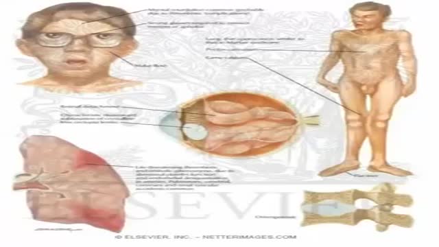
Homocystinuria is an inherited disorder that affects the metabolism of the amino acid methionine. Amino acids are the building blocks of life. Causes Homocystinuria is inherited in families as an autosomal recessive trait. This means that the child must inherit a non-working copy of the gene from each parent to be seriously affected. Homocystinuria has several features in common with Marfan syndrome, including joint and eye changes. Symptoms Newborn infants appear healthy. Early symptoms, if present, are not obvious. Symptoms may occur as mildly delayed development or failure to thrive. Increasing visual problems may lead to diagnosis of this condition. Other symptoms include: Chest deformities (pectus carinatum, pectus excavatum) Flush across the cheeks High arches of the feet Intellectual disability Knock knees Long limbs Mental disorders Nearsightedness Spidery fingers (arachnodactyly) Tall, thin build
The dog bite victim needs to be taken to a safe place away from the assailant dog to prevent further attack and injury. Since dog bites can cause significant damage beneath the skin, a type of injury that cannot always easily be appreciated, medical care should be accessed by a health care practitioner. Wounds should be kept elevated and, if possible, washing the wound with tap water may be attempted. Information should be obtained from the dog's owner about the dog's rabies immunization status, but if this is not possible, hospital, animal control centers, or law enforcement personnel will help gather any required information.
A torn meniscus is one of the most common knee injuries. Any activity that causes you to forcefully twist or rotate your knee, especially when putting the pressure of your full weight on it, can lead to a torn meniscus. Each of your knees has two menisci — C-shaped pieces of cartilage that act like a cushion between your shinbone and your thighbone. A torn meniscus causes pain, swelling and stiffness. You also might have trouble extending your knee fully. Conservative treatment — such as rest, ice and medication — is sometimes enough to relieve the pain of a torn meniscus and give the injury time to heal on its own. In other cases, however, a torn meniscus requires surgical repair.
Stuck with an Embroidery Needle
A lipoma is a growth of fat cells in a thin, fibrous capsule usually found just below the skin. Lipomas aren't cancer and don't turn into cancer. They are found most often on the torso, neck, upper thighs, upper arms, and armpits, but they can occur almost anywhere in the body. One or more lipomas may be present at the same time.
Dieta Iposodica Per Acufeni, Agopuntura Efficacia Contro Acufeni, Ronzio Orecchie Nel Silenzio---- http://acufeni-cura.plus101.com/ --- I sintomi dell'acufene, I sintomi dell'acufene possono causare molti fastidi e problemi a chi ne soffre. Inoltre anche i sintomi possono causare confusione. Prendiamo un esempio per capire meglio. Stai conducendo una vita perfetta, vai al lavoro e torni a casa ogni giorno, ma improvvisamente incominci a sentire strani rumori all'orecchio. Ovviamente credi che ci sia una sorgente di tali rumori. Stranamente nessun altro sembra sentirli. Puoi spaventarti e pensare che quei rumori provengano dal tuo corpo, così vai dal dottore. Dopo qualche esame il dottore conferma che è tutto a posto. Il problema è che tu i rumori continui a sentirli. Incominciano a darti sui nervi e ti influenzano nel lavoro. E ancora peggio, i rumori sembrano farsi più forti la notte, privandoti del tuo prezioso sonno. Si tratta di acufene. I rumori che senti sono i sintomi principali e possono essere di vario tipo. Puoi sentire strani scricchilii, ronzii, brusii, fischi o sibili all'orecchio. Non è mai lo stesso per tutti. Alcuni li sentono di tanto in tanto, altri per tutto il tempo. alcuni raccontano di forti attacchi. Ed altri sono stremati da trapanamenti costanti. Ma una cosa è certa - molte persone negli USA e altrove hanno gli acufeni. Quindi non sei davvero l'unico in queste condizioni. La maggior parte dei sintomi dell'acufene non sono altro che rumori fantasma Molte persone sono confuse se non riescono ad indivuare la fonte del rumore alle orecchie. Alcuni si spaventano. Spesso queste persone sono messe in ridicolo da chi non sente quei rumori. Ma per chi soffre di acufeni i suoni sono assolutamente reali. Di fatto sono rumori fantasma, una percezione delle orecchie. Vi sono eccezioni. In almeno un caso il rumore può essere reale. In altre parole c'è una effettiva fonte del rumore che senti. Questo è l'acufene pulsatile. In questo caso sei in grado di sentire il battito del tuo cuore e ciò ti può far impazzire perchè lo senti in continuazione. Vi è un'altra differenza. C'è sicuramente più di una persona che sente questi rumori e questo è il tuo dottore. Avrà bisogno di uno strumento di ascolto per sentirli. Prendi nota - non è come sentire il cuore con uno stetoscopio. in questo caso il dottore utilizza uno strumento di ascolto per sentire il rumore all'orecchio, non nel petto. Che cosa causa l'acufene pulsatile? Potresti sentire il tuo cuore in caso di pressione alta, danni alle arterie e anche cambiamenti della circolazione del sangue. A volte, una grande dose di stress, ansia o depressione può causare questa situazione. Non Importa Quale Sia La Causa, Tipo O Gravità Del Tuo Acufene, Puoi Iniziare Ad Utilizzare Questo Efficare Sistema PROPRIO ORA Ed Ottenere Un Sollivo E Una Liberazione Permanente ISTANTANEA Dai Tuoi Acufeni! inserisci ora: http://acufeni-cura.plus101.com/

Surprising Facts About High Blood PressureMust
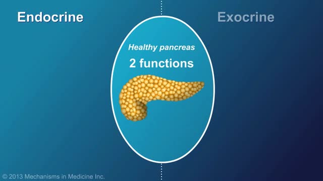
Chronic pancreatitis is a long-standing inflammation of the pancreas that alters the organ's normal structure and functions. It can present as episodes of acute inflammation in a previously injured pancreas, or as chronic damage with persistent pain or malabsorption.

What is an Aneurysm? A cerebral or intracranial aneurysm is an abnormal focal dilation of an artery in the brain that results from a weakening of the inner muscular layer (the intima) of a blood vessel wall. The vessel develops a "blister-like" dilation that can become thin and rupture without warning. The resultant bleeding into the space around the brain is called a subarachnoid hemorrhage (SAH). This kind of hemorrhage can lead to a stroke, coma, and/or death. Aneurysms are usually found at the base of the brain just inside the skull, in an area called the subarachnoid space. In fact, 90 percent of SAHs are attributed to ruptured cerebral aneurysms and the two terms are often used synonymously.
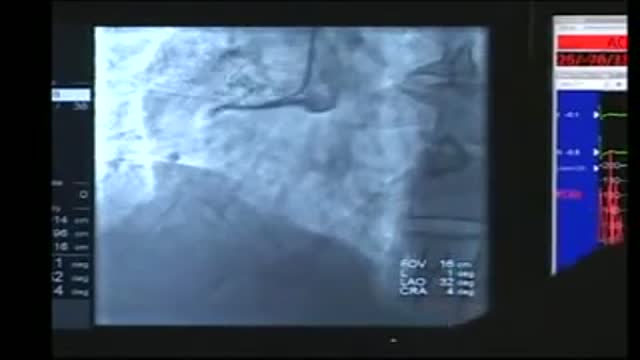
Cardiac catheterization (kath-uh-tur-ih-ZAY-shun) is a procedure used to diagnose and treat cardiovascular conditions. During cardiac catheterization, a long thin tube called a catheter is inserted in an artery or vein in your groin, neck or arm and threaded through your blood vessels to your heart. Using this catheter, doctors can then do diagnostic tests as part of a cardiac catheterization. Some heart disease treatments, such as coronary angioplasty, also are done using cardiac catheterization. Usually, you'll be awake during cardiac catheterization, but given medications to help you relax. Recovery time for a cardiac catheterization is quick, and there's a low risk of complications.

20 Reasons to Drink Lemon Water

Fibromyalgia syndrome (FMS) is a form of fibromyalgia where pain and stiffness occurs in muscles, tendons, and ligaments throughout the body, accompanied by other generalized symptoms such as fatigue, sleep disruption or unrefreshing sleep, mood disorder, and cognitive difficulties such as poor memory or mental ...
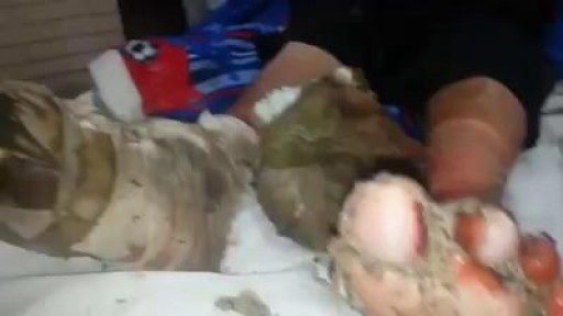
A patient suffering from Diabetic gangrene and maneged by "myiasis"

nkylosing spondylitis (pronounced ank-kih-low-sing spon-dill-eye-tiss), or AS, is a form of arthritis that primarily affects the spine, although other joints can become involved. It causes inflammation of the spinal joints (vertebrae) that can lead to severe, chronic pain and discomfort


http://hidradenitis-suppurativa-cure.plus101.com --- What Is Hs, How To Get Rid Of Hidradenitis Suppurativa, Hidradenitis Suppurativa Treatment Options. What is Hidradenitis Suppurativa? Hidradenitis Suppurativa is a non-contagious skin disease that is also known as Acne Inversa. This condition affects areas of the body where there is skin to skin contact and where sweat or oil glands are present; common areas are the underarms, breasts, buttocks, anal region, and groin. It affects between 1 to 4% of the world's population, and is more likely to occur in females. Symptoms Hidradenitis Suppurativa is characterized by persistent abscesses, cysts (epidermoid, sebaceous, and pilonidal) and infections. The condition is chronic and often goes through alternating periods of remission and flare-ups. During flare-ups, the inflammation tends to be severe and patients may develop fever and be very fatigued. The pain can be unbearable and the person's movements will be very limited. The abscesses often drain pus and leave open wounds that may not heal. Eventually, abscesses may become interconnected through tunnels under skin and this makes the condition harder to treat. Causes The immediate cause of Hidradenitis Suppurativa is clogging of the apocrine glands, due to dead skin cells become trapped in the gland, over production of oil, or bacterial accumulation. This will cause the plug to swell with pus formation. What causes this simple blockage to progress into a full blow Hidradenitis Suppurativa case is still debated, however, possible theories include an auto-immune reaction, hormone imbalances and genetic disorders. It is also known that excessive sweating and being overweight will increase the risk of developing the condition. Furthermore, wearing tight clothing, excessive shaving, using lithium medications and hot humid climates have been identified as triggering factors. For a complete guide on curing Hidradenitis Suppurativa through a natural and holistic approach, visit http://hidradenitis-suppurativa-cure.plus101.com

Generic minoxidil is known to treat hair-fall issues in men and women, it is best for hair growth, hair re-development, etc. it is available in the strength of 5mg and easily available at online pharmacy store. For more information visit to http://www.medstorerx.com/generic-minoxidil.aspx

Aumentar Gluteos, Como Hacer Crecer Los Gluteos En Una Semana, Eliminar Celulitis Gluteos.--- http://aumente-gluteos.plus101.com/ --- Para conseguir aumentar tu cola sin ejercicio, debes utilizar algunas cremas naturales caseras, sencillas y fáciles de preparar en la zona de tus glúteos, estas cremas le darán a tu cola una buena forma, firmeza y además una piel libre de manchas, estrías y celulitis. 1. Crema Con Omega 3, Vitamina E y Vitamina C Esta crema es bastante fácil de realizar y sumamente efectiva, solo necesitas tres capsulas de omega 3, dos de vitamina E y dos de vitamina C, los resultados se harán notar en cuestión de días. Con una aguja saca en contenido de cada una de las capsulas y mézclalas en un recipiente, una vez que estén bien mezcladas aplícala sobre tu cola dando masajes circulares que abarquen todo el trasero. Para mejores resultados envuelve la zona de tus glúteos en plástico y déjala actuar durante 30 minutos, luego retira el plástico, colócate un bóxer levanta cola y duerme con esta crema casera, por la mañana la puedes retirar con agua tibia y repetir este proceso todas las noches hasta que consigas los resultados que deseas. 2. Crema Con Aceite De Almendras, Pepino y Avena Esta crema es maravillosa, le dará a tu cola una atractiva apariencia, solo necesitas 3 cucharadas de aceite de almendras, un pepino pequeño y tres cucharadas de avena en hojuelas. El procedimiento es bastante sencillo, debes triturar muy bien el pepino, agregar la avena y luego el aceite de almendras, mezcla hasta que los ingredientes se compacten y listo. Aplica esta crema con suavidad sobre tu cola dando masajes de abajo hacia arriba para vencer los efectos de la gravedad, déjala actuar durante 45 minutos y retírala con agua fría, esta crema casera cuidará muy bien tu piel y le dará a tus glúteos una gran firmeza. 3. Vitamina C, Ciruelas y Aceite De Girasol Una crema divina que te dará una cola perfecta en poco tiempo, ayuda a regenerar la piel de la zona de tus glúteos, tonificándolos y moldeándolos, solo necesitas 3 capsulas de vitamina C, 5 ciruelas grandes y 3 cucharadas de aceite de girasol. Cientos de mujeres en todo el mundo están aumentando el tamaño de sus glúteos gracias a los conocimientos secretos de esta guía, Ingresa ya a: http://aumente-gluteos.plus101.com/
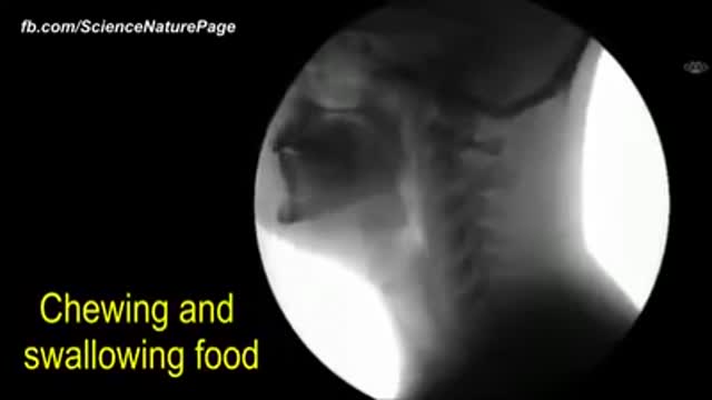
The human body as seen with MRI and X-RAY
