- Physical Examination
- Surgical Examination
- Ophthalmology
- Clinical Skills
- Orthopedics
- Surgery Videos
- Laparoscopy
- Pediatrics
- Funny Videos
- Cardiothoracic Surgery
- Nursing Videos
- Plastic Surgery
- Otorhinolaryngology
- Histology and Histopathology
- Neurosurgery
- Dermatology
- Pediatric Surgery
- Urology
- Dentistry
- Oncology and Cancers
- Anatomy Videos
- Health and Fitness
- Radiology
- Anaesthesia
- Physical Therapy
- Pharmacology
- Interventional Radiology
- Cardiology
- Endocrinology
- Gynecology
- Emergency Medicine
- Psychiatry and Psychology
- Childbirth Videos
- General Medical Videos
- Nephrology
- Physiology
- Diet and Food Health
- Diabetes Mellitus
- Neurology
- Women Health
- Osteoporosis
- Gastroenterology
- Pulmonology
- Hematology
- Rheumatology
- Toxicology
- Nuclear Medicine
- Infectious Diseases
- Vascular Disease
- Reproductive Health
- Burns and Wound Healing
- Other
Top videos
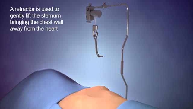
The cause of pectus excavatum is not known however it can run in families, with up to 25 percent of affected patients reporting chest wall abnormalities in other family members. Pectus excavatum occurs in approximately 1 out of 400–1000 children and is three to five times more common in males than females.

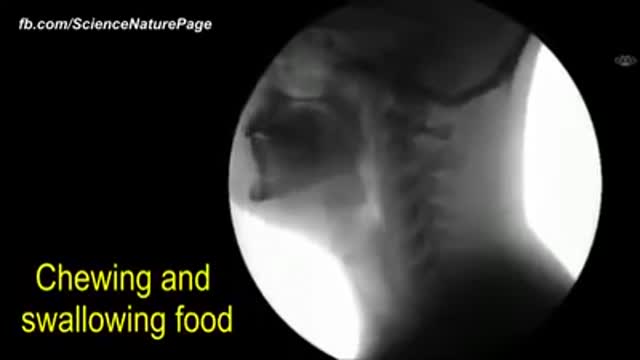
The human body as seen with MRI and X-RAY

The hips are one of the hardest places to loose fat. Liposuction can be done on this area to dramatically help loose inches. This area is also one of the most successful areas to show visible improvement after liposuction is done. Liposuction of the hips can help patients to reduce dress and pant sizes. Disclaimer. The photographs on these pages illustrate typical results of some liposuction surgery procedures and may contain some nudity. Viewer discretion is advised. In providing the photos and statements on this web site, Liposuction.com does not state or imply any guarantee.
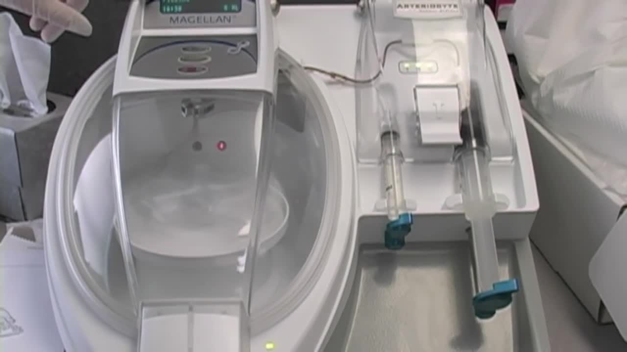
Stem Cell Injection Treatment - Stem Cell Therapy

Hereditary hemochromatosis (he-moe-kroe-muh-TOE-sis) causes your body to absorb too much iron from the food you eat. Excess iron is stored in your organs, especially your liver, heart and pancreas. Too much iron can lead to life-threatening conditions, such as liver disease, heart problems and diabetes.
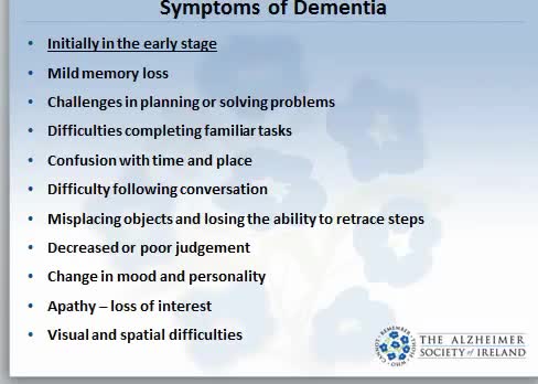
Dementia is not a specific disease. It's an overall term that describes a wide range of symptoms associated with a decline in memory or other thinking skills severe enough to reduce a person's ability to perform everyday activities. Alzheimer's disease accounts for 60 to 80 percent of cases. Vascular dementia, which occurs after a stroke, is the second most common dementia type. But there are many other conditions that can cause symptoms of dementia, including some that are reversible, such as thyroid problems and vitamin deficiencies.
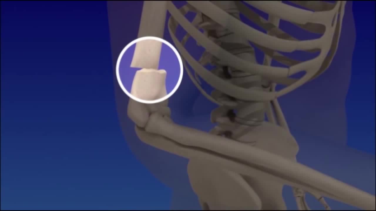
Distal Humerus Fractures of the Elbow. A distal humerus fracture is a break in the lower end of the upper arm bone (humerus), one of the three bones that come together to form the elbow joint. A fracture in this area can be very painful and make elbow motion difficult or impossible.

Lupus hair loss and alopecia explained
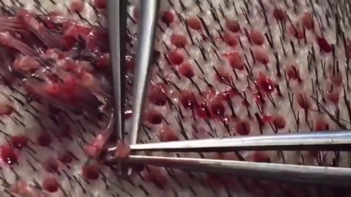
What Is a Hair Transplant? It's a type of surgery that moves hair you already have to fill an area with thin or no hair. Doctors have been doing these transplants in the U.S. since the 1950s, but techniques have changed a lot in recent years. You usually have the procedure in the doctor's office. First, the surgeon cleans your scalp and injects medicine to numb the back of your head. Your doctor will choose one of two methods for the transplant: follicular unit strip surgery (FUSS) or follicular unit extraction (FUE). With FUSS, the surgeon removes a 6- to 10-inch strip of skin from the back of your head. He sets it aside and sews the scalp closed. This area is immediately hidden by the hair around it. Next, the surgeon’s team divides the strip of removed scalp into 500 to 2,000 tiny grafts, each with an individual hair or just a few hairs. The number and type of graft you get depends on your hair type, quality, color, and the size of the area where you’re getting the transplant. If you’re getting the FUE procedure, the surgeon’s team will shave the back of your scalp. Then, the doctor will remove hair follicles one by one from there. The area heals with small dots, which your existing hair will cover. After that point, both procedures are the same. After he prepares the grafts, the surgeon cleans and numbs the area where the hair will go, creates holes or slits with a scalpel or needle, and delicately places each graft in one of the holes. He’ll probably get help from other team members to plant the grafts, too. Depending on the size of the transplant you’re getting, the process will take about 4 to 8 hours. You might need another procedure later on if you continue to lose hair or decide you want thicker hair. Expectations and Recovery After the surgery, your scalp may be very tender. You may need to take pain medications for several days. Your surgeon will have you wear bandages over your scalp for at least a day or two. He may also prescribe an antibiotic or an anti-inflammatory drug for you to take for several days. Most people are able to return to work 2 to 5 days after the operation. Within 2 to 3 weeks after surgery, the transplanted hair will fall out, but you should start to notice new growth within a few months. Most people will see 60% of new hair growth after 6 to 9 months. Some surgeons prescribe the hair-growing drug minoxidil (Rogaine) to improve hair growth after transplantation, but it’s not clear how well it works. Risks and Costs of Treatment The price of a hair transplant will depend largely on the amount of hair you’re moving, but it generally ranges from $4,000 to $15,000. Most insurance plans don’t cover it.
This video may contain images of a medical doctor providing emergency care for a patient.
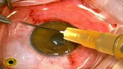
Eye cancers can be primary (starts within the eye) and metastatic cancer (spread to the eye from another organ). The two most common cancers that spread to the eye from another organ are breast cancer and lung cancer. Other less common sites of origin include the prostate, kidney, thyroid, skin, colon and blood or bone marrow. Melanomas (choroidal, ciliary body and uveal) -Early stages has no symptoms (the person does not know there is a tumor until an ophthalmology examination). As the tumor grows, symptoms can be blurred vision, decreased vision, double vision, eventual vision loss and if they continue to grow the tumor can break past the retina causing retinal detachment.
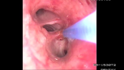
Bronchoscopy Procedure - See inside the lungs!
Shigellosis is a diarrheal disease caused by a group of bacteria called Shigella. Shigella causes about 500,000 cases of diarrhea in the United States annually 1. There are four different species of Shigella:

Tummy-tuck surgery or abdominoplasty, can flatten your abdomen by removing loose, excess fat and skin and tightening muscles in the abdominal wall. It can also remove some if not all of the stretch marks in your lower abdomen. It is popular following pregnancy, massive weight loss or whenever a flabby abdomen with weak muscles impairs body contour. Most patients report improved self-esteem as a result of this procedure.
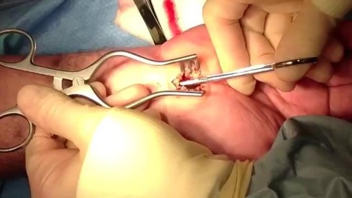
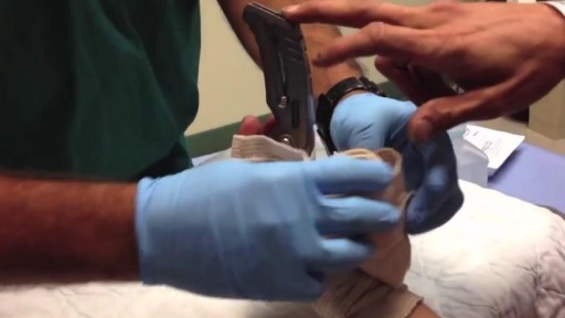
Carpal tunnel release is a surgery used to treat and potentially heal the painful condition known as carpal tunnel syndrome. Doctors used to think that carpal tunnel syndrome was caused by an overuse injury or a repetitive motion performed by the wrist or hand, often at work. They now know that it’s most likely a congenital predisposition (something that runs in families) – some people simply have smaller carpal tunnels than others. Carpal tunnel syndrome can also be caused by injury, such as a sprain or fracture, or repetitive use of a vibrating tool. It's also been linked to pregnancy, diabetes, thyroid disease, and rheumatoid arthritis.

http://cure-insomnio-para-siempre.good-info.co/ Paralisis Del Sueño, Como Combatir El Insomnio, Insomnio Tratamiento, Consejos Para Dormir Bien. https://youtu.be/kYvaazgJMv4 Seguramente después de haber leído usted se habrá hecho una pregunta clave: ¿Comer mal afecta al insomnio? La respuesta es sí. Una mala dieta es razón de suficiente para padecer de insomnio y es por ello que es importante que usted modifique sus conductas alimenticias si quiere disfrutar de un sueño reparador. La ingesta exagerada de carbohidratos y grasas puede llegar a perturbar nuestro metabolismo y dificulta nuestra digestión, haciendo de este modo, le sea imposible conciliar el sueño. ¿Se Puede Combatir El Insomnio Con Una Dieta? Lo primero que debe saber acerca de la dieta y su relación con el insomnio es que el mejor régimen para terminar con es la prevención y la planificación: haga una rutina firme, coma siempre a la misma hora y evite ingerir alimentos media hora antes dormir. También le recomiendo que no abuse de la cafeína ni de los carbohidratos. Evite además los alimentos que no sean fáciles de digerir o que le provocan algún tipo de alergia. ¿Existen Alimentos Que Combatan El Insomnio? Hay algunos elementos que pueden ayudarlo a conciliar el sueño y sentirse relajado y distendido a la hora de prepararse para el descanso. Tenga en cuenta, fundamentalmente, si usted padece de insomnio, que lo ideal es optar por una cena frugal y reposada a fin de evitar sobresaltos y condiciones que no le permitan dormir profundamente. ¿Qué Alimentos Debe Considerar Para Evitar El Insomnio? Hay una serie de alimentos concretos que usted puede considerar en si dieta para irse a la cama con la expectativa de un mejor sueño. Estos son alguno de los alimentos que debe evitar: Paralisis Del Sueño, Como Combatir El Insomnio, Insomnio Tratamiento, Consejos Para Dormir Bien

Meet Christian, an incredible man born with no arms or legs who lives life to the fullest
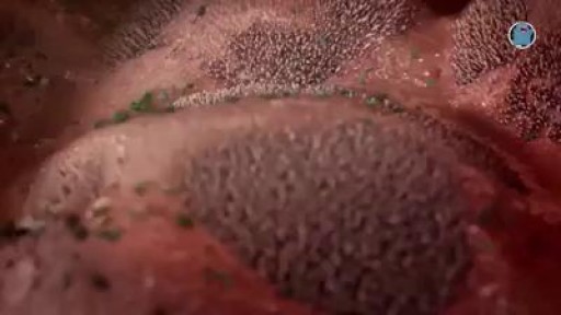
Watch that video to know What Causes Trypophobia?

Watch that video of The Most Amazing Plastic Surgeries
