- Physical Examination
- Surgical Examination
- Ophthalmology
- Clinical Skills
- Orthopedics
- Surgery Videos
- Laparoscopy
- Pediatrics
- Funny Videos
- Cardiothoracic Surgery
- Nursing Videos
- Plastic Surgery
- Otorhinolaryngology
- Histology and Histopathology
- Neurosurgery
- Dermatology
- Pediatric Surgery
- Urology
- Dentistry
- Oncology and Cancers
- Anatomy Videos
- Health and Fitness
- Radiology
- Anaesthesia
- Physical Therapy
- Pharmacology
- Interventional Radiology
- Cardiology
- Endocrinology
- Gynecology
- Emergency Medicine
- Psychiatry and Psychology
- Childbirth Videos
- General Medical Videos
- Nephrology
- Physiology
- Diet and Food Health
- Diabetes Mellitus
- Neurology
- Women Health
- Osteoporosis
- Gastroenterology
- Pulmonology
- Hematology
- Rheumatology
- Toxicology
- Nuclear Medicine
- Infectious Diseases
- Vascular Disease
- Reproductive Health
- Burns and Wound Healing
- Other
Top videos
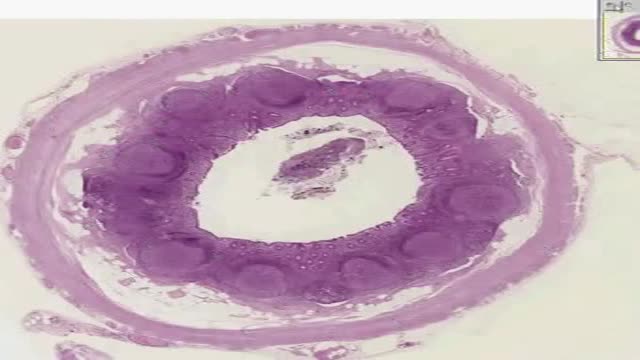
Histology of Appendix

Histology of Thymus
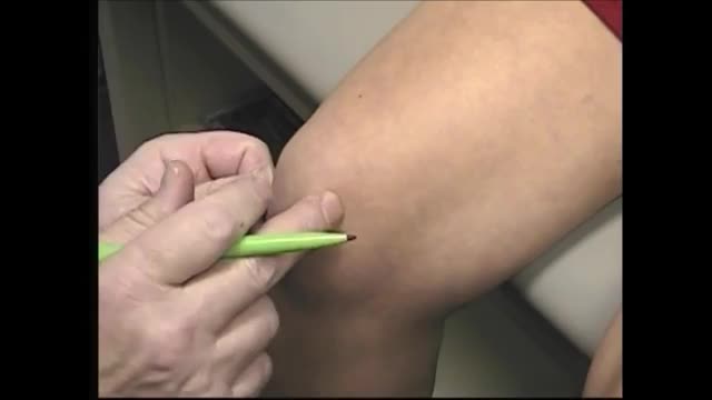
When oral medications do not relieve knee pain, but you're not to the point of pursuing knee surgery, one of the following injections or procedures may help. Hyaluronic acid supplements – Although not technically medications, these substances are injected into knee joints to supplement naturally occurring hyaluronic acid. In healthy joints hyaluronic acid acts as a shock absorber and lubricant, allowing joints to move smoothly over each other. However, the acid appears to break down in people with osteoarthritis. Injecting it into a joint may lessen pain and inflammation. The injections are given weekly for three or five weeks, depending on the product (examples are Synvisc and Hyalgan). A small amount of joint fluid is removed first to make room for the hyaluronic acid. Corticosteroid Injections – Doctors sometimes inject corticosteroids directly into the knee joint for quick relief of pain and inflammation. Their benefits may last anywhere from a few days to more than six months. While the injections bring targeted relief to the joint and lack many of the side effects of oral corticosteroid medications, they are not without risks. Repeated knee injections may actually contribute to cartilage breakdown. For that reason your doctor will likely put a limit on the number of injections you can receive. Read a report from the British Medical Journal on corticosteroid injections for knee osteoarthritis. Arthrocentesis – Also called joint fluid aspiration, arthrocentesis is removal of joint fluid through a hollow needle inserted into the joint space of the knee. Although the purpose of removing joint fluid from the knee is usually so that it can be tested in the lab, removing excess fluid can also quickly ease pain and swelling. Often after withdrawing fluid, doctors use the same puncture site where the fluid was removed to inject a corticosteroid preparation and/or anesthetic into the knee joint to further relieve pain and inflammation.

To learn more about licensing this video for content marketing or patient education purposes, visit: http://www.nucleushealth.com/?utm_source=youtube&utm_medium=video-description&utm_campaign=stroke-071411
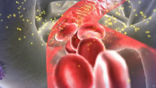
This video, created by Nucleus Medical Media, gives a thorough explanation of stroke, covering anatomy and physiology, different types of stroke, and treatment.
ANH11048
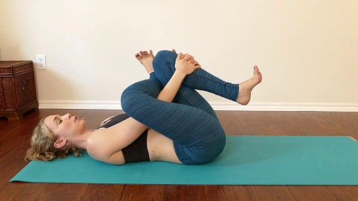
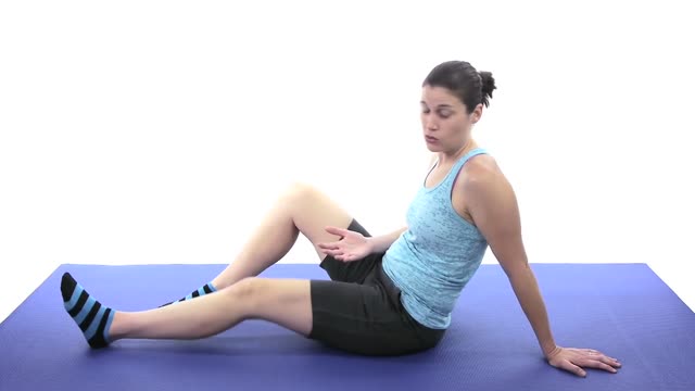
Fast Lower Back Pain & Sciatica Pain Relief – Beginners Yoga Stretches and Poses

This video contains some graphic images of an ingrown toenail procedure. Ingrown toenails are a common and painful issue that many people experience. If you're suffering from chronic ingrown toenails that make it difficult to do the things you love

This video: Polycystic ovary syndrome (PCOS) is a common endocrine disorder among women of reproductive age. Women with PCOS may have enlarged ovaries that contain small collections of fluid — called follicles — located in each ovary as seen during an ultrasound exam. Infrequent or prolonged menstrual periods, excess hair growth, acne, and obesity can all occur in women with polycystic ovary syndrome. In adolescents, infrequent or absent menstruation may raise suspicion for the condition. The exact cause of polycystic ovary syndrome is unknown. Early diagnosis and treatment along with weight loss may reduce the risk of long-term complications, such as type 2 diabetes and heart disease.
Here are 10 lifestyle changes you can make to lower your blood pressure and keep it down. Lose extra pounds and watch your waistline. Blood pressure often increases as weight increases. ... Exercise regularly. ... Eat a healthy diet. ... Reduce sodium in your diet. ... Limit the amount of alcohol you drink.

Tampon for The First Time

LASIK eye surgery has been popular for more than 20 years, with an estimated 20 million Americans undergoing the procedure to correct nearsightedness and improve distance vision. But some patients says the surgery has ruined their eyesight. Now an expert who once backed LASIK is campaigning to get it off the market. Dr. Tara Narula reports.
Watch "CBS This Morning" HERE: http://bit.ly/1T88yAR
Download the CBS News app on iOS HERE: https://apple.co/1tRNnUy
Download the CBS News app on Android HERE: https://bit.ly/1IcphuX
Like "CBS This Morning" on Facebook HERE: http://on.fb.me/1LhtdvI
Follow "CBS This Morning" on Twitter HERE: http://bit.ly/1Xj5W3p
Follow "CBS This Morning" on Instagram HERE: http://bit.ly/1Q7NGnY
Get new episodes of shows you love across devices the next day, stream local news live, and watch full seasons of CBS fan favorites anytime, anywhere with CBS All Access. Try it free! http://bit.ly/1OQA29B
Each weekday morning, "CBS This Morning" co-hosts Gayle King, Anthony Mason and Tony Dokoupil deliver two hours of original reporting, breaking news and top-level newsmaker interviews in an engaging and informative format that challenges the norm in network morning news programs. The broadcast has earned a prestigious Peabody Award, a Polk Award, four News & Documentary Emmys, three Daytime Emmys and the 2017 Edward R. Murrow Award for Best Newscast. The broadcast was also honored with an Alfred I. duPont-Columbia Award as part of CBS News division-wide coverage of the shootings at Sandy Hook Elementary School in Newtown, Connecticut. Check local listings for "CBS This Morning" broadcast times.
The ACL is one of the four main ligaments within the knee that connect the femur to the tibia. The knee is essentially a hinged joint that is held together by the medial collateral (MCL), lateral collateral (LCL), anterior cruciate (ACL) and posterior cruciate (PCL) ligaments.
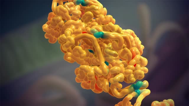
Amyloidosis (am-uh-loi-DO-sis) is a rare disease that occurs when a substance called amyloid builds up in your organs. Amyloid is an abnormal protein that is usually produced in your bone marrow and can be deposited in any tissue or organ. Amyloidosis can affect different organs in different people, and there are different types of amyloid. Amyloidosis frequently affects the heart, kidneys, liver, spleen, nervous system and digestive tract. Severe amyloidosis can lead to life-threatening organ failure.

Your body is a brilliant machine with many important parts. Watch movies to learn more
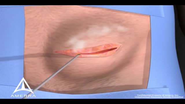
During surgery to repair the hernia, the bulging tissue is pushed back in. Your abdominal wall is strengthened and supported with sutures (stitches), and sometimes mesh. This repair can be done with open or laparoscopic surgery. You and your surgeon can discuss which type of surgery is right for you.

Cosmetic Eye and Eyelid Surgery
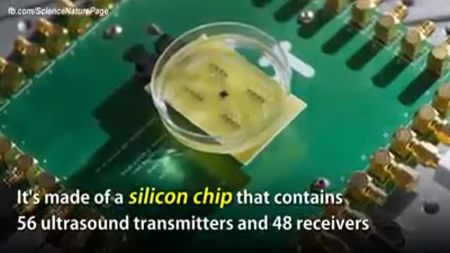
The term "miniaturization" is widely accepted in our vernacular as a positive step in product development. Reducing components to create less space, product footprint and more affordable medical devices are ongoing objectives for manufacturers today. Jabil strives to integrate new innovative technologies into product design and manufacturing as continual miniaturization of medical devices is a focus of the healthcare thought process. Miniaturization is a constantly moving target. Once a novel, new technology sets a higher bar for miniaturization standards, the next ambitious goal is to achieve an even thinner and smaller device. Industry trends, including minimally invasive surgical devices and home health care delivery, demand more sophisticated medical portable devices and easy-to-use electronics which may not be a core competency of medical device manufacturers.
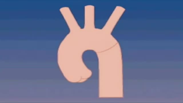
A narrowing of the major artery (the aorta) that carries blood to the body. This narrowing affects blood flow where the arteries branch out to carry blood along separate vessels to the upper and lower parts of the body. CoA can cause high blood pressure or heart damage.
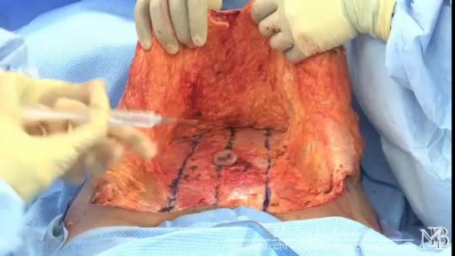
Tummy tuck surgery, also known as abdominoplasty, removes excess fat and skin and, in most cases, restores weakened or separated muscles creating an abdominal profile that is smoother and firmer. A flat and well-toned abdomen is something many of us strive for through exercise and weight control. Sometimes these methods cannot achieve our goals. Even individuals of otherwise normal body weight and proportion can develop an abdomen that protrudes or is loose and sagging. The most common causes of this include: Aging Heredity Pregnancy Prior surgery Significant fluctuations in weight What tummy tuck surgery can't do A tummy tuck is not a substitute for weight loss or an appropriate exercise program. Although the results of a tummy tuck are technically permanent, the positive outcome can be greatly diminished by significant fluctuations in your weight. For this reason, individuals who are planning substantial weight loss or women who may be considering future pregnancies would be advised to postpone a tummy tuck. A tummy tuck cannot correct stretch marks, although these may be removed or somewhat improved if they are located on the areas of excess skin that will be excised.

This type of gait is most often seen in peripheral nerve disease where the distal lower extremity is most affected. Because the foot dorsiflexors are weak, the patient has a high stepping gait in an attempt to avoid dragging the toe on the ground.
