- Physical Examination
- Surgical Examination
- Ophthalmology
- Clinical Skills
- Orthopedics
- Surgery Videos
- Laparoscopy
- Pediatrics
- Funny Videos
- Cardiothoracic Surgery
- Nursing Videos
- Plastic Surgery
- Otorhinolaryngology
- Histology and Histopathology
- Neurosurgery
- Dermatology
- Pediatric Surgery
- Urology
- Dentistry
- Oncology and Cancers
- Anatomy Videos
- Health and Fitness
- Radiology
- Anaesthesia
- Physical Therapy
- Pharmacology
- Interventional Radiology
- Cardiology
- Endocrinology
- Gynecology
- Emergency Medicine
- Psychiatry and Psychology
- Childbirth Videos
- General Medical Videos
- Nephrology
- Physiology
- Diet and Food Health
- Diabetes Mellitus
- Neurology
- Women Health
- Osteoporosis
- Gastroenterology
- Pulmonology
- Hematology
- Rheumatology
- Toxicology
- Nuclear Medicine
- Infectious Diseases
- Vascular Disease
- Reproductive Health
- Burns and Wound Healing
- Other
Top videos

Lung Sounds - Rales, Rhonchi, Wheezes
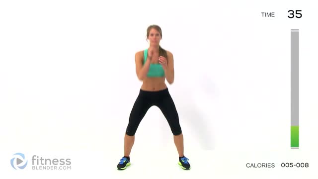
5 Minute Butt and Thigh Workout for a Bigger Butt - Exercises to Lift and Tone Your Butt and Thighs
Your body is a brilliant machine with many important parts. Watch movies to learn more
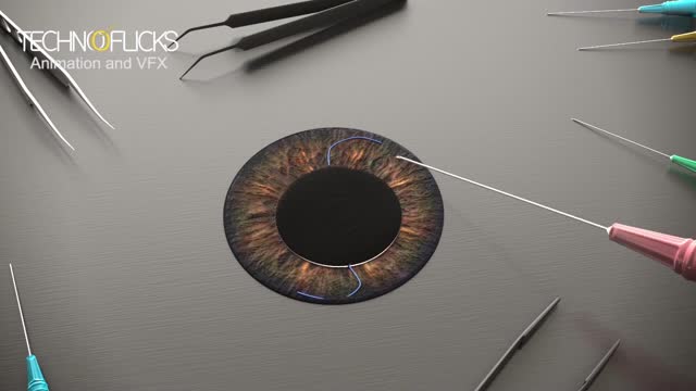
Cosmetic iris implants have not been evaluated by any U.S. regulatory agency or tested for safety in clinical trials. While the implants are not approved by the U.S. Food and Drug Administration, it has been reported in the media this month that the surgery is being performed overseas. During iris implant surgery, an artificial iris made of silicone is folded and inserted into a slit that has been cut into the cornea. Then the iris is unfolded and adjusted to cover the natural iris. Local anesthesia is used.

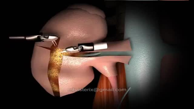
Ventral Hernia Repair
The robotic approach to renal surgery, particularly partial nephrectomy, has some inherent challenges, and some familiarity with the da Vinci robotic system is necessary. The surgeon must gain an understanding of the robotic arm movements and range of motion, especially in relation to the clutch and camera. The advent of robotically assisted prostatectomy in 2001 [23] paved the way for widespread accessibility to the da Vinci robotic unit and its application to renal surgery. Since that time, at least one multi-institutional survey has demonstrated superiority of the robotic approach when compared to laparoscopic for outcomes of blood loss, hospital stay and a substantially shorter warm ischemia time, while maintaining equivalence in positive margin rate, operative time and complications. [11] A transperitoneal approach is most commonly used. Prior abdominal operation is not necessarily a contraindication to this procedure, but access should be approached with regard for previous operation(s) by an experienced team.
Our specialists treat conditions that are recurrent and hard to treat. Simply put, TPIAT a procedure that lets surgeons remove the pancreas, take out islet cells – the cells in the pancreas that make insulin – and put those islet cells into the liver. Patients then take pancreatic enzymes to help them digest food.
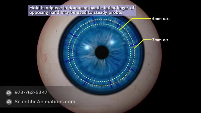
Keratoplasty is the procedure whereby abnormal corneal tissue is replaced by a healthy donor cornea.
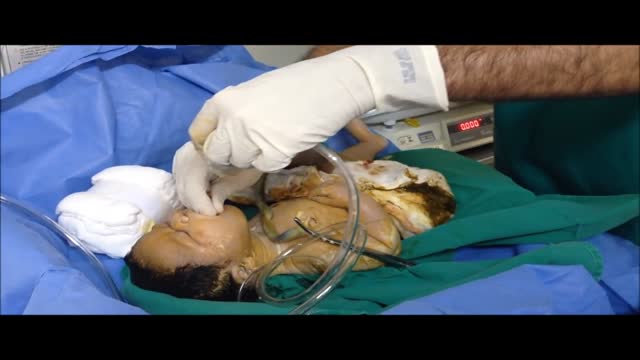
The following guidelines are an interpretation of the evidence presented in the 2010 International Consensus on Cardiopulmonary Resuscitation and Emergency Cardiovascular Care Science With Treatment Recommendations1). They apply primarily to newly born infants undergoing transition from intrauterine to extrauterine life, but the recommendations are also applicable to neonates who have completed perinatal transition and require resuscitation during the first few weeks to months following birth. Practitioners who resuscitate infants at birth or at any time during the initial hospital admission should consider following these guidelines. For the purposes of these guidelines, the terms newborn and neonate are intended to apply to any infant during the initial hospitalization. The term newly born is intended to apply specifically to an infant at the time of birth.

Wilson's disease is a rare inherited disorder that causes too much copper to accumulate in your liver, brain and other vital organs. Symptoms typically begin between the ages of 12 and 23. Copper plays a key role in the development of healthy nerves, bones, collagen and the skin pigment melanin. Normally, copper is absorbed from your food, and any excess is excreted through bile — a substance produced in your liver.
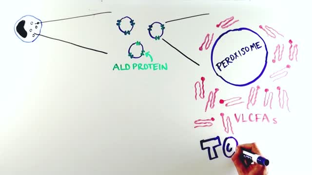
X-linked adrenoleukodystrophy is a genetic disorder that occurs primarily in males. It mainly affects the nervous system and the adrenal glands, which are small glands located on top of each kidney. In this disorder, the fatty covering (myelin) that insulates nerves in the brain and spinal cord is prone to deterioration (demyelination), which reduces the ability of the nerves to relay information to the brain. In addition, damage to the outer layer of the adrenal glands (adrenal cortex) causes a shortage of certain hormones (adrenocortical insufficiency). Adrenocortical insufficiency may cause weakness, weight loss, skin changes, vomiting, and coma.
Thyroid cancer is a disease that you get when abnormal cells begin to grow in your thyroid gland . The thyroid gland is shaped like a butterfly and is located in the front of your neck. It makes hormones that regulate the way your body uses energy and that help your body work normally.

Cataplexy is a sudden, temporary loss of muscle tone that can result in collapse. It is often caused by intense emotions, including laughter

http://control-blood-sugar.good-info.co Low Blood Sugar Symptoms, Low Sugar Symptoms, Normal Sugar Range, Blood Sugar Levels Chart get rid of their high and uncontrollable blood sugar in as little as 3 weeks. No matter how old you are or the severity of your blood sugar condition. Even if your doctor has told you that your blood sugar condition is permanent and incurable (Which really isn’t true) Even if your blood sugar hasn’t changed a bit from past treatments and methods. You can now finally say goodbye to the countless trips to your doctor. You can now say good bye to the depression and tiredness that uncontrollable blood sugar brings to you. You can now completely dodge dangerous diabetes surgeries and weight loss treatments which not only make your condition worse,but can also kill you. Just imagine. The shock on your friends and families faces when they see a slimmer and fitter you. Imagine the shock on your doctor’s face when your blood sugar readings are normal at any time of the day. Imagine finally being able to do the physical activities you once couldn’t do because of your uncontrollable blood sugar. Imagine how much more happier you will be waking up in the morning and eager to begin your day. destroy your high blood sugar! click here. http://control-blood-sugar.good-info.co

Watch that video of Horrifying Creatures Found Living Inside Human Body
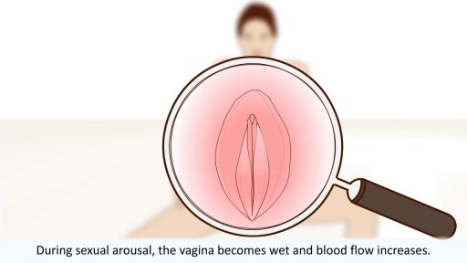
All you need to know about the female orgasm

Réduire La Cellulite, Meilleure Creme Anti Cellulite, Programme Anti Cellulite, Cuisse Cellulite http://perdre-sa-cellulite.plus101.com Une Bonne Alimentation Pour Lutter Contre la Cellulite. Certains aliments ont des composants naturels antioxydants et draineurs qui éliminent naturellement la cellulite. Parmi eux se trouvent le céleri branche. Il s’agit d’un légume un peu amère mais qui aide beaucoup à accélérer le métabolisme des graisses afin de débarrasser la cellulite. Coupé en bâtonnet, il peut être consommé en apéro ou en plat de crudités. Le poireau figure également dans la liste des meilleurs aliments anticellulite. Légume anti-rétention d’eau, il chasse les toxines tout en luttant contre la cellulite. Enfin, n’oubliez pas de consommer de l’ananas si vous voulez combattre votre cellulite. Il a pour principal mission de réduire la rétention d’eau. Selon des experts en physiologie, les femmes ont 90 muscles dans les membres inférieurs et en les stimulant doucement, ces muscles des fesses, jambes, hanches et cuisses, 76,3% des femmes peuvent inverser la cause de la peau d’orange et des capitons pour avoir une peau tonifiée et lisse. CLIQUEZ ICI: http://perdre-sa-cellulite.plus101.com
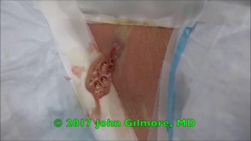
Watch that video of a Huge Cyst Infection Popping

Aumentar Gluteos, Como Hacer Crecer Los Gluteos En Una Semana, Eliminar Celulitis Gluteos.--- http://aumente-gluteos.plus101.com/ --- Para conseguir aumentar tu cola sin ejercicio, debes utilizar algunas cremas naturales caseras, sencillas y fáciles de preparar en la zona de tus glúteos, estas cremas le darán a tu cola una buena forma, firmeza y además una piel libre de manchas, estrías y celulitis. 1. Crema Con Omega 3, Vitamina E y Vitamina C Esta crema es bastante fácil de realizar y sumamente efectiva, solo necesitas tres capsulas de omega 3, dos de vitamina E y dos de vitamina C, los resultados se harán notar en cuestión de días. Con una aguja saca en contenido de cada una de las capsulas y mézclalas en un recipiente, una vez que estén bien mezcladas aplícala sobre tu cola dando masajes circulares que abarquen todo el trasero. Para mejores resultados envuelve la zona de tus glúteos en plástico y déjala actuar durante 30 minutos, luego retira el plástico, colócate un bóxer levanta cola y duerme con esta crema casera, por la mañana la puedes retirar con agua tibia y repetir este proceso todas las noches hasta que consigas los resultados que deseas. 2. Crema Con Aceite De Almendras, Pepino y Avena Esta crema es maravillosa, le dará a tu cola una atractiva apariencia, solo necesitas 3 cucharadas de aceite de almendras, un pepino pequeño y tres cucharadas de avena en hojuelas. El procedimiento es bastante sencillo, debes triturar muy bien el pepino, agregar la avena y luego el aceite de almendras, mezcla hasta que los ingredientes se compacten y listo. Aplica esta crema con suavidad sobre tu cola dando masajes de abajo hacia arriba para vencer los efectos de la gravedad, déjala actuar durante 45 minutos y retírala con agua fría, esta crema casera cuidará muy bien tu piel y le dará a tus glúteos una gran firmeza. 3. Vitamina C, Ciruelas y Aceite De Girasol Una crema divina que te dará una cola perfecta en poco tiempo, ayuda a regenerar la piel de la zona de tus glúteos, tonificándolos y moldeándolos, solo necesitas 3 capsulas de vitamina C, 5 ciruelas grandes y 3 cucharadas de aceite de girasol. Cientos de mujeres en todo el mundo están aumentando el tamaño de sus glúteos gracias a los conocimientos secretos de esta guía, Ingresa ya a: http://aumente-gluteos.plus101.com/

Pigmentflecken, Vitiligo Komplett Geheilt, Pigmentflecken Oberlippe, Vitiligo Heilung, Vitiligo--- http://vitiligo-heilung.info-pro.co/ --- Was ist Vitiligo? Vitiligo ist ein medizinischer Zustand, der die Haut befällt. Die Haut entwickelt dabei an unterschiedlichen Körperstellen nach und nach weiße Flecken, weshalb die Hautstörung im Deutschen auch als "Weißfleckenkrankheit" bekannt ist. Vitiligo macht keine Unterschiede bei Geschlecht oder Rasse und kann jeden befallen. Es wird geschätzt, dass gegenwärtig mehr als 100 Millionen Menschen weltweit in mehr oder minderem starken Maße an Vitiligo leiden. In den Vereinigten Staaten ist die Prävalenzrate etwa 1 % der Bevölkerung; Europa weist ähnliche Raten auf. Was verursacht den Zustand? Vitiligo tritt auf, wenn die Melanozyten in der Haut aus irgendeinem Grund zerstört werden oder ihre Funktion einstellen. Als Melanozyten bezeichnet man jene Hautzellen, die für die Hautfarbe verantwortlich sind. Werden sie zerstört oder anderweitig kompromittiert, stellen produzieren sie kein Melanin mehr, das Hautpigment. Das führt dann zu weißen Hautflecken. Es gibt verschiedene Faktoren, welche die Melanozyten zerstören oder beeinträchtigen können. Dennoch bleibt die Ursache bei den meisten Fällen von Vitiligo ungeklärt. Es wird außerdem angenommen, dass es sich bei Vitiligo um eine Autoimmunerkrankung handelt, welche das Immunsystem die Melanozyten angreifen lässt. Vitiligo kann zudem das Resultat einer Melanozytenstörung sein, welche diese Zellen praktisch veranlasst, "Sebstmord" zu begehen. Laut mancher Forscher kann Vitiligo aber auch durch chronischen Stress und Sonnenbrand hervorgerufen werden. Was sind die Symptome? Das offensichtlichste Symptom von Vitiligo sind die weißen Hautflecken, die sich im Laufe der Zeit vergrößern und auch weiter ausbreiten können. Die Rate, mit der die Hautstörung voranschreitet, unterscheidet sich dabei von Patient zu Patient. "Gratis-Präsentation enthüllt einen ziemlich ungewöhnlichen Tipp zur Beseitigung von Vitiligo für alle Zeiten und in nur 45-60 Tagen - Garantiert!" http://vitiligo-heilung.info-pro.co
