- Physical Examination
- Surgical Examination
- Ophthalmology
- Clinical Skills
- Orthopedics
- Surgery Videos
- Laparoscopy
- Pediatrics
- Funny Videos
- Cardiothoracic Surgery
- Nursing Videos
- Plastic Surgery
- Otorhinolaryngology
- Histology and Histopathology
- Neurosurgery
- Dermatology
- Pediatric Surgery
- Urology
- Dentistry
- Oncology and Cancers
- Anatomy Videos
- Health and Fitness
- Radiology
- Anaesthesia
- Physical Therapy
- Pharmacology
- Interventional Radiology
- Cardiology
- Endocrinology
- Gynecology
- Emergency Medicine
- Psychiatry and Psychology
- Childbirth Videos
- General Medical Videos
- Nephrology
- Physiology
- Diet and Food Health
- Diabetes Mellitus
- Neurology
- Women Health
- Osteoporosis
- Gastroenterology
- Pulmonology
- Hematology
- Rheumatology
- Toxicology
- Nuclear Medicine
- Infectious Diseases
- Vascular Disease
- Reproductive Health
- Burns and Wound Healing
- Other
Top videos

Developmental Psychology Documentary on Brain and Intelligence Development in Babies SHOW MORE

Heart failure can be ongoing (chronic), or your condition may start suddenly (acute). Heart failure signs and symptoms may include: Shortness of breath (dyspnea) when you exert yourself or when you lie down Fatigue and weakness Swelling (edema) in your legs, ankles and feet Rapid or irregular heartbeat Reduced ability to exercise Persistent cough or wheezing with white or pink blood-tinged phlegm Increased need to urinate at night Swelling of your abdomen (ascites) Sudden weight gain from fluid retention Lack of appetite and nausea Difficulty concentrating or decreased alertness Sudden, severe shortness of breath and coughing up pink, foamy mucus Chest pain if your heart failure is caused by a heart attack

Debulking epithelial ovarian cancer. The other important goal of surgery is to remove as much of the tumor as possible − this is called debulking. Debulking is very important in any patient with ovarian cancer that has already spread widely throughout the abdomen at the time of surgery.
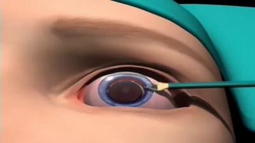
Most cataracts are associated with the aging process and are common among older Americans. In fact, according to the National Eye Institute (NEI), 68.3 percent of Americans 80 and older had cataracts in 2010. And the prevalence of cataracts in the U.S. is expected to grow significantly in the years ahead, due in part to the aging of the population. In 2010, roughly 24.4 million Americans had cataracts, and that number is projected to grow to 50.2 million by the year 2050, according to NEI.
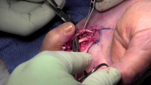
Flexor Tendon Repair

Your First Baby
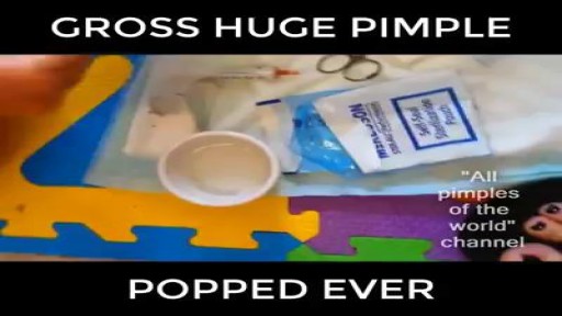
This is so gross Huge pimple
This video shows how the hypodermic needles are made

30 yr old man presented to ER after Motor Vehicle Crash..blunt chest trauma...

: Intrinsic muscles of the back, vertebral, spinal muscle & spinal cord

http://control-blood-sugar.good-info.co Low Blood Sugar Symptoms, Low Sugar Symptoms, Normal Sugar Range, Blood Sugar Levels Chart get rid of their high and uncontrollable blood sugar in as little as 3 weeks. No matter how old you are or the severity of your blood sugar condition. Even if your doctor has told you that your blood sugar condition is permanent and incurable (Which really isn’t true) Even if your blood sugar hasn’t changed a bit from past treatments and methods. You can now finally say goodbye to the countless trips to your doctor. You can now say good bye to the depression and tiredness that uncontrollable blood sugar brings to you. You can now completely dodge dangerous diabetes surgeries and weight loss treatments which not only make your condition worse,but can also kill you. Just imagine. The shock on your friends and families faces when they see a slimmer and fitter you. Imagine the shock on your doctor’s face when your blood sugar readings are normal at any time of the day. Imagine finally being able to do the physical activities you once couldn’t do because of your uncontrollable blood sugar. Imagine how much more happier you will be waking up in the morning and eager to begin your day. destroy your high blood sugar! click here. http://control-blood-sugar.good-info.co

Watch that video of Horrifying Creatures Found Living Inside Human Body
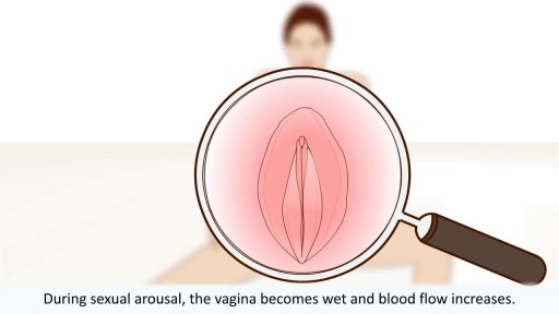
All you need to know about the female orgasm

Réduire La Cellulite, Meilleure Creme Anti Cellulite, Programme Anti Cellulite, Cuisse Cellulite http://perdre-sa-cellulite.plus101.com Une Bonne Alimentation Pour Lutter Contre la Cellulite. Certains aliments ont des composants naturels antioxydants et draineurs qui éliminent naturellement la cellulite. Parmi eux se trouvent le céleri branche. Il s’agit d’un légume un peu amère mais qui aide beaucoup à accélérer le métabolisme des graisses afin de débarrasser la cellulite. Coupé en bâtonnet, il peut être consommé en apéro ou en plat de crudités. Le poireau figure également dans la liste des meilleurs aliments anticellulite. Légume anti-rétention d’eau, il chasse les toxines tout en luttant contre la cellulite. Enfin, n’oubliez pas de consommer de l’ananas si vous voulez combattre votre cellulite. Il a pour principal mission de réduire la rétention d’eau. Selon des experts en physiologie, les femmes ont 90 muscles dans les membres inférieurs et en les stimulant doucement, ces muscles des fesses, jambes, hanches et cuisses, 76,3% des femmes peuvent inverser la cause de la peau d’orange et des capitons pour avoir une peau tonifiée et lisse. CLIQUEZ ICI: http://perdre-sa-cellulite.plus101.com
Watch that video of a Huge Cyst Infection Popping

Aumentar Gluteos, Como Hacer Crecer Los Gluteos En Una Semana, Eliminar Celulitis Gluteos.--- http://aumente-gluteos.plus101.com/ --- Para conseguir aumentar tu cola sin ejercicio, debes utilizar algunas cremas naturales caseras, sencillas y fáciles de preparar en la zona de tus glúteos, estas cremas le darán a tu cola una buena forma, firmeza y además una piel libre de manchas, estrías y celulitis. 1. Crema Con Omega 3, Vitamina E y Vitamina C Esta crema es bastante fácil de realizar y sumamente efectiva, solo necesitas tres capsulas de omega 3, dos de vitamina E y dos de vitamina C, los resultados se harán notar en cuestión de días. Con una aguja saca en contenido de cada una de las capsulas y mézclalas en un recipiente, una vez que estén bien mezcladas aplícala sobre tu cola dando masajes circulares que abarquen todo el trasero. Para mejores resultados envuelve la zona de tus glúteos en plástico y déjala actuar durante 30 minutos, luego retira el plástico, colócate un bóxer levanta cola y duerme con esta crema casera, por la mañana la puedes retirar con agua tibia y repetir este proceso todas las noches hasta que consigas los resultados que deseas. 2. Crema Con Aceite De Almendras, Pepino y Avena Esta crema es maravillosa, le dará a tu cola una atractiva apariencia, solo necesitas 3 cucharadas de aceite de almendras, un pepino pequeño y tres cucharadas de avena en hojuelas. El procedimiento es bastante sencillo, debes triturar muy bien el pepino, agregar la avena y luego el aceite de almendras, mezcla hasta que los ingredientes se compacten y listo. Aplica esta crema con suavidad sobre tu cola dando masajes de abajo hacia arriba para vencer los efectos de la gravedad, déjala actuar durante 45 minutos y retírala con agua fría, esta crema casera cuidará muy bien tu piel y le dará a tus glúteos una gran firmeza. 3. Vitamina C, Ciruelas y Aceite De Girasol Una crema divina que te dará una cola perfecta en poco tiempo, ayuda a regenerar la piel de la zona de tus glúteos, tonificándolos y moldeándolos, solo necesitas 3 capsulas de vitamina C, 5 ciruelas grandes y 3 cucharadas de aceite de girasol. Cientos de mujeres en todo el mundo están aumentando el tamaño de sus glúteos gracias a los conocimientos secretos de esta guía, Ingresa ya a: http://aumente-gluteos.plus101.com/

Pigmentflecken, Vitiligo Komplett Geheilt, Pigmentflecken Oberlippe, Vitiligo Heilung, Vitiligo--- http://vitiligo-heilung.info-pro.co/ --- Was ist Vitiligo? Vitiligo ist ein medizinischer Zustand, der die Haut befällt. Die Haut entwickelt dabei an unterschiedlichen Körperstellen nach und nach weiße Flecken, weshalb die Hautstörung im Deutschen auch als "Weißfleckenkrankheit" bekannt ist. Vitiligo macht keine Unterschiede bei Geschlecht oder Rasse und kann jeden befallen. Es wird geschätzt, dass gegenwärtig mehr als 100 Millionen Menschen weltweit in mehr oder minderem starken Maße an Vitiligo leiden. In den Vereinigten Staaten ist die Prävalenzrate etwa 1 % der Bevölkerung; Europa weist ähnliche Raten auf. Was verursacht den Zustand? Vitiligo tritt auf, wenn die Melanozyten in der Haut aus irgendeinem Grund zerstört werden oder ihre Funktion einstellen. Als Melanozyten bezeichnet man jene Hautzellen, die für die Hautfarbe verantwortlich sind. Werden sie zerstört oder anderweitig kompromittiert, stellen produzieren sie kein Melanin mehr, das Hautpigment. Das führt dann zu weißen Hautflecken. Es gibt verschiedene Faktoren, welche die Melanozyten zerstören oder beeinträchtigen können. Dennoch bleibt die Ursache bei den meisten Fällen von Vitiligo ungeklärt. Es wird außerdem angenommen, dass es sich bei Vitiligo um eine Autoimmunerkrankung handelt, welche das Immunsystem die Melanozyten angreifen lässt. Vitiligo kann zudem das Resultat einer Melanozytenstörung sein, welche diese Zellen praktisch veranlasst, "Sebstmord" zu begehen. Laut mancher Forscher kann Vitiligo aber auch durch chronischen Stress und Sonnenbrand hervorgerufen werden. Was sind die Symptome? Das offensichtlichste Symptom von Vitiligo sind die weißen Hautflecken, die sich im Laufe der Zeit vergrößern und auch weiter ausbreiten können. Die Rate, mit der die Hautstörung voranschreitet, unterscheidet sich dabei von Patient zu Patient. "Gratis-Präsentation enthüllt einen ziemlich ungewöhnlichen Tipp zur Beseitigung von Vitiligo für alle Zeiten und in nur 45-60 Tagen - Garantiert!" http://vitiligo-heilung.info-pro.co
Watch that video of Mystery of Ice Frozen woman came back to life
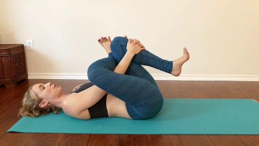
Fast Lower Back Pain & Sciatica Pain Relief – Beginners Yoga Stretches and Poses

Como Aumentar La Testosterona, Testosterona Masculina, Como Aumentar El Nivel De Testosterona --- http://aumentar-testosterona.good-info.co/ --- ¿Baja Testosterona En Hombres Es Peligrosa? ¿Cual es la hormona en los hombres que les permite construir músculo magro, quemar más grasa, ser más fertíil y aumentar la energía de manera general? es la TESTOSTERONA La testosterona es una hormona tan esencial para los hombres porque en realidad define lo que es la virilidad masculina. Y la pérdida de esta hormona tan importante resulta en: * Aumentos de peso * Falta de deseo * Falta de energía y motivación * Pérdida de músculo magro * Depresión * Grasa de pecho Lo que sucede hoy en dia es que más hombres que nunca están perdiendo la testosterona. De hecho, segun el famoso (experto de hormonas masculinas) el nivel de testosterona en los hombres disminuye tan rapidamente que es una verdadera CRISIS para los hombres. Si tu o otra persona experimenta la baja testosterona que sepa que NO es por tu culpa! ¿Cansado Con Tus Bajos Niveles De Testosterona, Tu Sobrepeso y Tu Falta de Libido? Haga click aqui http://aumentar-testosterona.good-info.co/
