- Physical Examination
- Surgical Examination
- Ophthalmology
- Clinical Skills
- Orthopedics
- Surgery Videos
- Laparoscopy
- Pediatrics
- Funny Videos
- Cardiothoracic Surgery
- Nursing Videos
- Plastic Surgery
- Otorhinolaryngology
- Histology and Histopathology
- Neurosurgery
- Dermatology
- Pediatric Surgery
- Urology
- Dentistry
- Oncology and Cancers
- Anatomy Videos
- Health and Fitness
- Radiology
- Anaesthesia
- Physical Therapy
- Pharmacology
- Interventional Radiology
- Cardiology
- Endocrinology
- Gynecology
- Emergency Medicine
- Psychiatry and Psychology
- Childbirth Videos
- General Medical Videos
- Nephrology
- Physiology
- Diet and Food Health
- Diabetes Mellitus
- Neurology
- Women Health
- Osteoporosis
- Gastroenterology
- Pulmonology
- Hematology
- Rheumatology
- Toxicology
- Nuclear Medicine
- Infectious Diseases
- Vascular Disease
- Reproductive Health
- Burns and Wound Healing
- Other
Top videos

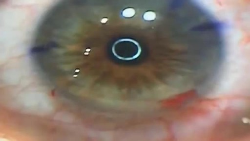
Unedited Cataract Surgery 3

Hammer toes and bunions are common foot problems in the western population. An Iraqi patient chose medical tourism to get this treatment in India.

Tampa body sculpting is the specialty of Dr. Thomas Su of the Artistic Lipo Sculpting Center. Dr. Su’s dedication to body contouring has allowed him to finely hone his craft over the years, making him the most trusted Tampa lipo surgeon. To learn more about Tampa fat removal procedures, visit http://www.artlipo.com/liposuction/liposuction-body-areas/lipo-abdomen.html.

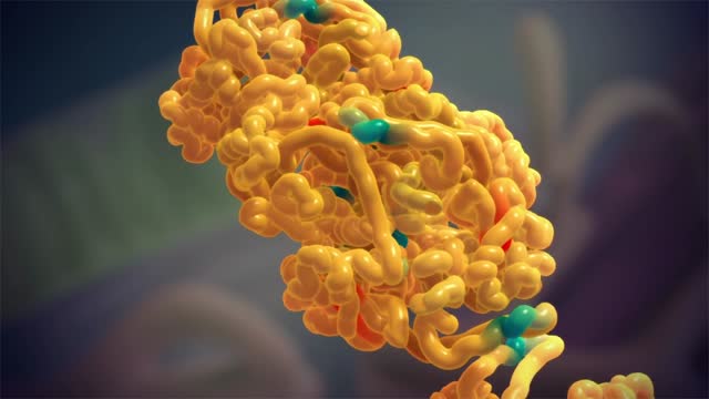
Gene Causing Breast Cancer Resistance to Chemotherapy
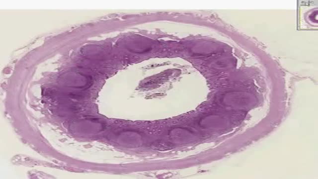
Histology of Appendix
Histology of Vocal Cords

Histology of Thymus

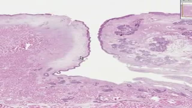
Histology of Palatine Tonsil
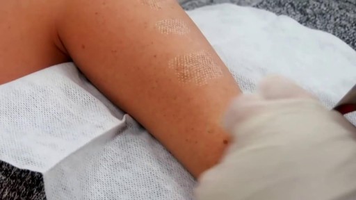
Birthmark Removal with Laser

http://destructeur-de-poids.info-pro.co Perdre Des Cuisses, Astuces Pour Maigrir, Objectif Ventre Plat, Exercice Pour Perdre Du Poids. Boire de l’eau est relié au gain de poids Ceci peut sembler vraiment étrange... Mais boire de l'eau serait la raison pourquoi la plupart d'entre nous avons des problèmes à perdre du poids ? Une découverte récente démontre que c'est vrai et que pour perdre du poids... il faut connaître exactement la quantité d'eau à boire... basé sur votre propre corps. En utilisant ce truc simple, les gens ont perdu 15, 20, 25 kilos même, en moins de 2 mois. Découvrez combien d'eau vous avez besoin et commencez à perdre du poids. http://destructeur-de-poids.info-pro.co http://astuce-ventre-plat.blogspot.com/

This video: Polycystic ovary syndrome (PCOS) is a common endocrine disorder among women of reproductive age. Women with PCOS may have enlarged ovaries that contain small collections of fluid — called follicles — located in each ovary as seen during an ultrasound exam. Infrequent or prolonged menstrual periods, excess hair growth, acne, and obesity can all occur in women with polycystic ovary syndrome. In adolescents, infrequent or absent menstruation may raise suspicion for the condition. The exact cause of polycystic ovary syndrome is unknown. Early diagnosis and treatment along with weight loss may reduce the risk of long-term complications, such as type 2 diabetes and heart disease.

Tampon for The First Time
A new study from Mayo Clinic finds the use of the drug therapy etanercept ineffective in treating alcoholic hepatitis, an acute inflammation of the liver caused by excessive consumption of alcohol. Alcoholic hepatitis is a major cause of morbidity and mortality worldwide. Severe alcohol-related liver disease carries a poor prognosis. Several research studies have worked to find a successful treatment for alcoholic hepatitis, but no consensus has been reached on the most effective treatment regimen.
Amyloidosis (am-uh-loi-DO-sis) is a rare disease that occurs when a substance called amyloid builds up in your organs. Amyloid is an abnormal protein that is usually produced in your bone marrow and can be deposited in any tissue or organ. Amyloidosis can affect different organs in different people, and there are different types of amyloid. Amyloidosis frequently affects the heart, kidneys, liver, spleen, nervous system and digestive tract. Severe amyloidosis can lead to life-threatening organ failure.
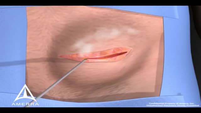
During surgery to repair the hernia, the bulging tissue is pushed back in. Your abdominal wall is strengthened and supported with sutures (stitches), and sometimes mesh. This repair can be done with open or laparoscopic surgery. You and your surgeon can discuss which type of surgery is right for you.
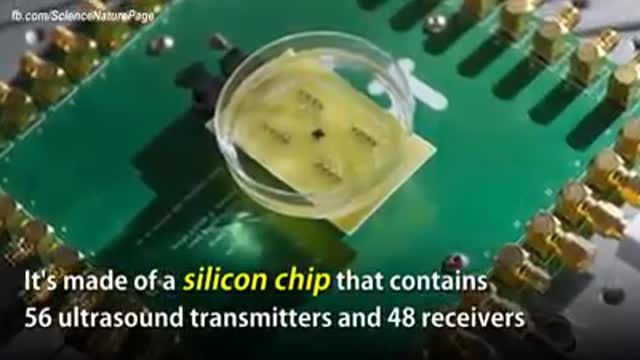
The term "miniaturization" is widely accepted in our vernacular as a positive step in product development. Reducing components to create less space, product footprint and more affordable medical devices are ongoing objectives for manufacturers today. Jabil strives to integrate new innovative technologies into product design and manufacturing as continual miniaturization of medical devices is a focus of the healthcare thought process. Miniaturization is a constantly moving target. Once a novel, new technology sets a higher bar for miniaturization standards, the next ambitious goal is to achieve an even thinner and smaller device. Industry trends, including minimally invasive surgical devices and home health care delivery, demand more sophisticated medical portable devices and easy-to-use electronics which may not be a core competency of medical device manufacturers.
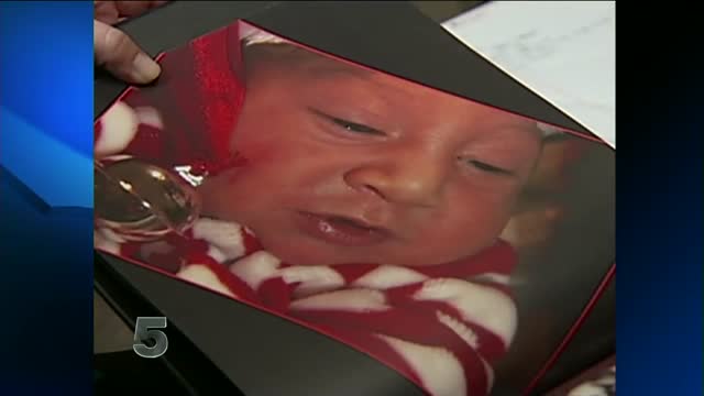
Baby born without brain

one of the best videos I've ever seen..

This type of gait is most often seen in peripheral nerve disease where the distal lower extremity is most affected. Because the foot dorsiflexors are weak, the patient has a high stepping gait in an attempt to avoid dragging the toe on the ground.
