- Physical Examination
- Surgical Examination
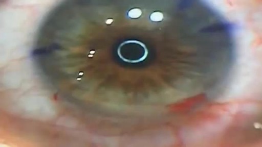
- Ophthalmology
- Clinical Skills
- Orthopedics
- Surgery Videos
- Laparoscopy
- Pediatrics
- Funny Videos
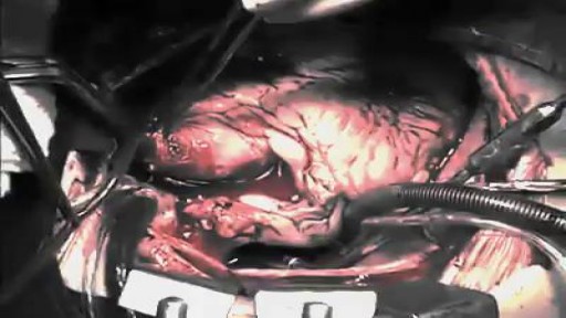
- Cardiothoracic Surgery
- Nursing Videos
- Plastic Surgery
- Otorhinolaryngology
- Histology and Histopathology
- Neurosurgery
- Dermatology
- Pediatric Surgery
- Urology
- Dentistry
- Oncology and Cancers
- Anatomy Videos
- Health and Fitness
- Radiology
- Anaesthesia
- Physical Therapy
- Pharmacology
- Interventional Radiology
- Cardiology
- Endocrinology
- Gynecology
- Emergency Medicine
- Psychiatry and Psychology
- Childbirth Videos
- General Medical Videos
- Nephrology
- Physiology
- Diet and Food Health
- Diabetes Mellitus
- Neurology
- Women Health
- Osteoporosis
- Gastroenterology
- Pulmonology
- Hematology
- Rheumatology
- Toxicology
- Nuclear Medicine
- Infectious Diseases
- Vascular Disease
- Reproductive Health
- Burns and Wound Healing
- Other
Top videos
The term "miniaturization" is widely accepted in our vernacular as a positive step in product development. Reducing components to create less space, product footprint and more affordable medical devices are ongoing objectives for manufacturers today. Jabil strives to integrate new innovative technologies into product design and manufacturing as continual miniaturization of medical devices is a focus of the healthcare thought process. Miniaturization is a constantly moving target. Once a novel, new technology sets a higher bar for miniaturization standards, the next ambitious goal is to achieve an even thinner and smaller device. Industry trends, including minimally invasive surgical devices and home health care delivery, demand more sophisticated medical portable devices and easy-to-use electronics which may not be a core competency of medical device manufacturers.
Baby born without brain

This type of gait is most often seen in peripheral nerve disease where the distal lower extremity is most affected. Because the foot dorsiflexors are weak, the patient has a high stepping gait in an attempt to avoid dragging the toe on the ground.
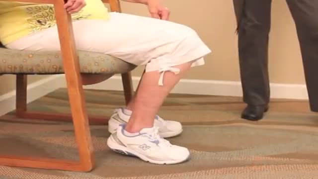
The "Get up and go" test is most commonly used to assess postural stability. In this test, the physician instructs the patient to stand up from a chair without assistance, walk a short distance, turn around, return, and sit down again. If the patient is unsteady or has difficulties during the test, further evaluation is necessary.

NEW WAVE Surgical Technique 3D Animation
Not all conditions that lead to heart failure can be reversed, but treatments can improve the signs and symptoms of heart failure and help you live longer. Lifestyle changes — such as exercising, reducing salt in your diet, managing stress and losing weight — can improve your quality of life.

Scientists have developed a gene editing technique which targets HIV-1 DNA. Through the technique, scientists successfully edited the virus out the genome in human cells; their study also showed the technique can prevent viral replication in cleared cells. Using CRISPR/Cas9 gene editing, the team tested patient cells grown in a lab which were no longer susceptible to HIV infection.
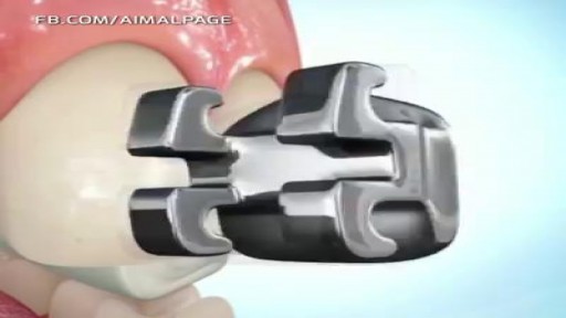
it takes about 1-2 hours. First, the orthodontist will thoroughly clean and dry your teeth. Next, he or she will apply the bonding glue to your teeth and...
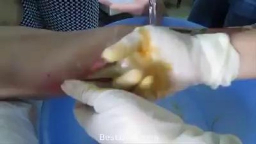
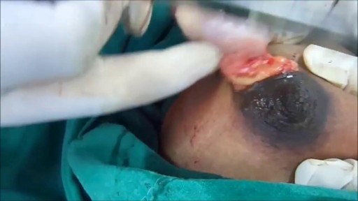
Infection leg gets cleaning inside
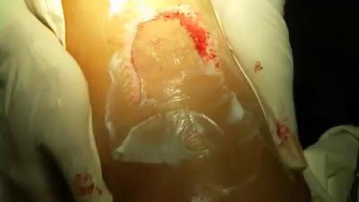
This surgery is usually done while you are under general anesthesia. That means you will be asleep and pain-free. Healthy skin is taken from a place on your body called the donor site. Most people who are having a skin graft have a split-thickness skin graft. This takes the two top layers of skin from the donor site (the epidermis) and the layer under the epidermis (the dermis). The donor site can be any area of the body. Most times, it is an area that is hidden by clothes, such as the buttock or inner thigh. The graft is carefully spread on the bare area where it is being transplanted. It is held in place either by gentle pressure from a well-padded dressing that covers it, or by staples or a few small stitches. The donor-site area is covered with a sterile dressing for 3 to 5 days. People with deeper tissue loss may need a full-thickness skin graft. This requires an entire thickness of skin from the donor site, not just the top two layers. A full-thickness skin graft is a more complicated procedure. Common donor sites for full-thickness skin grafts include the chest wall, back, or abdominal wall.
Wet dreams occur when you ejaculate during your sleep. The medical term for a wet dream is “nocturnal emission.“ Most wet dreams are reported in teenage boys and young men, and sometimes they occur well into adulthood.
The first operation is harvesting the heart from the donor. The donor is usually an unfortunate person who has suffered irreversible brain injury, called "brain death". Very often these are patients who have had major trauma to the head, for example, in an automobile accident. The victim's organs, other than the brain, are working well with the help of medications and other "life support" that may include a respirator or other devices. A team of physicians, nurses, and technicians goes to the hospital of the donor to remove donated organs once brain death of the donor has been determined. The removed organs are transported on ice to keep them alive until they can be implanted. For the heart, this is optimally less than six hours. So, the organs are often flown by airplane or helicopter to the recipient's hospital.

The most detailed explanation you'll ever hear on what makes some people's feet stink. (Not yours, obviously.)
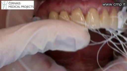
Tooth preparation for All-ceramic crown

Watch that video for a Boy Returns from the Beach with a Snail Inside His Knee

http://eliminar-celulite.plus101.com -- Como Tirar Celulite, Como Acabar Com A Celulite Das Pernas, Como Tirar Celulite Das Coxas. Esses 5 fatores contêm o segredo para conhecer algumas das causas dos furinhos, caroços e cavidades nas regiões problemáticas e pontos críticos da mulher comum. 1 – Muito Stress pode Causar Celulite ou Piorá-la Grande parte das mulheres nem ao menos sabe pelo nível de stress que elas têm passado em tempo integral. O ritmo da vida e sociedade moderna e a sobrecarga de fontes naturais estimulantes externas causa hiperatividade subconsciente. Isso se manifesta em vários níveis de preocupação, desassossego e uso desnecessário de energia mental e emocional. Mas pegue isso e adicione às ocorrências corriqueiras e intensas de stress que ocorrem em todas as nossas vidas em diversos momentos, em diferentes graus. Por exemplo: a doença de um ente querido, a morte de um amigo, divórcio, dificuldades no emprego, brigas familiares... Todos esses fatores de stress têm um impacto direto nos hormônios. Cortisol, epinefrina, e oxitocina, só para dar nome a alguns, são afetados pelo stress. Os níveis e frequência desses e de outros hormônios no corpo podem influenciar muitas características físicas. Uma delas sendo a sua integridade celular. Se as células de seu corpo estão sendo bombardeadas com níveis de hormônios que estão fora de sintonia, então os diversos resultados aparecerão. Músculos, tecido conectivo e células da pele ficarão moles, fracos e flácidos. Isto pode inclusive acontecer com mulheres que estão dando o seu melhor com exercícios e nutrição apropriados. Confira o vídeo abaixo e saiba como: http://eliminar-celulite.plus101.com
Watch that Massive Size Fibrodenoma Removal Under Local Anesthesia
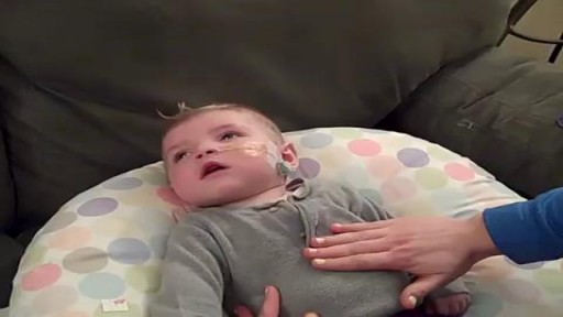
How to place an NG tube in a baby, plus some helpful tips!
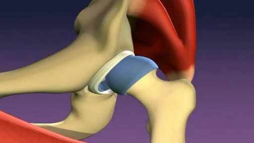
In a normal hip, the ball at the upper end of the thighbone (femur) fits firmly into the socket, which is part of the large pelvis bone. In babies and children with developmental dysplasia (dislocation) of the hip (DDH), the hip joint has not formed normally.
