- Physical Examination
- Surgical Examination
- Ophthalmology
- Clinical Skills
- Orthopedics
- Surgery Videos
- Laparoscopy
- Pediatrics
- Funny Videos
- Cardiothoracic Surgery
- Nursing Videos
- Plastic Surgery
- Otorhinolaryngology
- Histology and Histopathology
- Neurosurgery
- Dermatology
- Pediatric Surgery
- Urology
- Dentistry
- Oncology and Cancers
- Anatomy Videos
- Health and Fitness
- Radiology
- Anaesthesia
- Physical Therapy
- Pharmacology
- Interventional Radiology
- Cardiology
- Endocrinology
- Gynecology
- Emergency Medicine
- Psychiatry and Psychology
- Childbirth Videos
- General Medical Videos
- Nephrology
- Physiology
- Diet and Food Health
- Diabetes Mellitus
- Neurology
- Women Health
- Osteoporosis
- Gastroenterology
- Pulmonology
- Hematology
- Rheumatology
- Toxicology
- Nuclear Medicine
- Infectious Diseases
- Vascular Disease
- Reproductive Health
- Burns and Wound Healing
- Other
Top videos
During the examination, the doctor gently puts a lubricated, gloved finger of one hand into the rectum. He or she may use the other hand to press on the lower belly or pelvic area. A digital rectal exam is done for men as part of a complete physical examination to check the prostate gland .

The head-to-toe assessment in nursing is an important physical health assessment that you'll be performing as a nursing student and nurse.
Head-to-toe assessments allow nurses to assess the health status of patients by following a checklist of criteria.
On the job, your head-to-toe nursing assessment will be performed much faster, and it may be different or more specialized to accommodate the patients' needs within your nursing specialty.
This assessment represents a general assessment checklist (or cheat sheet) that you might encounter in nursing school. (Note: Always follow your instructor's requirements or your employer's assessment protocols).
This nursing head-to-toe examination video guide will focus on the following areas/skills:
-Vital Signs (pulse rate, respiration rate, temperature, oxygen saturation, blood pressure, pain assessment)
https://www.youtube.com/watch?v=gUWJ-6nL5-8
-Cranial Nerve examination
-Head assessment (hair, cranium, eyes, nose, mouth, ears, sinuses)
-Neck assessment (jugular vein, thyroid, trachea, carotid)
-Heart sounds assessment: https://www.youtube.com/watch?v=H48WsyIjFs0&t=73s
-Lung sounds assessment: https://www.youtube.com/watch?v=KNrcG077brQ
-Abdominal assessment
-Assessing extremities (arms, hands, legs, feet)
-Back assessment
-and more
While performing your comprehensive head-to-toe assessment, you'll want to record your findings in the documentation.
Nursing Gear: https://teespring.com/stores/registerednursern
Subscribe: http://www.youtube.com/subscri....ption_center?add_use
Notes: http://www.registerednursern.c....om/head-toe-assessme
Nursing School Supplies: http://www.registerednursern.c....om/the-ultimate-list
Visit our website RegisteredNurseRN.com for free quizzes, nursing care plans, salary information, job search, and much more: http://www.registerednursern.com
Check out other Videos: https://www.youtube.com/user/R....egisteredNurseRN/vid
All of our videos in a playlist: https://www.youtube.com/watch?v=pAhHxt663pU&list=PLQrdx7rRsKfXMveRcN4df0bad3ugEaQnk
Popular Playlists:
NCLEX Reviews: https://www.youtube.com/playli....st?list=PLQrdx7rRsKf
Fluid & Electrolytes: https://www.youtube.com/playli....st?list=PLQrdx7rRsKf
Nursing Skills: https://www.youtube.com/playli....st?list=PLQrdx7rRsKf
Nursing School Study Tips: https://www.youtube.com/playli....st?list=PLQrdx7rRsKf
Nursing School Tips & Questions" https://www.youtube.com/playli....st?list=PLQrdx7rRsKf
Teaching Tutorials: https://www.youtube.com/playli....st?list=PLQrdx7rRsKf
Types of Nursing Specialties: https://www.youtube.com/playli....st?list=PLQrdx7rRsKf
Healthcare Salary Information: https://www.youtube.com/playli....st?list=PLQrdx7rRsKf
New Nurse Tips: https://www.youtube.com/playli....st?list=PLQrdx7rRsKf
Nursing Career Help: https://www.youtube.com/playli....st?list=PLQrdx7rRsKf
EKG Teaching Tutorials: https://www.youtube.com/playli....st?list=PLQrdx7rRsKf
Dosage & Calculations for Nurses: https://www.youtube.com/playli....st?list=PLQrdx7rRsKf
Diabetes Health Managment: https://www.youtube.com/playli....st?list=PLQrdx7rRsKf

In this video I show the steps to give a woman a full body energy orgasm without even touching her.

Vaginal delivery is the most common and safest type of childbirth. When necessary in certain circumstances, forceps (instruments resembling large spoons) may be used to cup your baby's head and help guide the baby through the birth canal. Vacuum delivery is another way to assist delivery and is similar to forceps delivery. In vacuum delivery, a plastic cup is applied to the baby's head by suction and the health care provider gently pulls the baby from the birth canal.
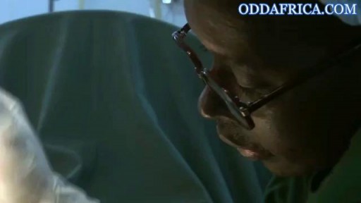
Correcting fgm https://oddafrica.com/videos/female-genital-mutilation-in-africa/
Part 2. Full Obstetric examination and normal delivery by Egyptian doctor Hussein Sulayman and the video is in English showing: Obstetric Examination Episiotomy Obstetric Forceps Obstetric Instruments
The big bang is the moment when the uterus, vagina, and anus contract simultaneously at 0.8-second intervals. A small orgasm may consist of three to five contractions; a biggie, 10 to 15. Many women report feeling different kinds of orgasms
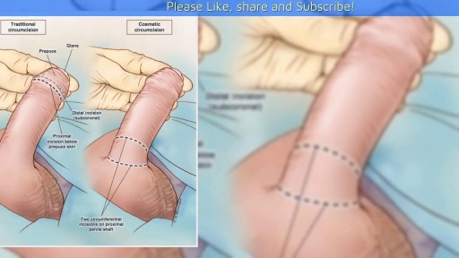
A video showing the circumcision of a male baby

Watch that video to learn How to Insert Enema
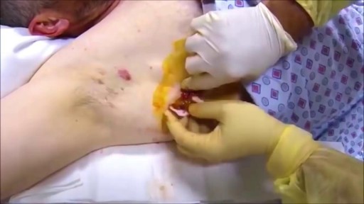
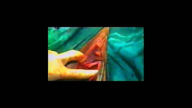
Armpit Abscess Drainage

CORRECTION: After review of this video, it is clear that this video is of a baby who is near full term (40 weeks) based on the size. Late trimester "abortions" are defined only to viability of a baby (24 weeks) A 24 week baby is much smaller than this baby shown and by definition this is not a late "abortion" procedure. The proper labeling of this video should be management of a deceased breech baby with "head entrapment" as this was almost certainly a naturally occuring delivery and an OB nightmare (Reviewed by Dr. Frederick Bright)

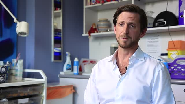
some knowledge

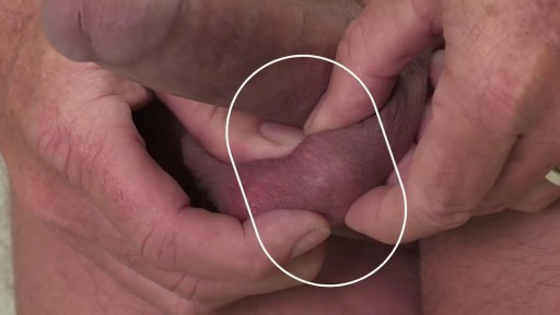
Morning erections have colloquially been termed as “morning wood” while scientifically it is called nocturnal penile tumescence. It is a normal and healthy physiological reaction and response that most men experience in their lives. Morning erections are really the ending of a series of erections that happen to men during the night. Healthy men can, on average, have anywhere between three to five erections in a full night of sleep, each of which lasts from 25-35 minutes.
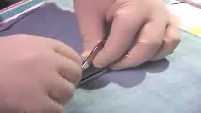
Watch that Female to Male Gender Changing Surgery
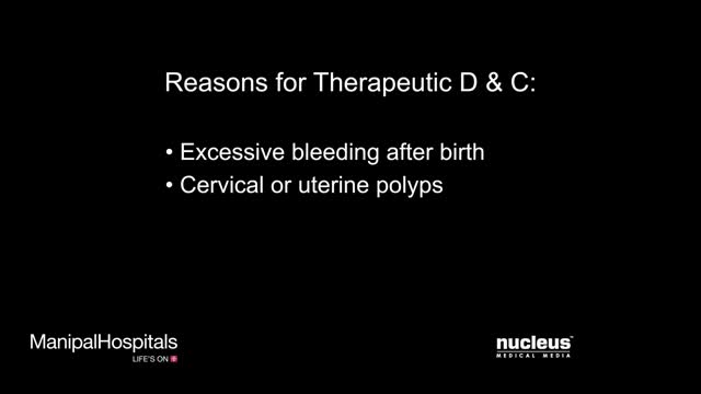
Dilation and curettage (D&C) is a procedure to remove tissue from inside your uterus. Doctors perform dilation and curettage to diagnose and treat certain uterine conditions — such as heavy bleeding — or to clear the uterine lining after a miscarriage or abortion. In a dilation and curettage — sometimes spelled "dilatation" and curettage — your doctor uses small instruments or a medication to open (dilate) your cervix — the lower, narrow part of your uterus. Your doctor then uses a surgical instrument called a curette to remove uterine tissue. Curettes used in a D&C can be sharp or use suction
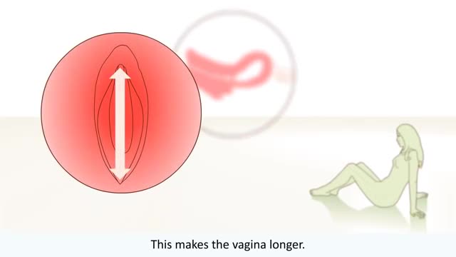
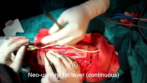
Ever wonder How Male to Female Trans'Gender Surgery works?

The cervix is fully dilated to about 10 cm,with the baby's head moving beyond the cervical opening , into the birth canal. The mother is encouraged to push during contractions,and rest in between them. In a normal delivery, the head rotates to face the mother's back
Penile implants are devices placed inside the penis to allow men with erectile dysfunction (ED) to get an erection. Penile implants are typically recommended after other treatments for ED fail. There are two main types of penile implants, semirigid and inflatable.
Recto-vaginal medical examination
