- Physical Examination
- Surgical Examination
- Ophthalmology
- Clinical Skills
- Orthopedics
- Surgery Videos
- Laparoscopy
- Pediatrics
- Funny Videos
- Cardiothoracic Surgery
- Nursing Videos
- Plastic Surgery
- Otorhinolaryngology
- Histology and Histopathology
- Neurosurgery
- Dermatology
- Pediatric Surgery
- Urology
- Dentistry
- Oncology and Cancers
- Anatomy Videos
- Health and Fitness
- Radiology
- Anaesthesia
- Physical Therapy
- Pharmacology
- Interventional Radiology
- Cardiology
- Endocrinology
- Gynecology
- Emergency Medicine
- Psychiatry and Psychology
- Childbirth Videos
- General Medical Videos
- Nephrology
- Physiology
- Diet and Food Health
- Diabetes Mellitus
- Neurology
- Women Health
- Osteoporosis
- Gastroenterology
- Pulmonology
- Hematology
- Rheumatology
- Toxicology
- Nuclear Medicine
- Infectious Diseases
- Vascular Disease
- Reproductive Health
- Burns and Wound Healing
- Other
Top videos
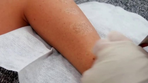
Birthmark Removal with Laser
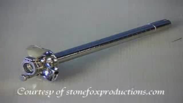
Proctoscope rectal examination
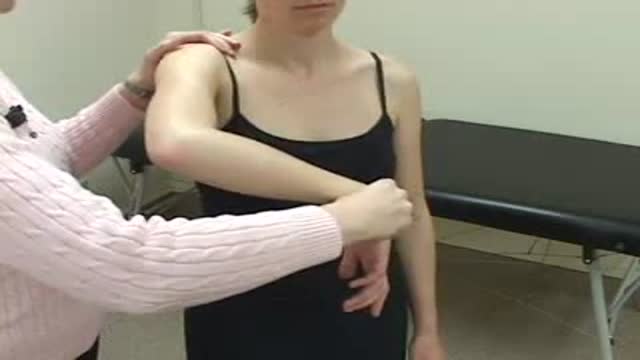
Hawkin's Test

http://www.landging.com/accident-animation-workers-compensation-lifting-box.html
This lifting box accident animation demonstrates the injury covered by workers compensation program.
A Pap smear (Papanicolau smear; also known as the Pap test) is a screening test for cervical cancer. The test itself involves collection of a sample of cells from a woman's cervix (the end of the uterus that extends into the vagina) during a routine pelvic exam
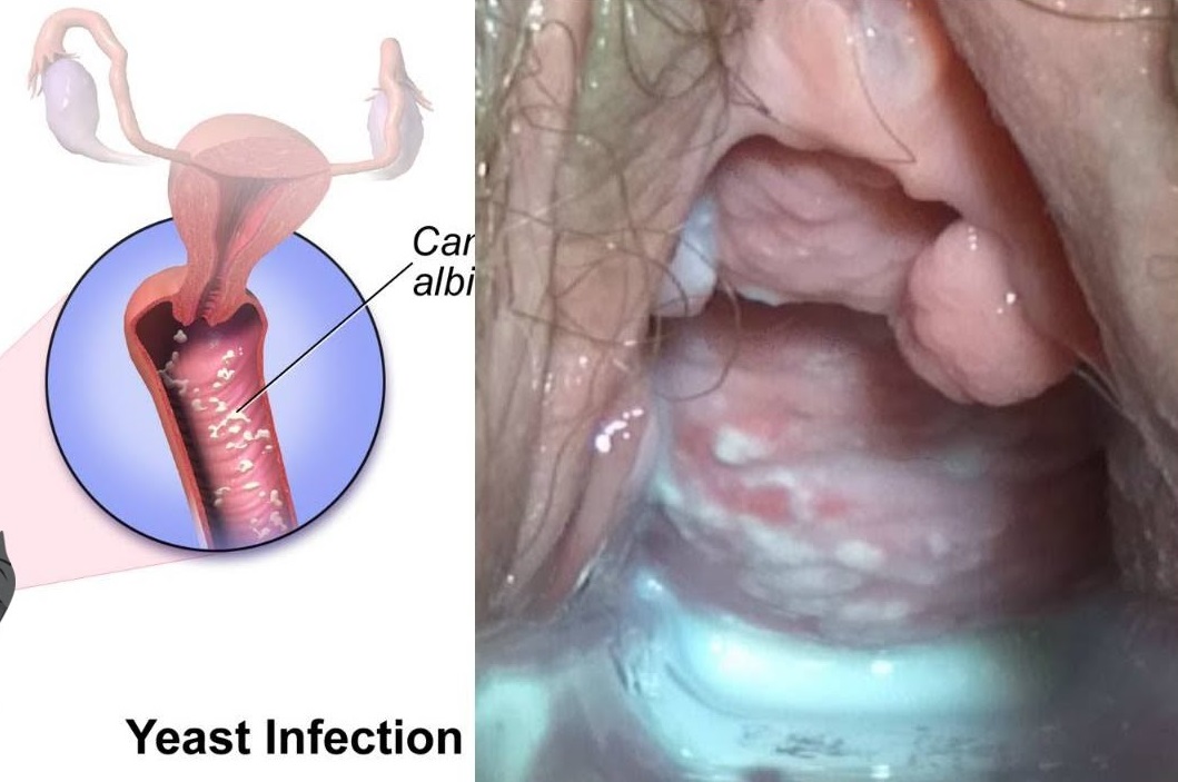
Watch that video to know Types and Causes of Vaginal Infection Yeast or Candidiasis, Trichomoniasis or Bacterial ?
During the examination, the doctor gently puts a lubricated, gloved finger of one hand into the rectum. He or she may use the other hand to press on the lower belly or pelvic area. A digital rectal exam is done for men as part of a complete physical examination to check the prostate gland .
all you need to know about the female orgasm
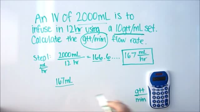
IV Dose Calculations
This video: Blisters caused by friction or minor burns do not require a doctor's care. New skin will form underneath the affected area and the fluid is simply absorbed. Do not puncture a blister unless it is large, painful, or likely to be further irritated. The fluid-filled blister keeps the underlying skin clean, which prevents infection and promotes healing.
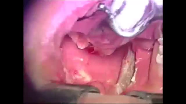
Microsurgical bipolar cautery tonsillectomy compares favorably with traditional techniques in terms of intraoperative bleeding, postoperative pain, otalgia, and hemorrhage. This technique combines the hemostatic advantage of cautery dissection, the excellent visualization achieved by a microscope, and, with the use of a video, greatly improves the physician's ability to teach how to perform a tonsillectomy.
This video shows the delivery of the placenta after delivery of the fetus

The Micturition Reflex
Use warm water and sea salt. Soak the wart for 10 to 15 minutes in warm salt water to moisten the skin. Scrape the dead skin layers off the wart using a nail file, pumice stone or mild sandpaper. You could also use your fingers, but wash them thoroughly before and after, as warts can easily spread.
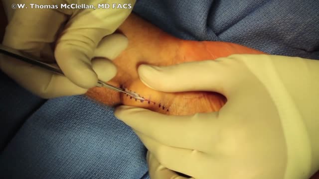
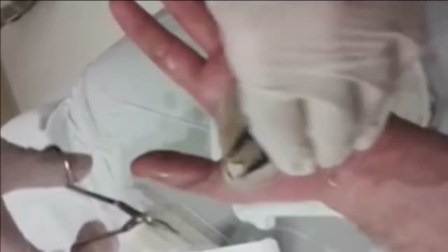
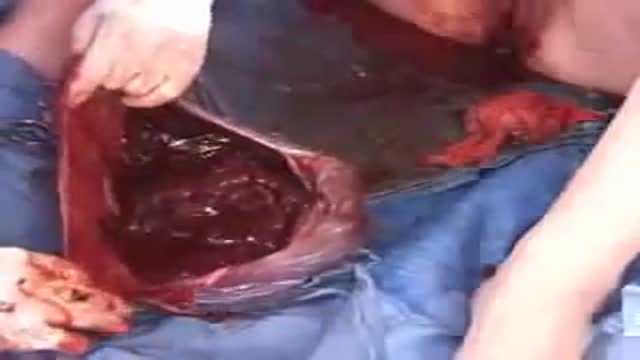
This is a surgical video that shows the removal of a volar ganglion cyst. This is a common surgical procedure and this video may help you better understand the steps that occur during the procedure.
ENT Physical Examination Lecture
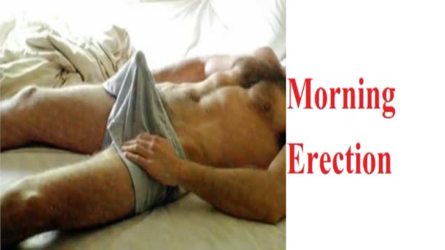
Morning erections have colloquially been termed as “morning wood” while scientifically it is called nocturnal penile tumescence. It is a normal and healthy physiological reaction and response that most men experience in their lives. Morning erections are really the ending of a series of erections that happen to men during the night. Healthy men can, on average, have anywhere between three to five erections in a full night of sleep, each of which lasts from 25-35 minutes.
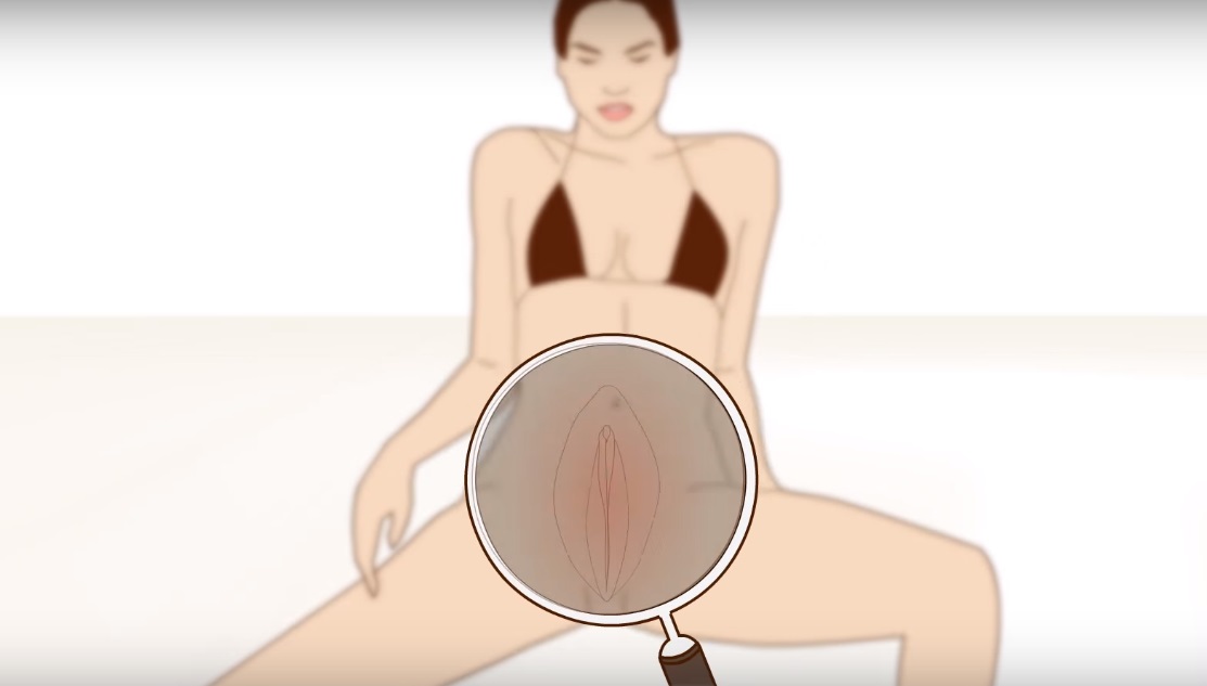
Watch that video to know What is Vaginal Discharge and how to Get Rid of it ?
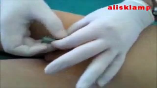
this video shows how the child circumcision is easy and safe with alisklamp
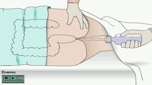
Enema how to apply Animation
