- Physical Examination
- Surgical Examination
- Ophthalmology
- Clinical Skills
- Orthopedics
- Surgery Videos
- Laparoscopy
- Pediatrics
- Funny Videos
- Cardiothoracic Surgery
- Nursing Videos
- Plastic Surgery
- Otorhinolaryngology
- Histology and Histopathology
- Neurosurgery
- Dermatology
- Pediatric Surgery
- Urology
- Dentistry
- Oncology and Cancers
- Anatomy Videos
- Health and Fitness
- Radiology
- Anaesthesia
- Physical Therapy
- Pharmacology
- Interventional Radiology
- Cardiology
- Endocrinology
- Gynecology
- Emergency Medicine
- Psychiatry and Psychology
- Childbirth Videos
- General Medical Videos
- Nephrology
- Physiology
- Diet and Food Health
- Diabetes Mellitus
- Neurology
- Women Health
- Osteoporosis
- Gastroenterology
- Pulmonology
- Hematology
- Rheumatology
- Toxicology
- Nuclear Medicine
- Infectious Diseases
- Vascular Disease
- Reproductive Health
- Burns and Wound Healing
- Other
Top videos

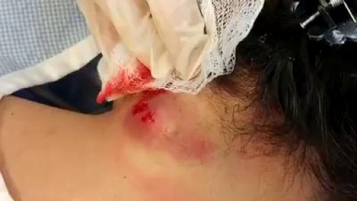
The brachial plexus is the network of nerves that sends signals from your spine to your shoulder, arm and hand. A brachial plexus injury occurs when these nerves are stretched, compressed, or in the most serious cases, ripped apart or torn away from the spinal cord. Minor brachial plexus injuries, known as stingers or burners, are common in contact sports, such as football. Babies sometimes sustain brachial plexus injuries during birth. Other conditions, such as inflammation or tumors, may affect the brachial plexus. The most severe brachial plexus injuries usually result from auto or motorcycle accidents. Severe brachial plexus injuries can leave your arm paralyzed, with a loss of function and sensation. Surgical procedures such as nerve grafts, nerve transfers or muscle transfers can help restore function.
To treat pregnancy acne, start with self-care: Wash problem areas with a gentle cleanser. Twice a day, use your hands to wash your face with a mild soap and warm water. ... Shampoo regularly. ... Don't pick or squeeze blemishes. ... Avoid irritants. ... Watch what touches your skin.

Instead, try these natural solutions and lifestyle changes, which may help you stop snoring. Change Your Sleep Position. ... Lose Weight. ... Avoid Alcohol. ... Practice Good Sleep Hygiene. ... Open Nasal Passages. ... Change Your Pillows. ... Stay Well Hydrated.
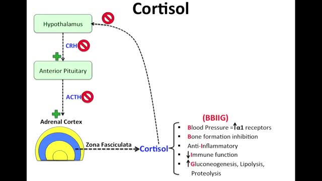
Cushing syndrome occurs when your body is exposed to high levels of the hormone cortisol for a long time. Cushing syndrome, sometimes called hypercortisolism, may be caused by the use of oral corticosteroid medication. The condition can also occur when your body makes too much cortisol on its own. Too much cortisol can produce some of the hallmark signs of Cushing syndrome — a fatty hump between your shoulders, a rounded face, and pink or purple stretch marks on your skin. Cushing syndrome can also result in high blood pressure, bone loss and, on occasion, type 2 diabetes. Treatments for Cushing syndrome can return your body's cortisol production to normal and noticeably improve your symptoms. The earlier treatment begins, the better your chances for recovery.
Dysentery is an infection of the intestines causing diarrhoea that contains blood or mucus. There are two main types of dysentery: Shigellosis, or bacillary dysentery, is the most common type experienced in the UK, caused by the shigella bacteria. Amoebic dysentery, also called amoebiasis, is caused by a single-celled parasite called Entamoeba histolytica. This form of dysentery is more common abroad in tropical countries. This article focuses on amoebic dysentery, This is usually caused by poor hygiene or contaminated food or water. Amoebic dysentery is a notifiable disease, so your GP must let the local health authority know if you have contracted it. Causes of amoebic dysentery Once inside the body, amoeba clump together to form a cyst that is protected by the stomach’s digestive acid. When the cyst passes through the intestines it breaks open infecting the body. The amoebae burrow into the intestinal wall and cause small ulcers or abscesses. Cysts exit the body via faeces but are still able to live outside, which is how many people become infected. Severe dysentery is more common in developing countries due to compromised hygiene. You can get sick in a number of ways including: Eating contaminated food Drinking contaminated water Contracting dysentery from another infected person. Symptoms of amoebic dysentery Symptoms can appear as many as 10 days after exposure and infection by the parasite. Signs of infection include: Watery diarrhoea with blood or pus in it Nausea or vomiting Stomach pain High temperature Chills Bleeding from back passage (rectum) Weight loss Loss of appetite. Complications of amoebic dysentery If the parasite gets into your bloodstream it can spread to other parts of your body, including the liver. When this happens you run the risk of developing a liver abscess. Symptoms include: High temperature Weakness Cough Jaundice Nausea Loss of appetite Weight loss
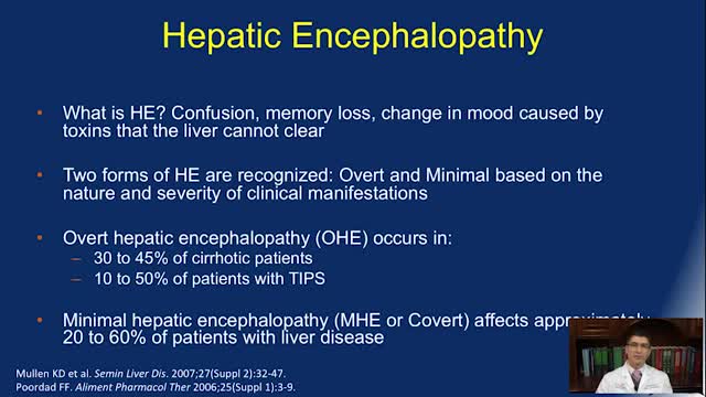
Symptoms of hepatic encephalopathy differ depending on the underlying cause of the liver damage. Symptoms and signs of hepatic encephalopathy may include: difficulty thinking. personality changes. poor concentration. problems with handwriting or loss of other small-hand movements. confusion. forgetfulness. poor judgment.
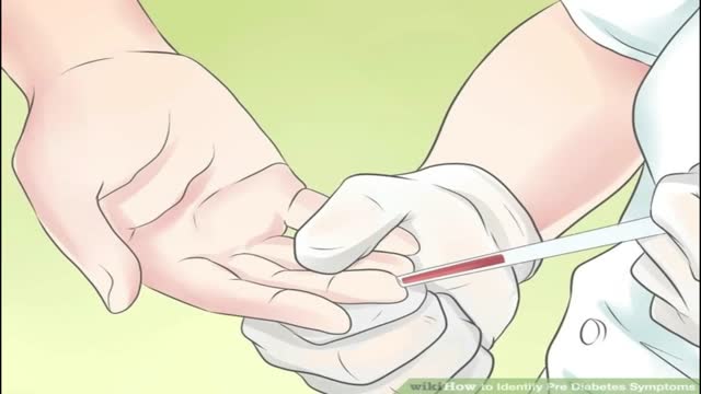
Prediabetes means that your blood sugar level is higher than normal but not yet high enough to be type 2 diabetes. Without lifestyle changes, people with prediabetes are very likely to progress to type 2 diabetes. If you have prediabetes, the long-term damage of diabetes — especially to your heart, blood vessels and kidneys — may already be starting. There's good news, however. Progression from prediabetes to type 2 diabetes isn't inevitable. Eating healthy foods, incorporating physical activity in your daily routine and maintaining a healthy weight can help bring your blood sugar level back to normal. Prediabetes affects adults and children. The same lifestyle changes that can help prevent progression to diabetes in adults might also help bring children's blood sugar levels back to normal.

Top 10 Foods that Can Kill You
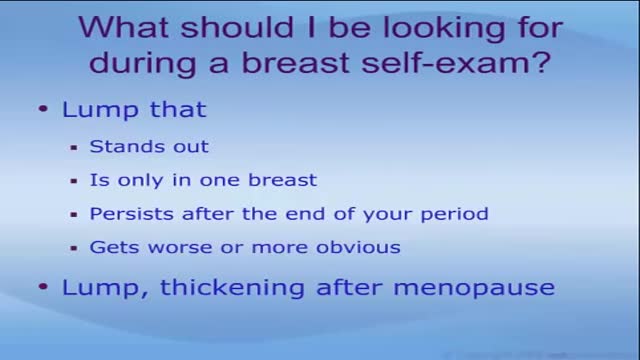
Breast lumps facts Breast lumps can be caused by infections, injuries, non-cancerous growths, and cancer. Breast cancer usually causes no pain in the breast. The symptoms of breast cancer include painless breast lumps, nipple discharge, and inflammation of the skin of the breast. The chances that a particular breast lump could be cancerous depends on many factors, including past medical history, physical examination, as well as genetic and other risk factors. The only way to be certain that a lump is not cancerous is to have a tissue sampling (biopsy). There are several ways to do the biopsy. The treatment of a breast lump depends on its cause.
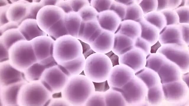
Cancer, also called malignancy, is an abnormal growth of cells. There are more than 100 types of cancer, including breast cancer, skin cancer, lung cancer, colon cancer, prostate cancer, and lymphoma. Symptoms vary depending on the type. Cancer treatment may include chemotherapy, radiation, and/or surgery.

Father & Mom feel their baby the same
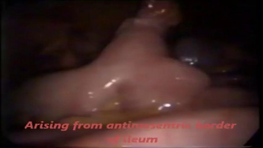
Meckel's Diverticulum is a vestigeal remnant of vitellointestinal duct. Its a true diverticulum as it contains all three layers of intestine. It is usually presents at anti mesenteric burder. Usually 2 cm (range 1- 12 cm ) in length, found in 2 % of population , and situated around 2 feet of Ileaocecal junction. 50 % cases it contains gastric mucosa , but may also contain colonic, duodenal or pancreatic mucosa .male : female ration in symptomatic cases is 3 : 1.It may mimic acute appendicitis, so in cases where one is going for surgery for appendicitis , must search for meckel's diverticulum........
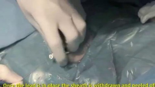
A pacemaker insertion is the implantation of a small electronic device that is usually placed in the chest (just below the collarbone) to help regulate slow electrical problems with the heart.
Huge Pimple Draining


Skin Whitening Tips, Vitamin C For Skin Whitening, Skin Whitening Before And After, Skin Whitening. http://skin-whitening.good-info.co Each and every person wants a clean and radiant skin. Some spend fortunes on cosmetic products that will lighten the skin and remove all the imperfections, others spend their money on esthetic operations in the hope that their skin will look perfect. Lastly, there are people trying to fake a healthy skin by using all kinds of makeup that will cover the imperfections and leave the impression that the skin is healthy and has no scars, wrinkles or spots. All these people are looking for a way through which they can make their skin look good. Yet, what they have not taken into consideration is the power of natural ingredients. Fruits, vegetables and products coming from animals are great sources of anti-oxidants and are rich in substances which can whiten the skin, moisturize it, attenuate the fine lines and wrinkles and give it elasticity. There are many natural ingredients which can be used in order to remove the dark spots and whiten the skin and as many reasons to start trying them. The first reason for which you should try the natural skin whitening ingredients is the fact that they have no side effects. Because most of the ingredients with which the homemade skin whitening recipes are made are natural, and are used in our everyday diet, the human body tolerates them very well and responds positively to the ingredients they contain. In addition to that, because you know what you put in that homemade recipe, you know if your skin will going to react negatively to it. There are certain products (fruits, vegetables, animal products) to which certain people are allergic. Exclude those ingredients from your recipes and you know you will obtain a 100% safe homemade product. Click Here. http://skin-whitening.good-info.co
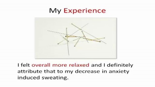
http://sweating-cure.info-pro.co/ --- Why Do I Sweat So Much, How To Stop Sweaty Armpits, How To Stop Your Hands From Sweating. The 4 Step Formula to Stop Sweating How to Stop Excessive Sweating in Minutes There is a simple 4 step formula you need to implement immediately if you want to finally end your excessive sweating and put a stop to the constant embarrassment. These 4 steps are absolutely essential if you’re having trouble controlling the endless perspiration. Don’t prolong the problem any longer. Take action with these simple steps right now. http://sweating-cure.info-pro.co/

http://cure-papiloma-humano.info-pro.co --- Sintomas Del Papiloma Humano, Sintomas De Papiloma Humano, Virus Papiloma Humano Cura. El Papiloma Humano Se Cura ¿El Papiloma Humano se Cura? Si te has encontrado recientemente con un diagnóstico positivo de VPH probablemente estas en busca de una solución para tratar este Virus. Seguramente tienes miedo de no encontrar una cura para las verrugas genitales, y que son muy difíciles de eliminar, amigo o amiga no te sientas avergonzado/a o preocupado el saber que estas infectado con este virus no es fácil, más aun ver cómo crecen verrugas en tu cuerpo, pero arriba los ánimos existen muchas cosas que puedes hacer para tratar este virus. Aparte de las verrugas genitales no hay otro síntoma que presente el Virus en tu cuerpo, puedes tratar las verrugas genitales con tratamientos naturales o los métodos actuales. Trata de no rascarse si sientes comezón en la zona afecta ya que puedes lastimarte o irritar más la piel, las verrugas genitales son altamente contagiosas, No debes tener relaciones sexuales con nadie hasta que hayas tenido tratamiento para el VPH. Hoy en Día existen varios tratamientos médicos diseñados para ayudarte a curar las verrugas genitales producidas por el papiloma humano, aunque debo aclararte que estos métodos son dolorosos y dejan cicatrices en la piel donde se encontraba la verruga. Crioterapia: Básicamente las verrugas genitales se congelan con nitrógeno líquido. Tratamiento a base de láser: se utilizan laser de CO2 para quemar las verrugas genitales, se aplica anestesia al área afectada para no sentir mucho dolor, aunque siempre existen molestias durante el procedimiento. Bisturí eléctrico: En esta técnica se utiliza una corriente eléctrica para destruir las verrugas, Se puede hacer en el consultorio con anestesia local, con este método se debe tener cierto cuidado ya que existe peligro de infección. La breve lista antes mencionada son los métodos médicos más comunes para eliminar las verrugas genitales, cuando se diagnostican verrugas genitales estos métodos son los primeros en que se piensan para curar las verrugas genitales. Aunque hay que decir la verdad, estos tratamientos no podrán eliminar el verdadero problema detrás de las verrugas genitales, el cual es el Virus del papiloma humano, aunque las verrugas se eliminan de la zona afectada el virus seguirá permaneciendo en el cuerpo de forma latente, ninguno de estos método puede garantizar que no volverá a haber otro brote de verrugas genitales. Descubre como mantener DESACTIVADO el VPH DE POR VIDA para permitirte una vida sin verrugas, sin frustraciones y sin molestias, ingresa ahora a: http://cure-papiloma-humano.info-pro.co

How To Help Your Child Learn To Read, Help My Child Learn To Read, Best Way To Teach Reading---- http://children-learning-reading.good-info.co -- how to help your child learn to read - Help My Child Learn to Read The ability to read is vital for success. It helps your child succeed in school, helps them build self-confidence, and helps to motivate your child. Being able to read will help your child learn more about the world, understand directions on signs and posters, allow them to find reading as an entertainment, and help them gather information. Learning to read is very different from learning to speak, and it does not happen all at once. There is a steady progression in the development of reading ability over time. The best time for children to start learning to read is at a very young age - even before they enter pre-school. Once a child is able to speak, they can begin developing basic reading skills. Very young children have a natural curiosity to learn about everything, and they are naturally intrigued by the printed texts they see, and are eager to learn about the sounds made by those letters. You will likely notice that your young child likes to look at books and thoroughly enjoys being read to. They will even pretend to behave like a reader by holding books and pretend to read them. As parents, you're the most important first step in your children's journey into the wonderful world of reading. It is up to you to create the most supportive environment that turns your child on to reading - such as reading aloud to them often during the day and before bedtime, and placing age appropriate books for children around the house, so that the child will have access to plenty of books. Reading often to your child will help develop their interest in books and stories, and soon they will want to read stories on their own. >>Teach your child to read and enable your child to become a fast and fluent reader! Click here to help your child learn to read http://children-learning-reading.good-info.co

Watch that video of Ingrown hair turns into Horrible 140-pound tumor in man’s stomach
