- Physical Examination
- Surgical Examination
- Ophthalmology
- Clinical Skills
- Orthopedics
- Surgery Videos
- Laparoscopy
- Pediatrics
- Funny Videos
- Cardiothoracic Surgery
- Nursing Videos
- Plastic Surgery
- Otorhinolaryngology
- Histology and Histopathology
- Neurosurgery
- Dermatology
- Pediatric Surgery
- Urology
- Dentistry
- Oncology and Cancers
- Anatomy Videos
- Health and Fitness
- Radiology
- Anaesthesia
- Physical Therapy
- Pharmacology
- Interventional Radiology
- Cardiology
- Endocrinology
- Gynecology
- Emergency Medicine
- Psychiatry and Psychology
- Childbirth Videos
- General Medical Videos
- Nephrology
- Physiology
- Diet and Food Health
- Diabetes Mellitus
- Neurology
- Women Health
- Osteoporosis
- Gastroenterology
- Pulmonology
- Hematology
- Rheumatology
- Toxicology
- Nuclear Medicine
- Infectious Diseases
- Vascular Disease
- Reproductive Health
- Burns and Wound Healing
- Other
Top videos
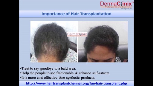
Hair transplant is the most commonly performed cosmetic surgical procedures today. Get the cost of Hair Transplant Surgery in Chennai at DermaClinix Chennai. At DermaClinix, we have a well experienced and skilled team of board certified hair transplant surgeons. For More Information Visit Here:- https://www.hairtransplantchennai.org/hair-transplant-results-chennai.php or call:- +91-8939636222

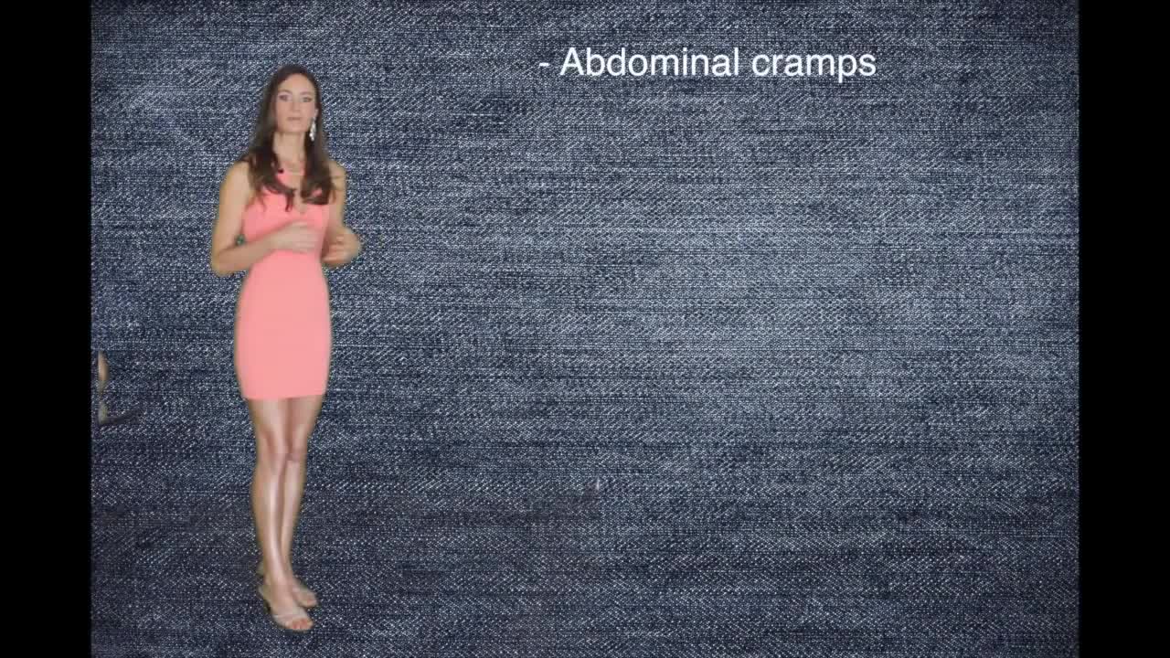
http://how-to-get-pregnant.info-pro.co ----- How To Get Pregnant, Ways To Get Pregnant, Best Days To Get Pregnant, Easiest Way To Get Pregnant. Signs of Infertility. What exactly is infertility? The problems with either conceiving a child, or with carrying out the pregnancy to its eventual fruitful end, fall under the definition of infertility. Infertility is the incapability of an individual to become pregnant, in case of females, or the incapability to induce pregnancy, in case of the males. The inability of an individual to carry out a pregnancy to its full term is also dubbed infertility. How does one recognize infertility? What are the signs of infertility? Signs of infertility are not always evident. Most people go through life without knowing there is a problem with their reproductive systems, attributing failed pregnancies to providence. In fact, miscarriages are the most common indicator of infertility. Signs of infertility in women: In women, the signs of infertility are more readily recognized as compared to men. Endometriosis causes the lining of the uterus to grow outside the uterus. Bacterial infections may begin around the uterus and spread to other reproductive organs, resulting in infertility. Fibroids in the uterus are indicative of infertility. Tumors in the cervix often cause stenosis, or narrowing of the cervix, which is a common indicator of infertility. Ovulating before the tenth day and after the twentieth day of one's monthly cycle, pre-menstrual spotting, menopausal symptoms, etc. are indicative of luteal phase defect, and thus in turn are signs too.
To treat pregnancy acne, start with self-care: Wash problem areas with a gentle cleanser. Twice a day, use your hands to wash your face with a mild soap and warm water. ... Shampoo regularly. ... Don't pick or squeeze blemishes. ... Avoid irritants. ... Watch what touches your skin.
Retropharyngeal abscess (RPA) produces the symptoms of sore throat, fever, neck stiffness, and stridor. RPA occurs less commonly today than in the past because of the widespread use of antibiotics for suppurative upper respiratory infections. The incidence of RPA in the United States is rising, however. Once almost exclusively a disease of children, RPA is observed with increasing frequency in adults. It poses a diagnostic challenge for the emergency physician because of its infrequent occurrence and variable presentation.
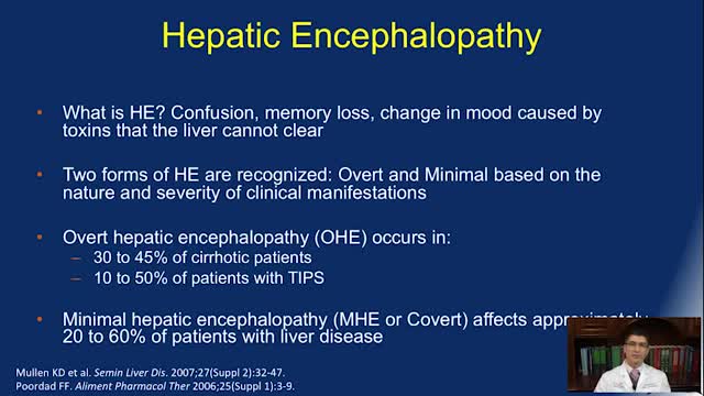
Symptoms of hepatic encephalopathy differ depending on the underlying cause of the liver damage. Symptoms and signs of hepatic encephalopathy may include: difficulty thinking. personality changes. poor concentration. problems with handwriting or loss of other small-hand movements. confusion. forgetfulness. poor judgment.
A blood transfusion is a routine medical procedure that can be lifesaving. During a blood transfusion, donated blood is added to your own blood. A blood transfusion may also be done to supplement various components of your blood with donated blood products. In some cases, a blood transfusion is done with blood that you've donated ahead of time before you undergo elective surgery. During a typical blood transfusion, certain parts of blood are delivered through an intravenous (IV) line that's placed in one of the veins in your arm. A blood transfusion usually takes one to four hours, though in an emergency it can be done much faster.
Ringworm On Chin, Best Way To Treat Ringworm, How Do I Get Rid Of Ringworm, How To Stop Ringworm --- http://ringworm-cure.plus101.com ---- I'm going to share with you how to cure Ringworm in less than 3 days by using these proven methods that have worked for thousands of people around the world with Ringworm. Not only that, but these same methods work for children, as well as adults, and even pets. Whether you have athlete's foot or jock it, this will work for you, as those are both types of Ringworm. This fungal infection can be cured without drugs or medications. It's important to understand that. In fact, there are many home remedies for Ringworm that have worked for thousands of years for treating many ailments and skin conditions. Below I will list a few. First, understand there are 3 primary methods to cure Ringworm. You must do all 3 if you want to know how to cure Ringworm as fast as possible. Over the 5 years I've spent studying and learning about Ringworm, I've tried almost every treatment and home remedy available. I've found this to be the most effective and fastest way to cure Ringworm. 1) Treat the rash with home remedies and treatments. There are many home remedies for Ringworm available, as well as natural treatments, that are good for the skin and can promote healing rapidly. Not only do they get rid of the itchiness, pain or discomfort, but they also heal the rash and prevent it from coming back again. 2) Use bathing procedures. Bathing procedures are a powerful way to cure Ringworm, especially when you add home remedies and ingredients to the bath. This again, promotes healing, and helps get rid of all symptoms associated with Ringworm. 3) Consume the right foods and supplements. A proper diet is important to cure Ringworm. Treating Ringworm externally is not enough - you need to provide the body with what it needs to fight off the infection and boost the immune system, so you never have to worry about it again. Nutrition, hydration, rest, and supplementation are all equally important. Follow these 3 methods and you will be cured from Ringworm within days. If you sit and wait, and do nothing, it will only get worse and you will suffer much longer than you need to. To find out more on how to cure Ringworm in 3 days or less, check out the Fast Ringworm Cure e-book program that goes into all the ways to cure Ringworm fast, along with a list of home remedies and treatments that you can apply right away. William Oliver is a nutritionist, medical researcher, and author of the Fast Ringworm Cure e-book. To find out how to cure Ringworm in 3 days or less, click below: http://ringworm-cure.plus101.com
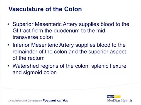
Ischemic colitis occurs when blood flow to part of the large intestine (colon) is reduced, usually due to narrowed or blocked blood vessels (arteries). The diminished blood flow doesn't provide enough oxygen for the cells in your digestive system. Ischemic colitis can cause pain and may damage your colon. Any part of the colon can be affected, but ischemic colitis usually causes pain on the left side of the belly area (abdomen). The condition can be misdiagnosed because it can easily be confused with other digestive problems. Ischemic colitis may heal on its own. But you may need medication to treat ischemic colitis or prevent infection, or you may need surgery if your colon has been damaged. Symptoms ShareTweet Oct. 13, 2015 References Products and Services Newsletter: Mayo Clinic Health Letter See also Abdominal pain Colonoscopy Color Blue Detects Colon Cancer CT scan CT scans: Are they safe? Diarrhea Ultrasound Advertisement Mayo Clinic does not endorse companies or products. Advertising revenue supports our not-for-profit mission. Advertising & Sponsorship PolicyOpportunitiesAd Choices Mayo Clinic Store Check out these best-sellers and special offers on books and newsletters from Mayo Clinic. NEW! – The Mayo Clinic Diet, Second Edition Treatment Strategies for Arthritis Mayo Clinic on Better Hearing and Balance Keeping your bones healthy and strong The Mayo Clinic Diet Online Ads by Swoop Psoriasis Treatment www.informationaboutpsoriasis.com Explore a Treatment Option for Moderate to Severe Plaque Psoriasis Immune Biomarker PD-L1 - Discover the Science iobiomarkers.bmsinformation.com Understanding Assay Results for PD-L1 is Crucial for Treatment Decisions. Biomarker PD-L1 Information - Easy to Download Resources iobiomarkers.bmsinformation.com Explore the Role of PD-L1 in Immuno-Oncology & the Evolving Biomarker Landscape.
Beta-blockers, also known as beta antagonists, beta-adrenergic blocking agents, or beta-adrenergic antagonists, are drugs that are prescribed to treat several different types of conditions, including hypertension (high blood pressure), angina, some abnormal heart rhythms, heart attack (myocardial infarction), anxiety, ...Jul 27, 2015

Master perfect plank form and you .ll strengthen your core in no time.

Symptoms Of Anxiety, Anxiety Disorder Symptoms, What Is Social Anxiety, Zoloft For Anxiety --- http://panic-attacks-anxiety.good-info.co --- Panic attacks and anxiety While there are times for doctors, I want you to consider this: MOST of your anxiety is under the radar… Masquerading as “just feeling a bit nervous”... or “just a tad irritable thanks to this diet plan”... or, “let’s skip the party and just stay home tonight.” And, I’m telling you, that anxiety not only destroys your fat burning power: It often leads to all-out panic disorder, if you just ignore it. Fortunately for you, there’s a 60-Second Solution that restores your calm, removes those anxious feelings, and allows you to keep burning body fat for energy. Now, this exact same technique works for all-out panic and even more serious anxiety issues, too… The man who delivers this presentation had panic attacks in “everyday” situations… and he too had feelings of anxiety whenever he tried to diet-off body fat… This Simple Trick Stops Panic Attacks And Anxiety Click Here: http://panic-attacks-anxiety.good-info.co

Whether you need to boost your energy or curb an afternoon craving, staying hydrated is made easy with these tips.

Gastroschisis is a birth defect that develops in a baby while a woman is pregnant. This condition occurs when an opening forms in the baby's abdominal wall. The baby's bowel pushes through this hole. It then develops outside of the baby's body in the amniotic fluid.
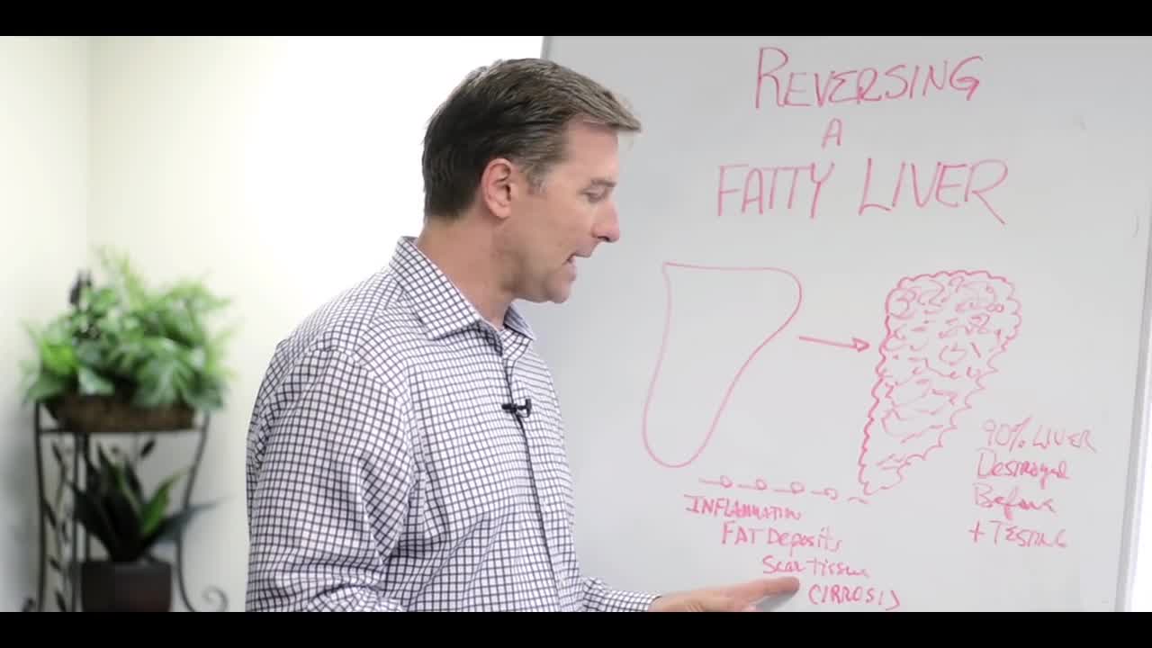
explains about fatty liver symptoms and fatty liver treatment. watch to learn more
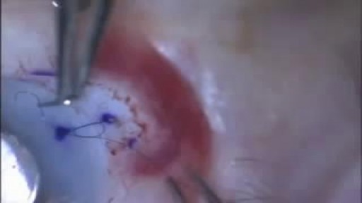
Video demonstrates the fundamental components of placing your first suture.

Reactive arthritis is a painful form of inflammatory arthritis (joint disease due to inflammation). It occurs in reaction to an infection by certain bacteria. Most often, these bacteria are in the genitals (Chlamydia trachomatis) or the bowel (Campylobacter, Salmonella, Shigella and Yersinia). Chlamydia most often transmits by sex. It often has no symptoms, but can cause a pus-like or watery discharge from the genitals. The bowel bacteria can cause diarrhea. If you develop arthritis within one month of diarrhea or a genital infection – especially with a discharge – see a health care provider. You may have reactive arthritis. - See more at: https://www.rheumatology.org/i-am-a/patient-caregiver/diseases-conditions/reactive-arthritis#sthash.PukaaQhj.dpuf
mply put, relapses, also known as flare ups, or (MS) attacks are new or worsening MS symptoms. But there is a concrete definition used by healthcare providers to identify MS attacks. To be considered an MS relapse: Old symptoms of MS must have become worse or new symptoms appeared.
What is peripheral neuropathy? Your peripheral nervous system connects the nerves from your brain and spinal cord, or central nervous system, to the rest of your body. This includes your: arms hands feet legs internal organs mouth face The job of these nerves is to deliver signals about physical sensations back to your brain.

Otitis media with effusion is inflammation and fluid buildup (effusion) in the middle ear without bacterial or viral infection. This may occur because the fluid buildup persists after an ear infection has resolved. It may also occur because of some dysfunction or noninfectious blockage of the eustachian tubes
